
Colon Cancer and Its Surgical Treatment: An Evidence-Based Review (Part 3)
Molecular classification and MMR/MSI testing, stage-specific adjuvant decisions and ctDNA, surgery for liver, lung and peritoneal metastases, a critical appraisal of HIPEC, where immunotherapy and targeted therapy meet the surgeon, recurrence and surveillance, Lynch syndrome and early-onset disease: what the evidence says today.
- Colon Cancer
- Surgical Oncology
- Liver Metastases
- Evidence-Based Medicine

Executive Summary
Part 1 of this series covered the elective resection and its quality standards; Part 2 covered the operation under pressure, from obstruction and perforation through the anastomosis, stomas and T4b disease to recovery and survivorship. This part covers the decisions that surround the operation and are increasingly made before it: molecular biology, staging and prehabilitation, stage-specific adjuvant therapy and circulating tumour DNA (ctDNA), the surgery of liver, lung and peritoneal metastases including cytoreduction and hyperthermic intraperitoneal chemotherapy (HIPEC), systemic therapy as the surgeon must understand it, quality metrics, recurrence and surveillance, and the older, the young, the hereditary and the pregnant patient. Roughly one patient in four develops liver metastases within five years of diagnosis [1], one in twelve peritoneal metastases [2], and one in three with stage III disease recurs within five years of resection [3]; the evidence reviewed here decides how many of them can still be cured.
Key evidence-based conclusions
- Mismatch repair (MMR) status must be known before the operation is booked. Universal testing finds Lynch syndrome in one patient in 35 [4]; fluoropyrimidine alone gives no benefit in deficient-MMR (dMMR) tumours (HR 1.10) and was associated with worse survival in stage II (HR 2.95)[5]; neoadjuvant ipilimumab–nivolumab produced a pathological complete response in 68% of locally advanced dMMR tumours [6]; adjuvant atezolizumab with FOLFOX raised three-year disease-free survival in stage III dMMR disease from 76.2% to 86.3% (HR 0.50)[7].
- Prehabilitation reliably improves fitness; its effect on complications is not secure. The Cochrane review found a 25 m gain in six-minute walk distance (moderate certainty) and no demonstrable effect on complications (RR 0.95, low certainty)[8]; PREHAB reduced severe complications from 29.7% to 17.1%[9], whereas prehabilitation did not reduce complications in frail patients with a mean age of 78 [10].
- In stage III disease, duration is decided by risk and regimen. In IDEA (12,834 patients), 3 months of CAPOX was non-inferior to 6 (HR 0.95) and sufficient for T1–3 N1 disease (83.1% vs 83.3%), whereas T4 or N2 disease did better with 6 months (62.7% vs 64.4%)[11]; the longer course more than doubled grade 2 or worse neuropathy (58% vs 25%)[12].
- ctDNA has so far improved only the decision not to treat. DYNAMIC halved chemotherapy in stage II (15% vs 28%) with identical five-year recurrence-free survival (88% vs 87%)[13,14]; in stage III neither ctDNA-guided de-escalation nor escalation met its endpoint [15], and trifluridine–tipiracil for ctDNA-positive patients failed in ALTAIR (HR 0.79)[16].
- Liver resection remains the reference and its boundaries have moved. Benchmark five-year survival is 45–58% for a solitary metastasis and at least 28% for more than three [17]; parenchymal-sparing resection matches extended hepatectomy with fewer complications [18]; thermal ablation was non-inferior to resection for lesions of 3 cm or less, with adverse events in 19% versus 46%[19]; transplantation for selected permanently unresectable disease gave five-year survival of 56.6% versus 12.6%[20].
- Lung metastasectomy rests on selection more than on proof. In the only randomised trial, five-year survival was 38% after metastasectomy and 29% with active monitoring (HR 0.82, 95% CI 0.43–1.56), not the under 5% historically assumed [21,22]; the observational advantage is largely selection [23].
- Cytoreduction is the active component of peritoneal surgery; HIPEC is not yet proven. Both arms of PRODIGE 7 reached a median survival of about 41 months after complete cytoreduction, and oxaliplatin HIPEC added 60-day morbidity (26% vs 15%) without survival (HR 1.00)[24]; prophylactic and adjuvant oxaliplatin HIPEC failed [25,26]; mitomycin C HIPEC improved locoregional control after a T4 primary (HR 0.21) without survival [27].
- The asymptomatic primary stays in place and surveillance should be targeted. Three randomised trials found no survival benefit from upfront resection of an asymptomatic primary with unresectable metastases, and one found 60-day mortality of 11% versus 3%[28–30]; intensive follow-up doubles salvage surgery without improving survival (HR 0.91)[31].
Abstract
Background. Colon cancer is now treated by molecular subtype and stage as much as by anatomy. Mismatch repair status decides whether chemotherapy, immunotherapy or neither follows resection; ctDNA measures residual disease more precisely than pathology; and the surgery of liver, lung and peritoneal metastases is now defined by randomised trials whose results overturn long-held assumptions. Recommendations on adjuvant duration, ctDNA, HIPEC and metastasectomy differ between societies.
Objective. To synthesise current evidence on the molecular classification of colon cancer and its surgical consequences, preoperative evaluation and prehabilitation, stage-specific adjuvant and neoadjuvant therapy, the management of liver, lung and peritoneal metastases including cytoreduction and HIPEC, systemic therapy as it affects surgical timing, quality metrics, recurrence and surveillance, and the management of older, early-onset, hereditary and pregnant patients.
Methods. Structured searches of PubMed/MEDLINE, Europe PMC, the Cochrane Library, publisher databases, guideline repositories and ClinicalTrials.gov through 27 October 2026, prioritising guidelines, systematic reviews, randomised trials and national registry cohorts published since 2016, plus landmark earlier studies. Quantitative claims were checked against the original abstracts or full texts.
Results. MMR testing identifies Lynch syndrome in 2.8% of patients [4], excludes fluoropyrimidine monotherapy in stage II [5] and defines eligibility for checkpoint inhibition, which produced a pathological complete response in 68% of locally advanced tumours [6] and a ten-point gain in three-year disease-free survival in stage III [7]. Three months of CAPOX suffices for T1–3 N1 stage III disease [11]. ctDNA-guided management halved chemotherapy in stage II without loss of recurrence-free survival [13], but neither de-escalation nor escalation guided by it succeeded in stage III [15]. Liver resection achieves benchmark five-year survival of 28–58% according to burden [17]; thermal ablation is non-inferior for small lesions [19]. Complete cytoreduction yields a median survival of about 40 months; oxaliplatin HIPEC adds morbidity without survival [24]. Upfront resection of an asymptomatic primary does not prolong life [30], and intensive surveillance doubles salvage surgery without a survival gain [31].
Conclusions. The molecular profile should be known from the biopsy, because it changes the first treatment step. Adjuvant therapy is shortest where risk is lowest and is intensified by biology rather than by stage alone. Metastasectomy should be planned against published benchmarks, with parenchymal sparing, ablation for small lesions and a strict limit on preoperative chemotherapy. Cytoreduction belongs in expert centres; HIPEC belongs in trials unless national policy has chosen otherwise. The main evidence gaps are the treatment of ctDNA-positive disease, perioperative versus adjuvant immunotherapy in dMMR disease, the long-term survival of ablation against resection, and the efficacy of mitomycin C HIPEC.
Methods
Design. Structured narrative review with quantitative synthesis of published pooled estimates. We did not perform a de novo PRISMA 2020 systematic review, risk-of-bias assessment of every primary study, or new meta-analysis; GRADE ratings are quoted from the source reviews where available, and otherwise given as the authors' judgement and labelled as such.
Sources and dates. PubMed/MEDLINE listings, Europe PMC, the Cochrane Library, publisher sites, the guideline repositories of NCCN, ESMO, ASCO, ASCRS, ESCP, PSOGI, EHTG, JSCCR and the Dutch Richtlijnendatabase, and ClinicalTrials.gov. Search terms included consensus molecular subtypes, mismatch repair deficiency, BRAF V600E, sidedness, malignant polyp, adjuvant chemotherapy duration, circulating tumour DNA, minimal residual disease, prehabilitation, frailty, germline testing, colorectal liver metastases, future liver remnant, parenchymal-sparing hepatectomy, thermal ablation, liver transplantation, disappearing liver metastases, pulmonary metastasectomy, peritoneal metastases, cytoreductive surgery, HIPEC, PIPAC, primary tumour resection asymptomatic, conversion therapy, surveillance colorectal cancer, early-onset colorectal cancer, Lynch syndrome colectomy and colorectal cancer in pregnancy, together with the acronyms of the trials named in this review. Last search: 27 October 2026.
Inclusion. Clinical practice guidelines and consensus statements; systematic reviews and meta-analyses; randomised trials; national audits, registry and pooled-database cohorts; prospective multicentre cohorts with defined outcomes; and, for molecular questions, large biomarker series. Priority was given to 2016–2026 publications, with landmark earlier trials (MOSAIC, QUASAR, EORTC 40983, the Dutch cytoreduction trial, CLOCC, SECA) retained where no newer evidence supersedes them. Exclusion. Single-centre retrospective series without comparison groups unless no better evidence existed, case reports, abstracts without extractable data, and studies whose numbers could not be traced to the source. Two conference reports are cited and flagged as such (the colorectal cohort of ICARuS; the closure of COBRA).
Verification and limitations. Each quantitative claim was checked against the source abstract or, where openly accessible, the full text, and the bibliographic record was confirmed through Europe PMC, PubMed or the publisher's DOI landing page; where a figure rests on the record alone or on a secondary source, the text says so. Several guidelines could not be read in full: the NCCN colon cancer guideline is cited from its journal record, with surveillance intervals and the ctDNA statement taken from the guideline text without line-by-line re-verification; the ESMO 2023 metastatic guideline is paywalled and its wording on cytoreduction and HIPEC was verified in the open-access Pan-Asian adaptation; the ESMO 2020 and hereditary cancer guidelines, the ASCRS guidelines, the US Multi-Society Task Force polyp recommendations and the Society of Thoracic Surgeons consensus are cited from records without abstracts, with positions summarised rather than quoted. Appendix A lists all such limitations.
Definitions and Terminology
| Term | Definition used in this review |
|---|---|
| Chromosomal instability (CIN) | Aneuploidy, loss of heterozygosity and mutations in APC, KRAS, TP53 and SMAD4; the commonest pattern of genomic instability in colon cancer [32]. |
| Microsatellite instability (MSI); dMMR and pMMR | Length changes in repetitive DNA caused by loss of mismatch repair (MMR), through germline mutation (Lynch syndrome) or, more often, somatic MLH1 promoter methylation. Deficient (dMMR) and proficient (pMMR) status is assigned by immunohistochemistry for MLH1, MSH2, MSH6 and PMS2, or by PCR or sequencing [33]. |
| CpG island methylator phenotype (CIMP) | Widespread promoter hypermethylation; encompasses almost all BRAF-mutant tumours and is the mechanism of sporadic MMR deficiency [34]. |
| Consensus molecular subtypes (CMS1–4) | Four transcriptional classes (MSI-immune, canonical, metabolic, mesenchymal); prognostic, but not yet treatment-directing outside trials [35]. |
| Circulating tumour DNA (ctDNA); molecular residual disease (MRD) | Tumour-derived DNA fragments detected in plasma. A positive result after resection, with no disease on imaging, defines MRD. Tumour-informed assays track a personalised mutation panel; tumour-naive assays use a fixed panel [36]. |
| Future liver remnant (FLR) | The liver volume remaining after the planned resection, standardised to body size. A remnant of 20% or less in a normal liver predicts postoperative liver insufficiency; about 30% is required after prolonged chemotherapy [37,38]. |
| Parenchymal-sparing hepatectomy | Non-anatomical resection removing the metastasis with a margin while preserving as much functioning liver as possible, in preference to anatomical or extended hepatectomy; it preserves the option of repeat resection [18,39]. |
| R1 vascular resection | Detachment of a metastasis from a major intrahepatic vessel with no parenchymal margin; distinguished from a parenchymal R1 margin, which carries a higher local recurrence rate [40]. |
| Two-stage hepatectomy; ALPPS | Two-stage hepatectomy: clearance of the future remnant, portal vein embolisation, then major resection after hypertrophy. ALPPS: portal vein ligation combined with parenchymal transection to force rapid hypertrophy before a second-stage resection [41,42]. |
| Peritoneal Cancer Index (PCI) | Sum of lesion-size scores (0 to 3) in 13 abdominopelvic and small-bowel regions, range 0–39 [43]. |
| Completeness of cytoreduction (CC) score | Residual disease after cytoreduction: CC-0 none visible; CC-1 under 2.5 mm; CC-2 2.5 mm to 2.5 cm; CC-3 larger. CC-0/1 is counted as complete [43]. |
| Cytoreductive surgery (CRS); HIPEC | CRS: peritonectomy and visceral resection to remove all macroscopic peritoneal disease. HIPEC: heated intraperitoneal chemotherapy delivered in theatre after cytoreduction, typically mitomycin C over 90 minutes or oxaliplatin over 30 minutes [44]. |
| PIPAC | Pressurised intraperitoneal aerosol chemotherapy: low-dose aerosolised drug delivered laparoscopically and repeatedly to unresectable peritoneal disease [45]. |
| Conversion therapy | Systemic therapy given to initially unresectable metastatic disease with the explicit aim of rendering it resectable [46]. |
| Oligometastatic disease | Metastatic disease limited enough in number and site for local treatment (surgery, ablation or stereotactic radiotherapy) to be chosen from a "toolbox" with curative or long-term disease-control intent [47]. |
| Early-onset colorectal cancer | Colorectal cancer diagnosed before the age of 50 [48]. |
Scope and Relationship to Parts 1 and 2
Part 1 of this series dealt with the planned operation in a fit patient: the planes of the mesocolon, complete mesocolic excision with central vascular ligation, the extent of lymphadenectomy, the choice of access, and the quality markers that define an adequate resection. Part 2 dealt with the operation that cannot follow the standard pathway: obstruction and perforation, the choice of emergency procedure, the anastomosis and its leakage, stomas, T4b disease, enhanced recovery, complications and survivorship. Both are assumed here; where this part refers to "a formal oncological resection" it means the standard of Part 1, and where it refers to obstruction, perforation or the randomised evidence on surveillance intensity it means the material of Part 2 and does not repeat it.
Section 6 covers molecular classification and the alterations that change surgical planning; Section 7 staging, frailty, prehabilitation, modifiable risks and genetic counselling; Section 8 the adjuvant decision from the malignant polyp through stage II and III disease to ctDNA-guided and immunotherapy-based strategies. Sections 9 and 10 address liver and lung metastases, including resectability, the remnant, sequencing, ablation, conversion and transplantation; Sections 11 and 12 peritoneal metastases, selection, cytoreduction and a critical appraisal of HIPEC; Section 13 systemic therapy as the surgeon needs it, including conversion, chemotherapy-associated liver injury, surgery after immunotherapy and the asymptomatic primary; Section 14 pathology, quality metrics and ctDNA as a surgical tool; Section 15 recurrence, salvage surgery and surveillance; and Section 16 older and frail patients, early-onset disease, hereditary syndromes and pregnancy.
Part 4 will conclude the series with emerging technologies, tabulated summaries of the landmark trials cited across all four parts, decision algorithms, a comparative treatment matrix, graded recommendations, a structured list of research gaps and a glossary. Topics introduced here and developed there include ctDNA in surveillance and metastasectomy decisions, organ preservation after neoadjuvant immunotherapy, and the place of mitomycin C HIPEC once GECOP-MMC reports.
Molecular Pathogenesis and Classification
Two routes to carcinoma
The classical model describes stepwise accumulation of genetic events, loss of APC in a normal crypt, activating KRAS mutation in the adenoma, and loss of TP53 and chromosome 18q genes at the transition to carcinoma, with accumulation mattering more than order [49]; this adenoma–carcinoma sequence still accounts for most colon cancers. The serrated pathway is estimated to produce 15–30% of colorectal cancers [50]. Its precursors are sessile serrated lesions and traditional serrated adenomas, its signature is BRAF (less often KRAS) mutation with widespread CpG island methylation, and its site is predominantly proximal [51]. Serrated-pathway cancers are the source of most sporadic mismatch-repair-deficient tumours, which is why molecular and anatomical variables are inseparable in this disease.
Three forms of genomic instability
Chromosomal instability (CIN), the commonest pattern, comprises aneuploidy, loss of heterozygosity and mutations in APC, KRAS, TP53 and SMAD4 [32]. Microsatellite instability (MSI) results from loss of DNA mismatch repair (MMR), through germline mutation (Lynch syndrome) or, far more often, somatic hypermethylation of the MLH1 promoter. The CpG island methylator phenotype (CIMP) encompasses almost all BRAF-mutant tumours (odds ratio 203) and is the mechanism of sporadic MMR deficiency [34]. In The Cancer Genome Atlas, 16% of 276 tumours were hypermutated, three-quarters of them MSI-high with MLH1 silencing and one-quarter carrying somatic MMR or polymerase ε (POLE) mutations [52]. The categories overlap: CIMP-high tumours may be MSI-high or microsatellite-stable, and the latter do badly. Across 33 studies (10,635 patients), CIMP was associated with shorter disease-free (HR 1.45, 95% CI 1.07–1.97) and overall survival (HR 1.43, 1.18–1.73), persisting in microsatellite-stable tumours (OS HR 1.37, 1.12–1.68)[53].
Consensus molecular subtypes
Six expression classifiers were reconciled into four consensus molecular subtypes: CMS1 (MSI-immune, 14%), hypermutated and immune-infiltrated; CMS2 (canonical, 37%), epithelial with WNT and MYC activation; CMS3 (metabolic, 13%); and CMS4 (mesenchymal, 23%), with transforming growth factor-β activation, stromal invasion and angiogenesis; 13% were mixed [35]. CMS4 tumours had the worst overall and relapse-free survival; CMS1 tumours rarely relapsed but did very poorly when they did [35,54]. CMS has shaped trial design but does not direct treatment outside trials: assignment is sensitive to tissue quality and stromal content, and no randomised study has shown benefit from CMS-directed therapy. Its value is conceptual: a right-sided BRAF-mutant dMMR tumour and a left-sided APC/TP53-mutant chromosomally unstable tumour are different diseases sharing a histological label.
Actionable alterations and their surgical relevance
Mismatch repair status. MMR status is the one molecular result every surgeon should know before planning treatment. Immunohistochemistry for MLH1, MSH2, MSH6 and PMS2 is the first-line test, PCR or sequencing the second [33], and testing of every newly diagnosed colorectal cancer is recommended by the US Multi-Society Task Force [55], NCCN [56] and ESMO [57]. It screens for Lynch syndrome: among 1,566 unselected patients, 2.8% (95% CI 2.1–3.8), one in 35, had Lynch syndrome, and restricting testing to revised Bethesda criteria would have missed 28%[4]. It is prognostic: across 32 studies, MSI carried a pooled hazard ratio for death of 0.65 (0.59–0.71)[58]. It predicts absence of benefit from fluoropyrimidine alone: adjuvant 5-FU improved disease-free survival in MMR-proficient (HR 0.67, 0.48–0.93) but not dMMR tumours (HR 1.10, 0.42–2.91), and in stage II dMMR disease was associated with worse overall survival (HR 2.95, 1.02–8.54)[5,59]. And it defines eligibility for checkpoint inhibition: first-line pembrolizumab in MSI-high metastatic disease doubled progression-free survival in KEYNOTE-177 (16.5 vs 8.2 months; HR 0.60, 0.45–0.80) with fewer severe adverse events [60], although final overall survival did not reach formal significance after 60% crossover (HR 0.74, 0.53–1.03)[61]; adjuvant atezolizumab with mFOLFOX6 raised three-year disease-free survival in stage III dMMR colon cancer from 76.2% to 86.3% (HR 0.50, 0.35–0.73) in ATOMIC [7]; and neoadjuvant nivolumab plus ipilimumab in NICHE-2 produced a major pathological response in 95% and a complete response in 68% of 115 locally advanced dMMR tumours, with no recurrence at a median of 26 months [6]. dMMR status should therefore be known before a bulky or cT4 tumour is booked for resection; FOxTROT found little benefit from preoperative chemotherapy in dMMR tumours [62]; later sections cover sequencing by stage.
RAS and BRAF. KRAS mutations occur in a median of 38% of colorectal cancers [63], NRAS in a few per cent more, and any RAS mutation abolishes anti-EGFR benefit: in PRIME, the 17% of KRAS exon 2 wild-type tumours with other RAS mutations gained nothing from panitumumab, whereas fully wild-type patients gained in progression-free (10.1 vs 7.9 months, HR 0.72) and overall survival (26.0 vs 20.2 months, HR 0.78)[64,65]. RAS mutation is modestly prognostic (OS HR 1.52, 1.26–1.84), BRAF V600E strongly so (OS HR 2.01, 1.49–2.71)[66]; BRAF mutation occurred in 8.2% of metastatic tumours, in 34.6% of dMMR versus 6.8% of pMMR cases, and drove the poor prognosis of dMMR metastatic disease [67]. Targeted therapy has altered this: encorafenib plus cetuximab improved overall survival from 5.9 to 9.3 months after prior therapy in BEACON (HR 0.61, 0.48–0.77)[68,69], and with mFOLFOX6 in the first line prolonged progression-free survival from 7.1 to 12.8 months (HR 0.53, 0.41–0.68) in BREAKWATER [70]. In localised disease, in a dMMR tumour with MLH1 loss, BRAF V600E or MLH1 methylation indicates sporadic disease and makes Lynch syndrome unlikely [55].
HER2, NTRK, KRAS G12C and POLE. HER2 amplification occurs in 2–3% of colorectal cancers, almost always RAS/BRAF wild-type and left-sided [71–73]; in refractory HER2-positive RAS wild-type disease, tucatinib plus trastuzumab produced responses in 38.1% (MOUNTAINEER)[74] and trastuzumab deruxtecan in 45.3%, with interstitial lung disease in 6% including two deaths (DESTINY-CRC01)[71]. Kinase fusions are rare (0.9%) but concentrated in MSI-high tumours (5% vs 0.4%) and especially MLH1-methylated RAS/BRAF wild-type cancers (42%)[75]; larotrectinib produced responses in 75% of TRK fusion-positive tumours [76]. KRAS G12C has a median prevalence of 3.1%[63]; adagrasib with cetuximab gave a 46% response rate [77], and sotorasib with panitumumab prolonged progression-free survival from 2.2 to 5.6 months (HR 0.49, 0.30–0.80)[78]. Pathogenic POLE proofreading mutations occurred in 1.0% of 6,517 cancers, were mutually exclusive with dMMR, and carried a comparable cytotoxic T-cell infiltrate and a recurrence hazard ratio of 0.34 (0.11–0.76)[79].
| Alteration | Frequency | Prognosis | Consequence | Key evidence |
|---|---|---|---|---|
| dMMR / MSI-high | 15% stage II–III; 5% metastatic | Better in stage II–III (OS HR 0.65) | Lynch screening; no 5-FU alone in stage II; checkpoint inhibition | [5–7,58,60] |
| BRAF V600E | 8% metastatic; 35% of dMMR | Strongly adverse (OS HR 2.01) | Encorafenib + cetuximab; marks sporadic dMMR | [66–68,70] |
| KRAS / NRAS | ~40–50% | Modestly adverse (OS HR 1.52) | No anti-EGFR antibody | [63–65] |
| RAS/BRAF wild-type, left-sided | ~35% metastatic | Best group | Anti-EGFR first line; more conversions | [80,81] |
| HER2 amplification | 2–3% | Not clearly prognostic | Tucatinib + trastuzumab or trastuzumab deruxtecan | [71,74] |
| NTRK / kinase fusion | 0.9%; 5% of MSI-high | Uncertain | TRK inhibitor; test MLH1-methylated RAS/BRAF wild-type | [75,76] |
| KRAS G12C | 3% | Possibly adverse | G12C inhibitor + anti-EGFR after chemotherapy | [63,77,78] |
| POLE proofreading | 1% | Favourable (recurrence HR 0.34) | Immunogenic despite pMMR | [79] |
Sidedness as a biological variable
Right-sided (caecum to splenic flexure) and left-sided tumours differ in embryology, microbiome and molecular profile. In PETACC-3 (3,045 patients), proximal tumours were more often mucinous, MSI-high, BRAF-mutant and serrated-signature, distal tumours more often chromosomally unstable and EGFR- or HER2-amplified; relapse risk did not differ, but survival after relapse was much worse for proximal stage III tumours (HR 1.95, 1.6–2.4)[72]. Across 66 studies and 1,437,846 patients, left-sided location carried a lower risk of death in all stages (HR 0.82, 0.79–0.84)[82]. Side is also predictive: across six randomised trials, anti-EGFR therapy improved overall survival in left-sided (HR 0.75, 0.67–0.84) but not right-sided RAS wild-type tumours (HR 1.12, 0.87–1.45)[80], and PARADIGM confirmed prospectively that panitumumab beat bevacizumab in left-sided disease (median OS 37.9 vs 34.3 months, HR 0.82; curative resection 18.3% vs 11.6%)[81]. The likelihood and timing of metastasectomy therefore depend on a biological assignment made at diagnosis.
The mandatory testing panel
NCCN recommends MMR or MSI testing for all newly diagnosed colon cancers and, in metastatic disease, extended RAS (KRAS and NRAS exons 2–4), BRAF V600E and HER2 testing, with NTRK fusion testing where other drivers are absent [56]. ESMO's metastatic guideline makes the same core requirement for RAS, BRAF V600E and MMR/MSI status at diagnosis of metastatic disease [47], and its localised guideline requires MMR status in all stage II tumours [57]. Testing belongs on the diagnostic biopsy (Figure), because the result may change the first treatment step.
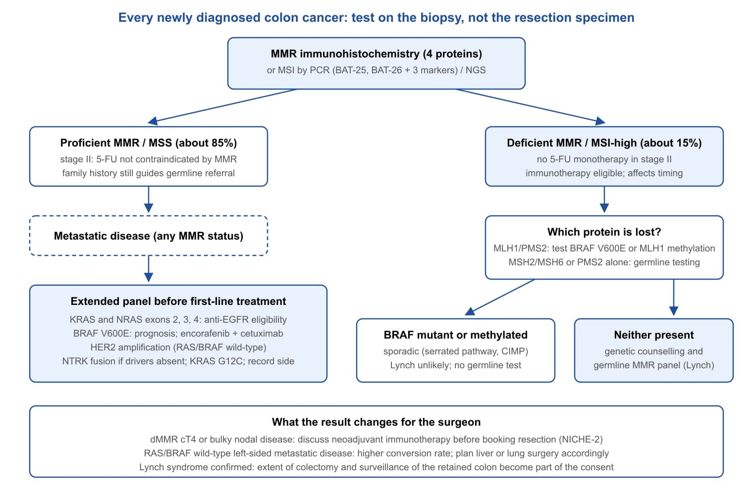
Molecular results to know before the operation is booked
- MMR/MSI status of every colon cancer, from the biopsy: it identifies Lynch syndrome (1 in 35), excludes fluoropyrimidine monotherapy in stage II and opens checkpoint inhibition.
- In a dMMR tumour with MLH1 loss, BRAF V600E or MLH1 methylation separates sporadic from hereditary disease before germline testing.
- In metastatic disease, extended RAS, BRAF V600E, HER2 and primary side decide the first-line regimen and so the timing of metastasectomy.
- CMS and CIMP explain prognosis but do not yet direct treatment.
Preoperative Evaluation, Staging, Prehabilitation and Genetic Testing
Staging investigations
Synchronous cancers occur in 3.9% of patients, a third of them in a different surgical segment [83], so the proximal colon must be examined before resection whenever possible: colonoscopy to the caecum, or CT colonography when the tumour is impassable (per-patient sensitivity for cancer 96.1%, 95% CI 93.8–97.7, comparable to colonoscopy at 94.7%)[84]; otherwise within three to six months of surgery.
Contrast-enhanced CT of the chest, abdomen and pelvis is the universal staging investigation, and its limits must be understood. Pooled sensitivity for invasion beyond the muscularis propria is 90% (95% CI 83–95) with specificity 69% (62–75); for deep invasion of 5 mm or more, 77% and 70%; for nodal involvement, 71% (59–81) and 67% (46–83)[85]. Clinical N-staging is little better than chance and should not decide the operation; clinical T4 staging is more reliable but carries a false-positive rate of roughly one in four for deep invasion, although FOxTROT found pT3 or deeper tumours in 98% of those going straight to surgery [86]. Preoperative CEA is prognostic (elevated: mortality HR 1.60, 1.46–1.76)[87], but its larger value is as a baseline: an elevation that normalises after resection carries no excess risk (HR 0.77, 0.45–1.30), whereas a persistently raised postoperative value doubles the recurrence hazard (HR 2.0, 1.1–3.5)[88].
Liver MRI and PET-CT are selective additions. Per-lesion sensitivity for liver metastases is 74.4% for CT, 80.3% for MRI and 81.4% for FDG-PET, with MRI superior below 10 mm [89], and gadoxetic-acid MRI is more sensitive again than CT (ratio 1.29, 1.18–1.40)[90]; MRI is indicated for indeterminate lesions, planned liver resection or a steatotic liver, not after a normal CT. In PETCAM, PET-CT before liver resection changed management in only 8.0% (5.0–11.9) of 263 patients and did not alter survival (HR 0.86, 0.60–1.21)[91]; it is reserved for equivocal lesions, suspected extrahepatic disease and a rising CEA with negative CT.
Cardiopulmonary assessment, risk scores and frailty
In 2.3 million NSQIP cases, ASA class independently predicted complications and death, with odds ratios rising from 2.05 at ASA 2 to 63.25 at ASA 5 [92]. The ACS NSQIP calculator gives patient-specific estimates from 21 variables (C-statistic 0.944 for mortality, 0.816 for morbidity)[93]. Cardiopulmonary exercise testing measures reserve directly: in 703 patients undergoing major colorectal surgery, an anaerobic threshold of 11.1 mL/kg/min or less carried an odds ratio for in-hospital morbidity of 7.56 (4.44–12.86; AUC 0.79)[94], and across 37 studies of intra-abdominal surgery the prognostic cut-point lay at 9–11 mL/kg/min [95]. CPET earns its place when fitness is uncertain and the result would change the plan.
Frailty predicts outcome independently of comorbidity. On the Clinical Frailty Scale, each one-category increase raised medium-term mortality by 21.2%[96]. The five-item modified frailty index correlates above 0.9 with the 11-item original and is calculable from routine data [97]; in 295,490 NSQIP colorectal resections, the 18% scoring 2 or more had adjusted odds of 2.00 (1.87–2.14) for death and 2.80 for discharge to a facility [98]. Pooled prevalence of frailty across 16 studies of colorectal cancer surgery was 31% (20–42)[99], and in older patients every study in a systematic review found more severe complications, longer stays and worse survival among the frail [100]. Frailty is not a contraindication: in GOSAFE (942 patients aged 70 or over, 68% colorectal), 30-, 90- and 180-day mortality were 3.7%, 6.3% and 9%, and quality of life was preserved at three months and improved at six [101]. The score should open a conversation about goals, not close one.
Prehabilitation: what the trials show
Prehabilitation has an attractive rationale and a mixed evidence base. The first colorectal cancer trial (77 patients) found that a trimodal programme started four weeks before surgery improved six-minute walk distance at eight weeks by 45.4 m (13.9–77.0) without changing complications or stay [102]. The 2022 Cochrane review of three such trials (250 participants) confirmed a clinically relevant preoperative gain of 24.91 m (11.24–38.57; moderate certainty) and found no demonstrable effect on complications (RR 0.95, 0.70–1.29; low certainty)[8].
The larger trials diverged. In frail patients (Fried index 2 or more, mean age 78), four weeks of multimodal prehabilitation within ERAS produced no difference in the 30-day Comprehensive Complication Index (adjusted difference −3.2, −11.8 to 5.3)[10], whereas a Dutch trial selecting 57 patients aged 60 or over by low aerobic fitness found that three weeks of community exercise reduced complications from 72.4% to 42.9% (RR 0.59, 0.37–0.96)[103]. PREHAB, stopped early by the pandemic at 251 patients, compared four weeks of supervised high-intensity exercise, nutrition, psychological support and smoking cessation with standard care, both within ERAS: severe complications fell from 29.7% to 17.1% (OR 0.47, 0.26–0.87), whereas six-minute walk distance four weeks after surgery, the co-primary endpoint, did not differ (15.6 m, −1.4 to 32.6)[9]. A 2025 network meta-analysis of 186 trials (15,684 participants) across major surgery found exercise alone (OR 0.50, 0.39–0.64), nutrition alone (OR 0.62, 0.50–0.77) and combined exercise, nutrition and psychosocial support (OR 0.64, 0.45–0.92) most likely to reduce complications, all at very low certainty [104].
Three conclusions follow (authors' judgement). Prehabilitation reliably improves preoperative function. Its effect on complications is plausible but not secure: the positive trials selected unfit patients or used supervised high-intensity programmes, and the negative trial in frail octogenarians suggests four weeks cannot reverse established frailty. Multimodal programmes are not demonstrably better than well-delivered exercise or nutrition alone, so the component that can actually be delivered should be chosen, without delaying resection beyond the three to four weeks the trials used.
| Study or tool | Population | Measure or intervention | Main result |
|---|---|---|---|
| Clinical Frailty Scale [96] | 2,305 older adults | Clinical judgement scale | Each category raised mortality by 21% |
| mFI-5 in NSQIP [98] | 295,490 colorectal resections | Score ≥2 (18%) | Death OR 2.00; non-home discharge OR 2.80 |
| CPET [94] | 703 major colorectal | Anaerobic threshold ≤11.1 mL/kg/min | Morbidity OR 7.56; AUC 0.79 |
| Gillis 2014 [102] | 77 colorectal cancer | Trimodal, 4 weeks pre vs post | 6MWD +45 m; complications unchanged |
| Carli 2020 [10] | 110 frail, mean age 78 | Multimodal, 4 weeks | CCI difference −3.2 (−11.8 to 5.3) |
| Berkel 2022 [103] | 57 unfit, age ≥60 | Community exercise, 3 weeks | Complications 42.9% vs 72.4% (RR 0.59) |
| PREHAB 2023 [9] | 251 non-metastatic CRC | Supervised multimodal, 4 weeks | Severe complications 17.1% vs 29.7% (OR 0.47) |
| Cochrane 2022 [8] | 3 RCTs, 250 | Multimodal vs none | Preop 6MWD +25 m; complications RR 0.95 |
| Network meta-analysis 2025 [104] | 186 RCTs, 15,684 | Components compared | Exercise OR 0.50; nutrition OR 0.62; trimodal OR 0.64 |
Nutrition, anaemia, smoking and alcohol
Malnutrition is a risk factor for complications; ESPEN recommends screening every patient, starting nutritional therapy as soon as risk is identified and, at severe risk, giving support before surgery even at the cost of delay [105].
About a third of patients with operable colorectal cancer are anaemic, mostly from iron deficiency [106], and transfusion is among the strongest operative associations with anastomotic leak (Part 2 of this series); consensus guidance is to identify anaemia at the first visit and treat iron deficiency, intravenously when time is short [107]. The trials temper expectations. In IVICA, intravenous iron raised haemoglobin more than oral iron (median 1.55 vs 0.50 g/dL) but did not reduce transfusion [108]; in FIT, haemoglobin had normalised by admission in only 17% after intravenous and 16% after oral iron, although by 30 days the intravenous group led (60% vs 21%) and only intravenous iron restored stores [106]; and in PREVENTT, 1,000 mg of intravenous iron 10–42 days before major open abdominal surgery did not reduce death or transfusion (29% vs 28%, RR 1.03, 0.78–1.37)[109]. Intravenous iron corrects iron deficiency but needs more time than most cancer pathways allow to correct anaemia; give it at diagnosis and do not expect it to prevent transfusion.
Six randomised trials of preoperative smoking cessation showed a 41% relative reduction in complications (95% CI 15–59), each additional week of abstinence adding 19%[110]; intensive support with nicotine replacement begun four to eight weeks before surgery reduced any complication (RR 0.42, 0.27–0.65), whereas brief interventions did not [111]. Preoperative alcohol consumption was associated with more morbidity (RR 1.56, 1.31–1.87) and, at clearly high intake, mortality (RR 2.68, 1.50–4.78)[112]; two small trials of intensive cessation reduced complications (OR 0.22, 0.08–0.61)[113]. The interval before resection is short, but long enough for both to start.
Genetic counselling and germline testing
Tumour MMR testing screens for Lynch syndrome; germline testing diagnoses it. Established triggers are a dMMR tumour without BRAF mutation or MLH1 methylation, a family history meeting Amsterdam or revised Bethesda criteria, diagnosis before 50, adenomatous or MUTYH-associated polyposis, and a known familial variant [55,114]. These criteria miss many carriers. In 1,058 unselected patients, 9.9% (8.2–11.9) carried a pathogenic variant in a 25-gene panel: 3.1% had Lynch syndrome, but 7.0% carried non-Lynch variants, including 1.0% in BRCA1/2, and neither age nor family history predicted them [115]. Among 450 patients diagnosed before 50, 16% carried a variant and a third of carriers met no established criterion for the gene involved [116]. Universal testing with a panel of more than 80 genes found pathogenic variants in 15.5% of 361 unselected patients, 9.4% of which would have escaped guideline-based testing; treatment changed in 11%, but only 16% of eligible relatives took up cascade testing [117].
The debate over universal germline panels therefore concerns what follows a finding: for, the one in three carriers who meet no criteria and the therapeutic relevance of DNA-repair variants; against, variants of uncertain significance, counselling capacity and low cascade uptake. ESMO [114] and the US Multi-Society Task Force [55] still anchor referral on tumour MMR status, age, polyposis and family history. The surgical consequences of a positive result, the extent of colectomy in Lynch syndrome and polyposis, surveillance of the retained bowel, should be settled before the operation; a germline diagnosis made after a segmental resection is a lost opportunity.
The preoperative dataset for an elective colon cancer resection
- Colonoscopy to the caecum or CT colonography, CT chest–abdomen–pelvis and baseline CEA; liver MRI for indeterminate lesions or planned liver surgery; PET-CT only when it would change the decision to operate.
- MMR/MSI status on the biopsy, with BRAF V600E or MLH1 methylation reflex testing for MLH1-lost tumours; extended RAS, BRAF and HER2 in metastatic disease.
- ASA class, a NSQIP estimate and a frailty score in every patient; CPET when reserve is uncertain or the operation is extended.
- Malnutrition, anaemia, smoking and alcohol addressed from the first visit; three to four weeks of exercise-based prehabilitation for unfit patients.
- Genetic counselling for dMMR tumours without a sporadic explanation, diagnosis before 50, polyposis or a suggestive family history, with the result before the extent of resection is fixed.
Stage-Specific Management and Adjuvant Decisions
Resection quality (Part 1 of this series) determines whether a colon cancer can be cured locally; the decisions in this section determine what, if anything, should be added to a complete resection. The evidence is uneven. For stage III disease, pooled trials of more than 12,000 patients settle regimen and duration to within a few percentage points. For stage II disease the absolute benefit is small, the high-risk features were defined by consensus rather than by trial, and circulating tumour DNA (ctDNA) is being tested as a replacement. For the malignant polyp the question is whether an operation can safely be omitted. Mismatch repair (MMR) status, introduced in Section 6, runs through every tier.
Malignant polyps and pT1 cancer
A malignant polyp is an adenoma in which carcinoma has crossed the muscularis mucosae into the submucosa (pT1); once it is removed endoscopically the only remaining question is the probability of nodal metastasis. Two classifications describe depth. Haggitt's levels 1–4 apply to pedunculated lesions, with level 4 (invasion below the stalk) the adverse category: in the original series of 129 carcinomas arising in adenomas, 7 of 28 level 4 lesions had an adverse outcome, against 1 of 36 at lower levels [118]. Kikuchi's sm1–sm3 thirds apply to sessile lesions; in 182 early invasive cancers followed for at least five years, none of the 17 patients with recurrence or nodal metastasis had sm1 disease [119]. Because thirds cannot be measured when the muscularis propria is absent from an endoscopic specimen, Japanese practice substituted an absolute depth of 1,000 µm, and this threshold entered most guidelines.
Pooled estimates show that lymphovascular invasion, budding and poor differentiation predict nodal disease more strongly than depth. In 17 studies (3,621 patients), lymphatic invasion (RR 5.2, 95% CI 4.0–6.8), budding (RR 5.1, 3.6–7.3) and poor differentiation (RR 4.8, 3.3–6.9) carried relative risks similar to submucosal invasion of 1 mm or more (RR 5.2, 1.8–15.4), but with far narrower intervals [120]. In 16 studies (10,181 patients, 1,307 with nodal metastases), the pooled odds ratios were 7.42 for lymphovascular invasion, 4.00 for budding, 3.75 for high grade and 3.53 for invasion ≥1,000 µm [121]. The decisive analysis pooled 67 studies (21,238 patients): deep submucosal invasion was associated with nodal metastasis in univariable analysis (OR 2.58) but not after adjustment (OR 1.73, 95% CI 0.96–3.12), and when depth was the only adverse feature the absolute risk was 2.6%[122]. A meta-analysis of 60 studies found poorly differentiated clusters (OR 14.61) and budding (OR 4.99) more informative than any depth cut-off (OR 2.76 at 1,000 µm)[123]. Budding is graded by the three-tier International Tumor Budding Consensus Conference system [124].
| Feature | Threshold used | Pooled estimate | Source |
|---|---|---|---|
| Lymphovascular invasion | Present | RR 5.2 (4.0–6.8); OR 7.42 | [120,121] |
| Tumour budding | ITBCC Bd2–3 (high grade) | RR 5.1 (3.6–7.3); OR 4.00–4.99 | [120,121,123] |
| Poor differentiation / high grade | G3, signet ring, mucinous | RR 4.8 (3.3–6.9); OR 3.75 | [120,121] |
| Poorly differentiated clusters | Present | OR 14.61 | [123] |
| Submucosal depth | ≥1,000 µm or sm2–3 | OR 2.76–3.53 univariable; OR 1.73 (0.96–3.12) adjusted; 2.6% absolute risk when sole feature | [121–123] |
| Vertical margin | <1 mm or involved | Local-risk criterion in all guidelines; not a nodal predictor | [125,126] |
Guidelines agree on the features and differ on the arithmetic. The 2022 ESGE guideline defines a low-risk (curative) resection as en bloc R0, sm1, well or moderately differentiated, without lymphovascular invasion or Bd2–3 budding; deeper invasion, lymphovascular invasion, undifferentiated histology, high-grade budding or a positive vertical margin is high risk and prompts staging and multidisciplinary discussion rather than automatic colectomy, and a positive horizontal margin alone is a local-risk finding managed by surveillance or endoscopic re-treatment [125]. The JSCCR guideline recommends additional surgery when any one of depth ≥1,000 µm, vascular invasion, poor, signet-ring or mucinous histology, or Bd2–3 budding is present, or the vertical margin is positive [126]. The US Multi-Society Task Force uses the same unfavourable-histology list with a margin of at least 1 mm [127], as does the ASCRS guideline, which treats a single adverse feature as an indication for oncological resection in a fit patient [128]. A 2026 comparison notes that Eastern guidelines act on any single feature whereas Western guidelines increasingly weigh cumulative risk against patient factors [129]; since only about 10% of pT1 cancers harbour nodal metastases, a single-feature rule sends roughly nine patients to surgery for each one who needs it [130].
Two facts make a staged approach defensible. First, endoscopic resection followed by completion surgery does not compromise outcome: in a Dutch multicentre cohort of 602 high-risk pT1 cancers, nodal metastasis (OR 0.97, 95% CI 0.49–1.93) and recurrence (HR 0.97, 0.41–2.34; 5.6% overall) were identical whether surgery was primary or followed endoscopic resection [131]. A diagnostic en bloc resection therefore costs nothing oncologically, provided the specimen is intact and reported against all five features. Second, risk models outperform the single-feature rule. A nomogram built on 4,673 endoscopically resected pT1 cancers from 27 Japanese institutions combined six factors with a concordance index of 0.784 (development) and 0.790 (validation)[132], and a machine-learning model trained on 3,134 patients achieved an area under the curve of 0.83 in 939 external patients against 0.73 for guideline criteria (0.84 vs 0.77 after endoscopic resection)[133]. Neither has been tested prospectively as a decision tool (authors' judgement). Completion colectomy, when chosen, is a formal oncological resection with central ligation as described in Part 1.
Stage I
Stage I disease (T1–2 N0) is treated by surgery alone; no trial has tested adjuvant therapy, and the recurrence rate leaves no room to detect a benefit. In the Swedish national cohort of 14,325 patients with stage I–III colon cancer operated on between 2007 and 2012, the five-year recurrence rate was 5% for stage I, 12% for stage II and 33% for stage III [3]. ASCO and ESMO recommend no adjuvant chemotherapy for stage I [57,134]; follow-up was reviewed in Part 2 of this series. The practical risk in stage I is understaging from an inadequate lymph node yield (Part 1).
Stage II
The baseline benefit. QUASAR randomised 3,239 patients, 91% with stage II colorectal cancer, to fluorouracil and folinic acid or observation; at a median of 5.5 years the relative risk of death was 0.82 (95% CI 0.70–0.95), an absolute gain of about 3.6% (1.0–6.0) at five years if untreated mortality is 20%[135]. The Cochrane review of stage II trials found a disease-free survival benefit (RR 0.83, 0.75–0.92) without an overall survival benefit (RR 0.96, 0.88–1.05)[136]. These are the numbers a patient should be given.
High-risk features. Both ASCO (2022) and ESMO (2020) list the same features that should move the conversation towards chemotherapy: pT4, obstruction or perforation at presentation, fewer than 12 nodes examined, lymphovascular or perineural invasion, poorly differentiated histology and high-grade budding [57,134]. The features predict recurrence; no trial has shown that they predict benefit. ASCO recommends against routine chemotherapy without high-risk features, recommends offering it for pT4, and allows it for the remaining features after discussion of the size of the effect [134]. The limitation is visible in the Swedish data: pT3N0 patients with no or one risk factor had five-year recurrence of 9% and 11%[3], so a single conventional factor barely changes risk.
Oxaliplatin in stage II. MOSAIC enrolled 899 stage II patients (330 low risk, 569 high risk). At six years there was no overall survival difference in stage II [137], and the dedicated subgroup analysis found a non-significant disease-free survival trend in high-risk stage II (HR 0.72, 95% CI 0.51–1.01), a significant reduction in time to recurrence (HR 0.62, 0.41–0.92) and no effect on survival (HR 0.91, 0.61–1.36)[138]; at ten years the absence of a stage II survival benefit was confirmed [139]. In the IDEA collaboration's high-risk stage II analysis (3,273 patients, 62% CAPOX), five-year disease-free survival was 80.7% with 3 months and 83.9% with 6 months of doublet chemotherapy (HR 1.17, 80% CI 1.05–1.31), failing the non-inferiority margin of 1.2; 3 months of CAPOX was close to equivalent (HR 1.02, 0.88–1.17) whereas 3 months of FOLFOX was clearly inferior (HR 1.41, 1.18–1.68)[140]. ASCO therefore allows oxaliplatin for pT4 and other high-risk stage II disease, with 3 months of CAPOX or 6 months of FOLFOX as the durations [134].
MMR-deficient stage II. Deficient MMR is present in 10–15% of non-metastatic colon cancers [6] and 9.4% of MOSAIC specimens [139]. In the pooled analysis of 457 patients from randomised trials of fluorouracil versus surgery alone, adjuvant therapy improved disease-free survival in proficient-MMR tumours (HR 0.67, 95% CI 0.48–0.93) but not in deficient-MMR tumours (HR 1.10, 0.42–2.91), and in stage II dMMR disease treated patients had worse overall survival (HR 2.95, 1.02–8.54)[5]. Good prognosis plus no demonstrable benefit is why every guideline recommends against single-agent chemotherapy for stage II dMMR disease and why MMR testing must precede any adjuvant decision [57,134]. Whether oxaliplatin restores a benefit in high-risk dMMR stage II is unproven; MOSAIC supported FOLFOX in dMMR stage III, not stage II [139].
ctDNA-guided decisions. Post-operative ctDNA detects minimal residual disease with a precision pathology cannot match. In the first prospective stage II cohort, 14 of 178 patients managed without chemotherapy were ctDNA-positive after surgery and 11 (79%) recurred, against 16 of 164 (9.8%) ctDNA-negative patients (HR 18, 95% CI 7.9–40)[141]. DYNAMIC then randomised 455 stage II patients 2:1 to ctDNA-guided management (chemotherapy only if ctDNA was detectable at week 4 or 7) or standard management. Chemotherapy use fell from 28% to 15% (relative risk 1.82, 95% CI 1.25–2.65); two-year recurrence-free survival was 93.5% versus 92.4% (difference 1.1 points, 95% CI −4.1 to 6.2), within the −8.5 point non-inferiority margin; three-year recurrence-free survival was 86.4% in treated ctDNA-positive patients and 92.5% in untreated ctDNA-negative patients [13]. At 59.7 months, five-year recurrence-free survival was 88% versus 87% (difference 1.1, −5.8 to 8.0), overall survival 93.8% versus 93.3%, and 35 of 40 treated ctDNA-positive patients (87.5%) cleared ctDNA [14]. The trial shows that ctDNA can safely halve chemotherapy use in stage II; it does not show that treating ctDNA-positive patients improves their outcome, because that arm had no untreated control.
CIRCULATE-Japan supplies the observational half of the missing comparison. In GALAXY's first 1,039 patients, ctDNA positivity at four weeks was the strongest predictor of recurrence (HR 10.0) and identified the patients who benefited from adjuvant chemotherapy (HR 6.59 for omission in ctDNA-positive disease)[142]; in 2,240 patients, ctDNA positivity carried a disease-free survival hazard ratio of 11.99, sustained clearance during chemotherapy was associated with 24-month disease-free survival of 89.0% against 3.3% for transient clearance, and spontaneous clearance without recurrence occurred in 1.9%[143]. The randomised halves are less encouraging. VEGA (observation versus 3 months of CAPOX in ctDNA-negative high-risk stage II and low-risk stage III) has not reported. ALTAIR gave trifluridine–tipiracil or placebo to 243 ctDNA-positive patients after standard treatment: median disease-free survival was 9.30 versus 5.55 months (HR 0.79, 95% CI 0.60–1.05; P = 0.107) and the primary endpoint was not met [16]. In the United States, COBRA (NRG-GI005), which randomised stage IIA patients without high-risk features to observation or ctDNA-directed chemotherapy, was closed for futility at its interim analysis of ctDNA clearance, a result the investigators attributed partly to assay specificity in a low-prevalence population (congress report, 2024; not independently verified in a peer-reviewed publication). CIRCULATE-US (NRG-GI008), a ctDNA-stratified stage III trial, continues to recruit [144].
Guidelines have moved slowly. The 2022 ESMO Precision Medicine Working Group found strong evidence that molecular residual disease predicts relapse but none that acting on it changes outcome, and did not recommend it outside trials [36]; the 2025 Japan Society of Clinical Oncology position paper supports perioperative MRD testing as a prognostic tool while listing cost, non-standardised assays and limited interventional evidence as barriers [145]. ASCO's stage II guideline predates DYNAMIC [134]. As of 2026 no guideline directs treatment on ctDNA alone (authors' judgement: ctDNA negativity is a reasonable additional argument for omitting chemotherapy in a borderline stage II candidate; ctDNA positivity is a strong prognostic signal with no proven treatment response).
| Trial | Population | Design | Primary result | Status |
|---|---|---|---|---|
| DYNAMIC [13,14] | 455 stage II | ctDNA-guided vs standard | Chemo 15% vs 28%; 2-yr RFS 93.5% vs 92.4%; 5-yr RFS 88% vs 87% | Non-inferior; de-escalation proven |
| DYNAMIC-III [15] | 968 stage III | ctDNA-negative: de-escalate; ctDNA-positive: escalate | Negative: 3-yr RFS 85.3% vs 88.1%, NI not met; positive: 2-yr RFS 51% vs 61% | Neither strategy supported |
| GALAXY [142,143] | 2,240 stage II–IV | Observational | DFS HR 11.99 for ctDNA positivity | Platform for VEGA/ALTAIR |
| ALTAIR [16] | 243 ctDNA-positive | FTD/TPI vs placebo | DFS HR 0.79 (0.60–1.05) | Negative |
| VEGA | ctDNA-negative high-risk II / low-risk III | Observation vs CAPOX 3 mo | Not reported | Recruiting / follow-up |
| COBRA (NRG-GI005) | Stage IIA, no risk features | Observation vs ctDNA-directed | Futility at interim (clearance endpoint) | Closed (congress report) |
| CIRCULATE-US (NRG-GI008)[144] | Stage III | ctDNA-stratified escalation / de-escalation | Not reported | Recruiting |
Stage III
Regimen. Oxaliplatin plus a fluoropyrimidine has been standard since MOSAIC. Against LV5FU2, FOLFOX4 improved five-year disease-free survival from 67.4% to 73.3% (HR 0.80, 95% CI 0.68–0.93) and six-year overall survival in stage III from 68.7% to 72.9% (HR 0.80, 0.65–0.97)[137]; at ten years stage III survival was 67.1% versus 59.0% (HR 0.80), with the benefit retained in dMMR and BRAF-mutant tumours [139]. Guidelines treat CAPOX and FOLFOX as interchangeable [57,146], though their duration-dependence differs.
Duration. The IDEA collaboration pooled six trials (12,834 patients) comparing 3 with 6 months of doublet chemotherapy, with a non-inferiority margin for the hazard ratio of 1.12. Overall, the hazard ratio for disease-free survival was 1.07 (95% CI 1.00–1.15), and because the upper limit exceeded 1.12 non-inferiority was formally not shown. The result was regimen- and risk-dependent: CAPOX HR 0.95 (0.85–1.06, non-inferior), FOLFOX HR 1.16 (1.06–1.26, inferior); in T1–3 N1 disease 83.1% versus 83.3% (HR 1.01, 0.90–1.12), in T4 or N2 disease 62.7% versus 64.4% (HR 1.12, 1.03–1.23; P = 0.01 for superiority of 6 months)[11]. Overall survival followed the same pattern: 82.4% versus 82.8% at five years (HR 1.02, 0.95–1.11)[147]. SCOT, the largest component (6,065 analysed), was itself non-inferior (3-year DFS 76.7% vs 77.1%; HR 1.006, 0.909–1.114; margin 1.13) and quantified the price of the longer course: grade 2 or worse neuropathy in 58% of patients after 6 months against 25% after 3 [12]. A statistically "failed" non-inferiority analysis thus changed practice, because a difference of about one percentage point in disease-free survival was bought with a more than doubled rate of neuropathy. ASCO recommends 6 months for T4 or N2 disease and offers 3 months of CAPOX for T1–3 N1 disease after shared decision-making [146]; ESMO's position is the same [57].
| Comparison | 3 months | 6 months | HR (95% CI) | Reading |
|---|---|---|---|---|
| All patients, 3-yr DFS | – | – | 1.07 (1.00–1.15) | NI margin 1.12 exceeded |
| CAPOX, 3-yr DFS | – | – | 0.95 (0.85–1.06) | Non-inferior |
| FOLFOX, 3-yr DFS | – | – | 1.16 (1.06–1.26) | Inferior |
| T1–3 N1, 3-yr DFS | 83.1% | 83.3% | 1.01 (0.90–1.12) | Non-inferior |
| T4 and/or N2, 3-yr DFS | 62.7% | 64.4% | 1.12 (1.03–1.23) | 6 months superior |
| All patients, 5-yr OS | 82.4% | 82.8% | 1.02 (0.95–1.11) | No difference |
| SCOT, neuropathy grade ≥2 | 25% | 58% | – | Main cost of 6 months |
Timing. Chemotherapy should start within eight weeks of surgery. In a meta-analysis of 10 studies (15,410 patients), each four-week delay was associated with worse overall (HR 1.14, 95% CI 1.10–1.17) and disease-free survival (HR 1.14, 1.10–1.18)[148]. The association is observational and confounded by the complications that cause delay, but it is the strongest argument for not letting a stoma or wound problem postpone oncological referral.
Older patients. Two pooled analyses reach slightly different conclusions. In four trials of XELOX or FOLFOX versus fluoropyrimidine alone, the disease-free survival benefit of oxaliplatin was present below 70 (HR 0.68) and smaller but significant at 70 or older (HR 0.77), with more serious adverse events but comparable oxaliplatin-specific toxicity in the older group [149]. In the ACCENT database (11,953 patients under 70, 2,575 aged 70 or older), the addition of oxaliplatin gave no significant benefit after 70 (DFS HR 0.94, 95% CI 0.78–1.13; OS HR 1.04, 0.85–1.27), with an interaction P of 0.05 for survival [150]; MOSAIC's own 70–75 subgroup showed no effect (DFS HR 0.93, 0.64–1.35; OS HR 1.10, 0.73–1.65)[138]. The reasonable practice is a fluoropyrimidine for every fit older patient with stage III disease, with oxaliplatin added according to physiological reserve, comorbidity and the patient's weighting of neuropathy against a benefit that is at best smaller than in younger patients (authors' judgement); where it is used, 3 months of CAPOX limits neuropathy [12].
Adjuncts. Celecoxib 400 mg daily for three years added to FOLFOX in 2,526 stage III patients (CALGB/SWOG 80702) did not significantly improve three-year disease-free survival (76.3% vs 73.4%; HR 0.89, 95% CI 0.76–1.03; P = 0.12) and increased hypertension [151]. Aspirin 200 mg daily for three years in ASCOLT (1,587 randomised) gave five-year disease-free survival of 77.0% versus 74.8% (HR 0.91, 0.73–1.13; P = 0.38)[152]. ALASCCA, restricted to the 37% of 2,980 patients with PI3K pathway alterations, is the exception: aspirin 160 mg daily for three years reduced three-year recurrence from 14.1% to 7.7% in PIK3CA exon 9/20-mutated tumours (HR 0.49, 95% CI 0.24–0.98) and from 16.8% to 7.7% in other PI3K alterations (HR 0.42, 0.21–0.83), with more severe adverse events (16.8% vs 11.6%)[153]. The ADD-ASPIRIN colorectal cohort has not reported. Vitamin D's phase II signal in metastatic disease (SUNSHINE: progression-free survival 13.0 vs 11.0 months; P = 0.07)[154] was not reproduced in the 455-patient SOLARIS phase III (11.8 vs 10.3 months; P = 0.25)[155], and the only adjuvant trial, AMATERASU, found no significant effect on five-year relapse-free survival in 417 digestive-tract cancers, 48% colorectal (77% vs 69%; HR 0.76, 0.50–1.14)[156]. By contrast, CHALLENGE's three-year structured exercise programme after adjuvant chemotherapy (889 patients) improved five-year disease-free survival from 73.9% to 80.3% (HR 0.72, 95% CI 0.55–0.94) and eight-year overall survival from 83.2% to 90.3% (HR 0.63, 0.43–0.94)[157]; survivorship was covered in Part 2 of this series.
Stage III adjuvant therapy in 2026
- Oxaliplatin plus fluoropyrimidine remains standard; the 10-year stage III survival gain over fluoropyrimidine alone is about eight percentage points [139].
- T1–3 N1: 3 months of CAPOX; T4 or N2: 6 months. Three months costs about one point of disease-free survival and halves grade ≥2 neuropathy [11,12].
- Start within 8 weeks; each 4-week delay carries a 14% relative increase in mortality in observational data [148].
- Deficient-MMR stage III: FOLFOX plus atezolizumab improved 3-year disease-free survival by ten percentage points in ATOMIC [7].
- ctDNA identifies risk precisely but neither escalation nor de-escalation guided by it has met its endpoint in stage III [15].
- Aspirin helps only in PI3K-altered tumours [153]; celecoxib, unselected aspirin and vitamin D do not; exercise does [157].
Neoadjuvant therapy for locally advanced resectable disease
Preoperative treatment of resectable locally advanced colon cancer was reviewed in Part 2 of this series (Section 12.4) and is only summarised here. FOxTROT randomised 1,053 patients with cT3–4 tumours to six weeks of oxaliplatin–fluoropyrimidine before surgery or to surgery first; two-year recurrence or residual disease fell from 21.5% to 16.9% (rate ratio 0.72, 95% CI 0.54–0.98), with more histologically complete resections (94% vs 89%), fewer serious postoperative complications, and little benefit in dMMR tumours [62]. NICHE-2 treated 115 patients with locally advanced dMMR colon cancer with one dose of ipilimumab and two of nivolumab: 98% had timely surgery, 4% had grade 3–4 immune-related events, and 98% of 111 evaluable patients had a pathological response, 68% complete [6]. Together they set the current rule: consider neoadjuvant chemotherapy for pMMR cT4 or bulky cT3 disease and neoadjuvant checkpoint inhibition for dMMR disease, which requires MMR status before the operation.
Immunotherapy in the adjuvant and perioperative setting for dMMR disease
ATOMIC answered the adjuvant question. In 712 patients with resected stage III dMMR colon cancer (53.9% T4 or N2), mFOLFOX6 with atezolizumab, continued as atezolizumab alone to 12 months, improved three-year disease-free survival over mFOLFOX6 alone from 76.2% (95% CI 70.9–80.6) to 86.3% (81.8–89.8), HR 0.50 (0.35–0.73; P < 0.001), at a median follow-up of 40.9 months; grade 3–4 adverse events rose from 71.9% to 84.1%[7]. This is the first randomised evidence that a checkpoint inhibitor improves cure in non-metastatic colon cancer, and it makes MMR testing before adjuvant planning mandatory. Overall survival, a secondary endpoint, is not reported in the primary analysis, and the trial does not address whether chemotherapy is needed at all in dMMR disease; the control arm's 76% three-year disease-free survival shows that stage III dMMR cancers do recur.
The perioperative alternative has longer follow-up. In the NICHE-2 long-term report (111 per-protocol patients, median post-operative follow-up 58.7 months), three-year disease-free survival was 100% (97.5% CI 100–100) and five-year overall survival 100%; baseline ctDNA was detectable in 92% and had cleared in 82% before surgery [158]. These are single-arm results from selected centres and cannot be compared directly with ATOMIC; what they establish is that a brief neoadjuvant course achieves, in dMMR disease, a recurrence rate no adjuvant strategy has matched. The randomised test is AZUR-2 (NCT05855200), a phase III trial of perioperative dostarlimab versus surgery and standard adjuvant care in untreated T4N0 or stage III dMMR colon cancer, with event-free survival as the primary endpoint; it is recruiting. Until it reports, the choice between the ATOMIC and NICHE-2 pathways depends on whether dMMR status is known before surgery, which argues for MMR testing on the diagnostic biopsy (authors' judgement; the comparison is indirect).
Decision table
The table condenses the preceding evidence; the evidence column is the authors' summary of the cited trial base, not a formal GRADE assessment.
| Stage | MMR | ctDNA (if tested) | Recommended approach | Evidence |
|---|---|---|---|---|
| pT1 (malignant polyp) | Any | – | Surveillance if en bloc R0, no LVI, no Bd2–3, well/moderate grade, sm1 or <1,000 µm; otherwise MDT, usually completion colectomy | Meta-analyses, guidelines; no RCT [122,125,126] |
| I | Any | – | Surgery alone | Consistent, low recurrence [3] |
| II, no risk features | pMMR | Negative or untested | Observation | RCT: benefit ≈3–4% absolute [13,135] |
| II, no risk features | pMMR | Positive | Discuss fluoropyrimidine ± oxaliplatin; prognosis strongly adverse | RCT without untreated control [13,142] |
| II, high risk (esp. pT4) | pMMR | Negative | Observation reasonable; chemotherapy acceptable | RCT (DYNAMIC) and guidelines [14,134] |
| II, high risk (esp. pT4) | pMMR | Positive or untested | CAPOX 3 months or FOLFOX 6 months; fluoropyrimidine alone if oxaliplatin unsuitable | Subgroup RCT data [138,140] |
| II, any | dMMR | Any | No fluoropyrimidine monotherapy; observation (pT4 dMMR: MDT, no proven regimen) | Pooled RCT [5] |
| III, T1–3 N1 | pMMR | Any | CAPOX 3 months (or FOLFOX 6) | Pooled RCT, 12,834 [11] |
| III, T4 or N2 | pMMR | Any | CAPOX or FOLFOX 6 months | Pooled RCT [11] |
| III, any | pMMR | Negative | Do not de-escalate below 3 months on ctDNA alone | RCT: NI not met [15] |
| III, any | pMMR | Positive | Standard duration; no proven escalation | RCT: escalation negative [15,16] |
| III, any | dMMR | Any | mFOLFOX6 + atezolizumab (12 months) | Phase III RCT [7] |
| cT4 / bulky cT3 pre-op | pMMR | – | Consider neoadjuvant FOLFOX/CAPOX 6 weeks | RCT [62] |
| cT3–4 pre-op | dMMR | – | Neoadjuvant ipilimumab–nivolumab in trial or expert centre | Single-arm [6,158] |
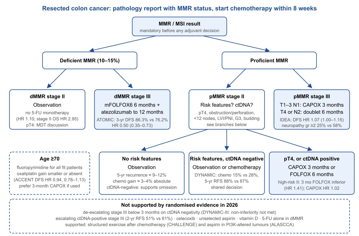
The pattern is consistent: the largest effects belong to interventions targeted by biology (checkpoint inhibition in dMMR disease, aspirin in PI3K-altered tumours), the smallest to those applied to a whole stage (fluoropyrimidine in stage II), and ctDNA, the most accurate prognostic test available, has so far improved only the decision not to treat.
Colorectal Liver Metastases
Epidemiology and what resection achieves
The liver is the commonest site of distant spread. In the Burgundy population registry, 14.5% of patients had synchronous liver metastases and the five-year cumulative rate of metachronous liver metastases was a further 14.5% (3.7% after stage I, 13.3% after stage II and 30.4% after stage III primaries)[159]. In a Stockholm population-based cohort diagnosed in 2008, liver metastases were detected in 26.5% within five years, more often after left-sided primaries but with more extensive liver involvement after right-sided tumours [1]. Resection reaches a minority: 6.3% of synchronous and 16.9% of metachronous liver metastases were resected for cure in Burgundy between 1976 and 2000 [159], and in the Dutch nationwide series metastasectomy in liver-only stage IV disease rose from 5% to 18% between 1996–1999 and 2008–2011, with median survival rising from 9 to 16 months [160].
For those resected, long-term survival is well documented. A review of 60 studies found median five- and ten-year survival of 38% (range 16–74%) and 26% (9–69%)[161]. Ten-year survival approximates cure: of 612 patients resected between 1985 and 1994, 102 were actual ten-year survivors, 97% of them disease-free [162]. In the same institution's later cohort of 1,211 patients, actual ten-year survival was 24.4% and observed cure 20.6%; extrahepatic disease, CEA above 200 ng/mL, a positive margin and more than ten tumours each carried cure rates below 10%[163]. The contemporary reference values come from LiverMetSurvey: among 12,154 patients completely resected at 43 high-volume centres between 2000 and 2022, 90-day mortality was below 5% in most subgroups and benchmark five-year survival was at least 45% for a solitary synchronous metastasis, 58% for a solitary metachronous one, 48% for two to three metastases, 28% for more than three and 29% for initially unresectable disease [17].
The modern definition of resectability
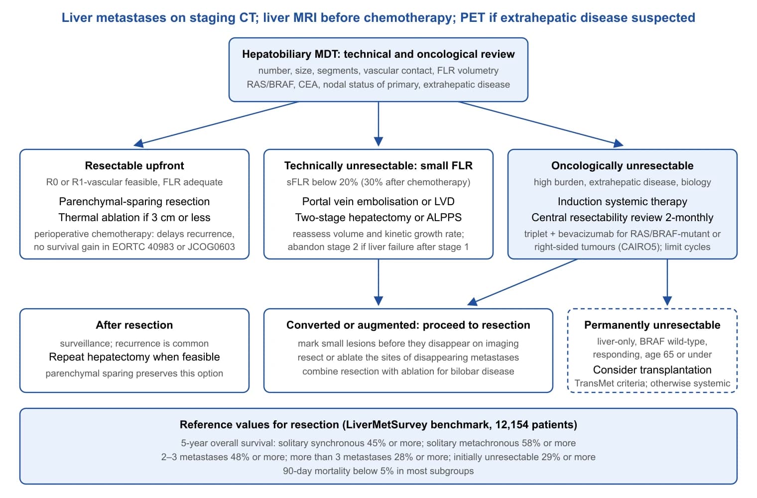
Resectability has two components. Technical resectability asks whether all disease can be removed with clear margins while preserving a future liver remnant (FLR) with intact inflow, outflow and biliary drainage; oncological resectability asks whether the biology justifies the operation. The EGOSLIM consensus stated that resection should not be denied to patients with stable disease after optimal chemotherapy provided an adequate remnant can be preserved [164]. Number, size and bilobar distribution are no longer absolute contraindications.
The remnant threshold is empirical. In 301 extended right hepatectomies, a standardised FLR of 20% or less independently predicted postoperative liver insufficiency (OR 3.18, 95% CI 1.34–7.54), outcomes with 20–30% and 30% or more were similar, and patients whose remnant was raised above 20% by portal vein embolisation did as well as those who started above it [37]. After prolonged chemotherapy most groups require about 30%, the threshold at which LIGRO randomised patients to augmentation [38]; the kinetic growth rate after embolisation predicts post-hepatectomy outcome better than static volume [165].
Oncological selection began with the clinical risk score: in 1,001 consecutive resections, five-year survival was 37% and ten-year 22%; node-positive primary, disease-free interval under 12 months, more than one metastasis, largest tumour above 5 cm and CEA above 200 ng/mL scored one point each, and no patient with a score of 5 survived long term [166]. The score predates modern chemotherapy and ignores biology. The GAME score adds KRAS mutation, CEA of 20 ng/mL or more, nodal status, a tumour burden score and extrahepatic disease; five-year survival was 73.4% with 0–1 points and 11% with 4 or more, with better discrimination than the Fong score in an external cohort of 747 patients [167]. A modified score built on RAS mutation, nodal status and size above 50 mm outperformed it in 608 validation patients [168]. The effect sizes are large: among 193 patients resected after modern chemotherapy, three-year survival was 81% with wild-type and 52.2% with mutant RAS (HR 2.3), and RAS mutation predicted lung but not liver recurrence [169]. BRAF mutation is rare in patients selected for hepatectomy (35 of 1,497, 2%), with median survival of 40 versus 81 months; V600E and non-V600E did not differ, and long-term survival remained possible with a node-negative primary, CEA at or below 200 ng/mL and a clinical risk score below 4 [170]. No single mutation contraindicates resection, but RAS and BRAF status belong in every resectability discussion, and BRAF mutation excluded patients from transplantation in TransMet [20].
Extent of resection and margins
Parenchymal-sparing hepatectomy is the default. A meta-analysis of 18 studies (7,081 patients, 56.1% parenchymal-sparing) found identical overall survival (HR 1.01, 95% CI 0.94–1.08) and recurrence-free survival (HR 1.00, 0.94–1.07), whereas anatomic or extended resection carried more complications (RR 1.39, 1.16–1.66) and more transfusion (RR 2.27, 1.60–3.23)[18], with no difference in R0 rates across 12 earlier studies [171]. The oncological argument is salvageability: among 300 patients with a solitary metastasis of 30 mm or less, liver-only recurrence was equally frequent after parenchymal-sparing and major resection (14% vs 17%), but repeat hepatectomy was possible in 68% versus 24%, five-year survival after liver-only recurrence was 73.6% versus 30.1%, and major resection independently predicted non-candidacy for repeat surgery (HR 8.18)[39].
The margin debate has narrowed. In 2,368 patients with high-resolution histological measurement, median survival was 32 months after R1 resection (tumour at the margin), 40 months with 0.1–0.9 mm, 53 months with 1–9 mm and 56 months with 10 mm or more; every width, including sub-millimetre clearance, was better than R1 after adjustment [172]. The "R1 vascular" concept separates two kinds of R1. In 226 consecutive patients (627 resection areas), detachment of a metastasis from a major intrahepatic vessel (R1Vasc) produced per-patient local recurrence of 4.3%, no different from R0 (5.3%), whereas a parenchymal R1 margin (R1Par) produced 19.6% and independently worsened survival [40]. A later single-centre analysis suggested that KRAS status modifies this effect, a finding awaiting confirmation [173]. The practical position (authors' judgement) is to aim for at least 1 mm, accept vessel detachment when the alternative is sacrificing the remnant or declining resection, and avoid parenchymal R1 wherever a wider plane exists.
Synchronous disease: sequencing the liver and the primary
Three strategies exist: simultaneous resection, primary-first (classical staged) and liver-first (reverse), the last introduced in Geneva for advanced synchronous disease, where 16 of 20 patients completed both resections after induction chemotherapy [174]. The EGOSLIM consensus favoured chemotherapy before surgery in most scenarios and liver-first resection when the primary is asymptomatic [164]. Randomised evidence is thin. METASYNC recruited 105 patients with resectable synchronous metastases and analysed 85: major complications within 60 days were 49% after simultaneous and 46% after delayed resection (adjusted OR 0.84, 95% CI 0.35–2.01), 8 of 46 patients in the delayed arm never reached liver resection (six because of progression), and survival tended to favour the simultaneous arm [175]. In LiverMetSurvey (7,360 patients: 4,415 primary-first, 552 liver-first, 2,393 simultaneous), strategy made no difference in solitary or unilobar disease, but in multiple bilobar metastases liver-first was an independent favourable factor (three-year survival 65.9% vs 60.4% primary-first and 54.4% simultaneous; after propensity matching HR 1.667 and 2.278 for the alternatives)[176]. A network meta-analysis found no significant differences among the three [177]. All comparisons are confounded by indication.
When metastases are unresectable and the primary asymptomatic, resecting the primary does not prolong life. The combined SYNCHRONOUS and CCRe-IV trials randomised 393 patients: median survival was 16.7 months with and 18.6 months without primary resection (adjusted HR 0.944, 95% CI 0.738–1.209), and 24.1% of the resection arm never received chemotherapy [30]. JCOG1007 was stopped for futility after 165 patients (median survival 25.9 vs 26.7 months; HR 1.10, 0.76–1.59), with three postoperative deaths [28]. Obstruction and perforation are covered in Part 2 of this series.
| Strategy | Typical indication | Key evidence | Finding |
|---|---|---|---|
| Simultaneous resection | Limited hepatic burden, minor hepatectomy, fit patient | METASYNC RCT, 85 analysed [175] | Major complications 49% vs 46% delayed (OR 0.84); 8/46 delayed never reached liver surgery |
| Primary-first (classical) | Symptomatic primary; liver disease resectable at diagnosis | LiverMetSurvey, 4,415 patients [176] | Survival equal to other strategies in solitary or unilobar disease |
| Liver-first (reverse) | Asymptomatic primary, high hepatic burden, rectal primary needing radiotherapy | LiverMetSurvey, 552 patients [176]; network meta-analysis [177] | 3-year survival 65.9% vs 60.4% and 54.4% in multiple bilobar disease; ranked first but no significant difference overall |
| Primary resection with unresectable metastases | Asymptomatic primary, unresectable disease | SYNCHRONOUS/CCRe-IV, 393 [30]; JCOG1007, 165 [28] | No survival benefit (HR 0.944 and 1.10); 24.1% never received chemotherapy after resection |
Increasing the remnant
Portal vein embolisation (PVE) is the reference technique: in 44 studies (1,791 patients), technical success was 99.3%, mean hypertrophy 37.9%, surgery was abandoned because of PVE failure in 3.9% and because of progression during the waiting period in 6.1%, major complications occurred in 2.5% and mortality was 0.1%[178]. Liver venous deprivation (simultaneous portal and hepatic vein embolisation) accelerates growth: remnant volume at day 21 rose by 52.6% versus 18.6% after PVE in a Montpellier cohort [179], and in the DRAGON collaborative (39 combined versus 160 PVE) hypertrophy was 59% versus 48%, resectability 90% versus 68% and 90-day mortality 3% versus 16% (P = 0.065)[180], with better three-year survival (HR 0.46, 95% CI 0.27–0.76) in a retrospective comparison [181]; the randomised HYPER-LIV01 trial was in progress when these cohorts were reported [179].
Two-stage hepatectomy (TSH) combines clearance of the remnant with PVE and a later major resection; across ten studies (459 patients), 77% completed both stages, R0 was achieved in 75%, second-stage morbidity and mortality were 40% and 3%, and median survival was 37 months in completers against 16 months in drop-outs [41]. ALPPS forces rapid hypertrophy by combining portal vein ligation with parenchymal transection [42], but in the first 320 registry patients 90-day mortality was 8.8%, 75% of deaths were from liver failure, and liver failure after stage 1 (14%) or a MELD score above 10 before stage 2 raised the odds of death four- to five-fold [182]. LIGRO randomised 97 patients with a standardised FLR below 30% between ALPPS and TSH: resection rates were 92% versus 57% (rate ratio 8.25, 95% CI 2.6–26.6), with no difference in Clavien–Dindo grade 3a or worse complications (43% vs 43%), 90-day mortality (8.3% vs 6.1%) or R0 rate [38]; by intention to treat, median survival was 46 versus 26 months (P = 0.028)[183]. The 6–8% mortality in both arms of this single trial marks remnant augmentation as a high-stakes strategy for a selected minority (authors' judgement).
| Technique | Hypertrophy | Completion of resection | Mortality | Source |
|---|---|---|---|---|
| Portal vein embolisation | Mean 37.9% | Abandoned in 3.9% (PVE failure) and 6.1% (progression) | 0.1% after PVE | Systematic review, 1,791 patients [178] |
| Liver venous deprivation (PVE + HVE) | 59% vs 48% for PVE; volume +52.6% vs +18.6% at day 21 | 90% vs 68% for PVE | 90-day 3% vs 16% (P = 0.065) | DRAGON cohort [180]; Montpellier cohort [179] |
| Two-stage hepatectomy | PVE in 76% | 77% completed stage 2; R0 75% | 3% after stage 2 | Systematic review, 459 patients [41] |
| ALPPS, registry | Median +74% in 9 days (first series) | Not stated | 90-day 8.8%; 75% liver failure | Registry, 320 [182]; first series [42] |
| ALPPS vs TSH, randomised | sFLR <30% at entry | 92% vs 57% (rate ratio 8.25); with rescue ALPPS 92% vs 80% | 90-day 8.3% vs 6.1% | LIGRO [38,183] |
Thermal ablation and stereotactic radiotherapy
For unresectable disease, ablation added to chemotherapy prolongs survival. The EORTC CLOCC trial randomised 119 patients with fewer than ten unresectable metastases and no extrahepatic disease to systemic therapy with or without radiofrequency ablation (with resection where needed); at 9.7 years, survival favoured local treatment (HR 0.58, 95% CI 0.38–0.88), with eight-year survival of 35.9% versus 8.9%[184]. A 2018 systematic review concluded that further randomised comparisons against chemotherapy alone would be unethical [185].
The question for resectable small lesions was addressed by COLLISION. In 14 centres, 300 patients with ten or fewer metastases of 3 cm or less and no extrahepatic disease were randomised to thermal ablation or resection. The trial was stopped at the pre-planned interim analysis because its stopping rules were met: the conditional likelihood of proving non-inferiority for overall survival was 90.5% (median not reached in either arm; HR 1.05, 95% CI 0.69–1.58), local control was non-inferior (HR 0.13, 0.02–1.06), and safety favoured ablation, with adverse events in 19% versus 46%, serious adverse events in 7% versus 20%, and no treatment-related deaths versus three after resection [19]. The caveats are a median follow-up of 28.9 months and an immature survival endpoint; the authors' reading, that ablation should no longer be reserved for unresectable disease, is fair. Stereotactic body radiotherapy (SBRT) rests on weaker evidence: across 18 studies (656 patients), two-year local control was 59.3%, two-year survival 56.5% and median survival 31.5 months, with severe liver toxicity in 8.7%[186]. No randomised comparison of SBRT with ablation or resection was identified for this review.
| Study | Design and population | Comparison | Main result |
|---|---|---|---|
| CLOCC [184] | Phase II RCT, 119 unresectable, <10 metastases | Systemic therapy ± RFA (± resection) | OS HR 0.58 (0.38–0.88); 8-year OS 35.9% vs 8.9% |
| COLLISION [19] | Phase III non-inferiority RCT, 300 resectable, ≤10 metastases ≤3 cm | Thermal ablation vs resection | OS HR 1.05 (0.69–1.58); local control HR 0.13; adverse events 19% vs 46%; deaths 0 vs 3 |
| SBRT pooled analysis [186] | 18 studies, 656 patients | Single-arm | 2-year local control 59.3%; median OS 31.5 months; severe liver toxicity 8.7% |
| Systematic review [185] | 48 studies incl. 2 RCTs | RFA/MWA vs chemotherapy vs resection | Ablation superior to chemotherapy alone; resection superior to RFA alone but not to RFA + resection or MWA |
Perioperative chemotherapy, conversion and disappearing metastases
Perioperative chemotherapy for resectable disease delays recurrence without a demonstrated survival gain. EORTC 40983 randomised 364 patients with up to four metastases to perioperative FOLFOX4 or surgery alone; at 8.5 years, survival did not differ (HR 0.88, 95% CI 0.68–1.14; five-year 51.2% vs 47.8%) despite the earlier progression-free benefit [187]. JCOG0603 randomised 300 patients to hepatectomy with or without adjuvant mFOLFOX6: five-year disease-free survival improved from 38.7% to 49.8% (HR 0.67, 0.50–0.92), but five-year survival was 71.2% with and 83.1% without chemotherapy at the first report [188] and remained no better at 7.7 years (HR 1.07, 0.73–1.57)[189]. Chemotherapy is better used to test biology in high-risk or borderline disease than added routinely to an operation already curative in low-risk patients (authors' judgement).
Conversion is a different proposition. In the Paul Brousse series, 138 of 1,104 initially unresectable patients (12.5%) were rescued by resection after chemotherapy, with five- and ten-year survival of 33% and 23% against 48% and 30% for primarily resectable disease and operative mortality of 0.7%[46]. In CELIM, cetuximab-based induction raised assessed resectability from 32% to 60%[190]. CAIRO5 defined the modern induction standard in 530 patients judged by a central panel every two months: FOLFOXIRI plus bevacizumab improved progression-free survival over doublet plus bevacizumab in right-sided or RAS/BRAF-mutated tumours (10.6 vs 9.0 months; HR 0.76, 0.60–0.98) at the cost of more serious adverse events (52% vs 31%), whereas in left-sided wild-type tumours panitumumab added toxicity but no benefit over bevacizumab [191].
Effective chemotherapy creates disappearing metastases. In the original Paris series of 66 vanished metastases, residual macroscopic or microscopic disease or recurrence in situ within a year was found at 83% of sites [192]. A systematic review of 15 studies (479 patients) confirmed that size below 2 cm, more cycles and oxaliplatin predispose to disappearance, that contrast-enhanced MRI detects residual disease best, and that resecting the sites reduces local recurrence without a demonstrated survival benefit [193]. Baseline liver MRI and surgery planned before the target disappears are the practical safeguards.
Laparoscopic and robotic liver resection
Three randomised trials support minimally invasive resection. OSLO-COMET randomised 280 patients to laparoscopic or open parenchymal-sparing resection: 30-day complications were 19% versus 31% (difference 12 percentage points, 95% CI 1.67–21.8) and hospital stay 53 versus 96 hours, with no difference in blood loss or margins [194]; five-year survival was 54% versus 55% (HR 0.93, 0.67–1.30), the trial not being powered for survival [195]. LapOpHuva (193 analysed) found lower morbidity (11.5% vs 23.7%) and identical five-year survival (49.3% vs 47.4%)[196]. ORANGE II PLUS extended this to hemihepatectomy (332 operated): functional recovery took 4 versus 5 days, major complications were 14.5% versus 16.9%, R0 rates 77.9% versus 84.1% (P = 0.14), and adjuvant therapy started sooner after laparoscopy (46.5 vs 62.8 days)[197]. Randomised data comparing robotic with laparoscopic resection for colorectal liver metastases were not identified for this review.
Liver transplantation for unresectable disease
Transplantation for colorectal metastases was abandoned in the 1990s because of 18% five-year survival and revived in Oslo, where SECA-I gave five-year survival of 60% in 21 patients [198]. With stricter selection in SECA-II (at least 10% response to chemotherapy and more than one year from diagnosis), five-year survival was 83%[199]; at ten-year follow-up of 25 patients, median survival was 128.2 months, 48% had relapsed, and the investigators estimated that up to 15 of 25 might be cured [200]. The randomised evidence came from TransMet, in which 20 European centres randomised 94 patients aged 18–65 with permanently unresectable, liver-only, BRAF wild-type metastases responding to no more than three lines of chemotherapy to transplantation plus chemotherapy or chemotherapy alone. Five-year survival was 56.6% versus 12.6% by intention to treat (HR 0.37, 95% CI 0.21–0.65) and 73.3% versus 9.3% per protocol; serious adverse events occurred in 80% of transplanted patients, three were retransplanted and one of these died [20]. The International Hepato-Pancreato-Biliary Association consensus standardised selection and raised the allocation question directly: a graft given for metastatic cancer is a graft withheld from the waiting list [201]. Access therefore depends on organ supply and allocation policy.
Recurrence and repeat hepatectomy
Recurrence after resection is the rule, and the liver is the commonest site. In a meta-analysis of 15 cohorts (1,039 patients), median survival after repeat hepatectomy was 54.0 months when recurrence occurred three to six months after the first operation, 53.0 months at seven to twelve months and 60.0 months beyond a year (adjusted HR 0.70, 95% CI 0.53–0.92), so that a short interval is prognostic but not disqualifying [202]. Among the 250 observed cures in the Memorial Sloan Kettering cohort, 58 had undergone resection of a recurrence [163]; the policy that makes this possible is parenchymal sparing at the first operation [39].
Liver metastases: what the evidence supports
- Resection remains the reference: benchmark five-year survival is 45–58% for solitary disease and at least 28% for more than three metastases, with 90-day mortality below 5%[17].
- Resectability is defined by the remnant (20% in a normal liver, about 30% after chemotherapy) and by biology; RAS and BRAF status should be known before the decision [37,169,170].
- Spare parenchyma, aim for 1 mm, accept vessel detachment and avoid parenchymal R1 [18,40,172].
- Thermal ablation is non-inferior to resection for lesions of 3 cm or less with fewer complications; perioperative chemotherapy delays recurrence without improving survival [19,187,189].
- ALPPS raised resection rate and survival over two-stage hepatectomy in one trial, at 6–8% 90-day mortality in both arms [38,183].
- Transplantation for selected permanently unresectable, liver-only, BRAF wild-type disease gave five-year survival of 56.6% versus 12.6%; access is limited by organ supply [20,201].
Lung Metastases
Epidemiology and selection
Lung metastases are less common than liver metastases and more often rectal in origin. In the Burgundy registry, 11.0% of patients had synchronous lung metastases, the five-year cumulative risk of metachronous lung metastases was 5.8%, and rectal primaries carried higher odds of both (OR 2.80 and 2.63); only 4.1% of synchronous and 14.3% of metachronous lung metastases were resected for cure, with three-year relative survival of 53.0% and 59.2% after resection against 11.3% and 13.8% overall [203]. In the Netherlands the proportion with lung metastases at diagnosis rose from 1.7% to 5.0% between 1996 and 2011, largely a product of CT staging [160]. Single-arm surgical series reporting five-year survival above 50% were for two decades the basis of practice [204].
The PulMiCC trial
PulMiCC is the only randomised trial of pulmonary metastasectomy. Teams in 13 hospitals randomised patients with resectable lung metastases to metastasectomy or continued active monitoring; the trial closed after 65 participants because of poor and worsening recruitment. The hazard ratio for death within five years was 0.82 (95% CI 0.43–1.56), and five-year survival was 38% (23–62%) after metastasectomy and 29% (16–52%) in controls, against the widely assumed figure of under 5% without surgery [21]. An updated analysis of 93 randomised patients (one to six metastases, 29% with prior liver metastasectomy) gave median survival of 3.5 years after metastasectomy and 3.8 years in controls (HR 0.93, 0.56–1.56)[22]. The parallel observational cohort of 481 patients explains why surgical series look so good: 69% of electively operated patients had a solitary metastasis compared with 35% of those not operated, five-year survival was 47% versus 22%, and the investigators concluded that no inference about the effect of surgery could be drawn [23]. PulMiCC does not show that metastasectomy is ineffective; it shows that control survival is far better than historical comparisons implied and that any true benefit is likely to be modest.
Prognostic factors, radiotherapy and combined disease
A meta-analysis of 25 surgical series (2,925 patients) identified four factors associated with poorer survival after lung metastasectomy: short disease-free interval (HR 1.59, 95% CI 1.27–1.98), multiple metastases (HR 2.04, 1.72–2.41), hilar or mediastinal nodal involvement (HR 1.65, 1.35–2.02) and raised pre-thoracotomy CEA (HR 1.91, 1.57–2.32); previously resected liver metastases were not significant (HR 1.22, 0.91–1.64)[205].
| Factor | Pooled HR (95% CI) | Implication for selection |
|---|---|---|
| Short disease-free interval | 1.59 (1.27–1.98) | Early metachronous disease: consider interval observation or systemic therapy first |
| Multiple lung metastases | 2.04 (1.72–2.41) | Solitary lesions are the strongest surgical candidates |
| Hilar or mediastinal nodes | 1.65 (1.35–2.02) | Nodal sampling informs prognosis; positive nodes argue against further resection |
| Raised pre-thoracotomy CEA | 1.91 (1.57–2.32) | Marker of occult systemic disease |
| Prior liver metastasectomy | 1.22 (0.91–1.64), not significant | Not a contraindication |
Stereotactic ablative radiotherapy (SABR) competes with wedge resection for small peripheral lesions. In a single-centre comparison of 404 patients, three-year local control was 88.6% after surgery and 86.7% after SABR (P = 0.174)[206]; a two-centre Dutch comparison found similar median survival (58 vs 70 months) but more local recurrence after SABR (38.3% vs 12.5% of patients)[207]. No randomised comparison exists. Simultaneous liver and lung metastases do not preclude surgery: in LiverMetSurvey, adjusted five-year survival was 51.5% after resection of isolated liver metastases, 44.5% after resection of both (149 patients) and 14.3% when lung disease was left unresected (285 patients)[208], a difference that reflects selection as much as treatment.
Guideline positions
The Society of Thoracic Surgeons expert consensus endorses metastasectomy for selected patients with controlled primary disease, resectable pulmonary disease and adequate reserve, within multidisciplinary review [209]. The ESMO 2023 metastatic colorectal cancer guideline treats lung metastases within its oligometastatic framework, in which local treatment (surgery, SABR or ablation) is selected from a "toolbox" according to site, goal, morbidity and patient factors; metastasectomy is one option rather than a default [47].
Lung metastases: what PulMiCC changed
- Controls with resectable lung metastases survived five years in about 29%, not under 5%; the assumed 35-point absolute benefit was not observed (HR 0.82–0.93, confidence intervals including 1)[21,22].
- The advantage in observational series is largely selection: solitary metastases in 69% of operated versus 35% of non-operated patients [23].
- Metastasectomy remains reasonable for a solitary lesion, long disease-free interval, normal CEA and no nodal disease, provided the patient understands that the benefit is unproven and probably modest [47,205,209].
Peritoneal Metastases: Assessment, Cytoreductive Surgery and Patient Selection
Incidence, risk factors and natural history
Peritoneal metastases (PM) are the third most common site of colorectal spread and the one imaging detects worst. In the Stockholm population-based cohort of 11,124 patients, 8.3% developed PM, synchronous in 4.3% and metachronous in 4.2%; independent predictors of metachronous PM were T4 stage (HR 9.98, 95% CI 3.10–32.11), N2 disease with fewer than 12 nodes examined (HR 7.41), emergency surgery (HR 2.11) and R2 resection (HR 2.75)[2]. A Dutch nationwide cohort of 7,233 patients found synchronous PM in 5.7% and a 3-year cumulative incidence of metachronous PM of 5.5%, with signet-ring (OR 6.58) and mucinous (OR 2.72) histology, T4 (OR 4.82) and N2 (OR 3.27) stage as predictors [210], consistent with the peritoneal tropism of these histological subtypes in autopsy series [211].
The number that matters for the prophylactic trials in Section 12 is the risk after a pT4 primary. In 200 resected T4 cancers, synchronous PM was present in 23% and metachronous PM developed in 21% of those at risk, roughly one patient in five; no variable predicted metachronous PM specifically [212]. A systematic review of 16 low-quality studies (recurrent PM 8–75%) singled out resected synchronous PM, isolated ovarian metastases and a perforated primary as the situations with a real excess risk [213].
With systemic therapy alone the prognosis is worse than for other single-site disease. In the ARCAD pooled analysis of 10,553 patients in 14 first-line phase 3 trials, isolated non-peritoneal disease carried better survival than isolated PM (adjusted HR 0.75, 0.63–0.91; p=0.003), and PM with two or more other sites the worst (HR 1.40, 1.14–1.71)[214]. Surgical series show the same gradient: patients excluded from cytoreduction at laparoscopy for extensive disease had a median survival of 14 months (95% CI 10–18) against 36 months (27–45) in those deemed suitable [215].
Imaging, diagnostic laparoscopy and second-look surgery
CT systematically understages peritoneal disease: nodules under 0.5 cm were detected with a sensitivity of 11% against 94% for nodules over 5 cm, and radiological PCI significantly underestimated operative PCI (p<0.001)[216]. Adding MRI with diffusion-weighted sequences raised the sensitivity of two readers from 63% to 81% and from 44% to 81% and reduced the mean error in PCI from 4.89 to 3.96 (p=0.010)[217]. PET/MRI reached a sensitivity of 0.97 against 0.54 for standard imaging in a mixed prospective cohort [218].
Because no modality counts millimetre nodules, diagnostic laparoscopy has become the arbiter of resectability. In 184 consecutive Dutch patients it gave a good view of diaphragm, pelvis and small bowel in 75%, was converted in 13%, and excluded 50.5% from cytoreduction, most often because there was no PM (37% of exclusions) or the PCI was 20 or more (35%), with 2.7% major complications and no deaths [215].
Second-look surgery extends this reasoning to high-risk patients without visible disease. In the Gustave Roussy series, 29 high-risk patients underwent planned laparotomy at 13 months; PM was found and treated with complete cytoreduction and HIPEC in 55%, with 14% grade III–IV morbidity [219]. That yield justified PROPHYLOCHIP, whose negative result (Section 12.3) showed that finding disease early did not improve disease-free survival.
Scoring the disease: PCI, completeness of cytoreduction and the PSDSS
The Peritoneal Cancer Index (PCI) scores the largest lesion in each of 13 regions (nine abdominopelvic, four small-bowel) from 0 to 3 (none; up to 0.5 cm; up to 5 cm; over 5 cm or confluent), giving 0–39; the completeness-of-cytoreduction (CC) score records residual disease as CC-0 (none visible), CC-1 (under 2.5 mm), CC-2 (2.5 mm to 2.5 cm) or CC-3 (larger)[43]. CC-0/1 counts as complete because 2.5 mm is the assumed penetration depth of intraperitoneal chemotherapy.
Two French analyses set the thresholds in use. Among 180 patients taken to theatre with curative intent, complete cytoreduction was achieved in 139 (mean PCI 11) and not in 41 (mean PCI 23); 3-year survival was 52% versus 7%, but compared point by point the curves no longer separated above a PCI of 17 (HR 0.64, 0.38–1.09)[220]. In 173 uniformly treated patients the relationship was linear (HR 1.1 per PCI point; 5-year survival 41%): surgery is generally indicated below 12, contraindicated above 17, and individual between the two [221]. The Dutch guideline uses an upper limit of 20 [222], PRODIGE 7 enrolled up to 25 [24] and GECOP-MMC caps at 20 [223]; "PCI above 20 excluded" is consensus practice, not a validated cut-off. Guideline positions on cytoreduction and HIPEC (ESMO, NCCN, Dutch, PSOGI) are set out in Section 12.4.
The Peritoneal Surface Disease Severity Score (PSDSS) combines symptoms, radiological extent and primary histology into four preoperative stages [224]; in the ASPSM evaluation of 1,013 patients, median survival after cytoreduction and HIPEC was 86, 43, 29 and 28 months for stages I–IV, against 45, 19, 8 and 6 months without surgery [225]. It stratifies trials well but has not displaced operative PCI and CC score.
Outcomes of cytoreductive surgery
| Series | n | Complete CRS | Major morbidity | Mortality | Survival |
|---|---|---|---|---|---|
| French multicentre, 1990–2007 [226] | 523 | 84% | Grade 3–4 31% | 3% | Median 30.1 mo (33 if complete); 5-yr OS 27% |
| Dutch protocol, CRC subset [227] | 660 | 80% (whole cohort) | Grade III–V 34% | 3% | Median 33 mo; 5-yr 31%; PFS 15 mo |
| Gustave Roussy, complete CRS + HIPEC [221] | 173 | 100% by inclusion | — | — | 5-yr OS 41%; HR 1.1 per PCI point |
| Registry, 39 centres [228] | 2,093 | — | — | — | Median OS 43 mo; RFS 12 mo |
| PRODIGE 7, surgery-alone arm [24] | 132 | CC-0/1, PCI ≤25 | Grade ≥3 15% (60-day) | 2% | Median OS 41.2 mo (35.1–49.7) |
| CAIRO6, upfront CRS-HIPEC arm [229] | 178 | 154 complete or near-complete | Grade 3–5 26% (90-day) | 1% | Median OS 39 mo (31–46) |
The pattern over two decades is consistent: 80–85% of patients taken to theatre reach complete cytoreduction, one in four to one in three has a Clavien–Dindo grade III or worse complication, perioperative mortality is 1–3%, and median survival after complete cytoreduction is 33–43 months with 5-year survival of 27–41%. In 211 Dutch patients, 25.1% had grade ≥3 morbidity, predicted by extensive prior surgery (OR 4.3), smoking (OR 4.0) and poor performance status (OR 2.9)[230]. In NSQIP, 30-day mortality after 1,822 CRS/HIPEC procedures was 1.1%, against 2.5% after pancreaticoduodenectomy [231].
Does cytoreduction add to systemic therapy? The randomised evidence
One randomised trial has compared a surgical strategy with systemic therapy alone. The Dutch trial randomised 105 patients to fluorouracil–leucovorin with or without palliative surgery, or to cytoreduction with mitomycin C HIPEC followed by the same chemotherapy; median survival was 12.6 versus 22.3 months (p=0.032), with 8% treatment-related mortality [232]. At almost 8 years the difference held: median progression-free survival 7.7 versus 12.6 months (p=0.020), disease-specific survival 12.6 versus 22.2 months (p=0.028), and 5-year survival of 45% after complete resection [233]. Two caveats follow: the comparator was fluorouracil alone, before oxaliplatin, irinotecan or biological agents; and the experimental arm bundled cytoreduction and HIPEC, so the trial, routinely cited as evidence for HIPEC, isolated nothing.
No trial has repeated the comparison; equipoise has gone. A 2025 systematic review pooled two randomised and nine non-randomised studies (3,316 patients) and estimated a hazard ratio for overall survival of 0.447 (0.387–0.595) in favour of cytoreduction, with no heterogeneity and, by the authors' GRADE assessment, moderate certainty after downgrading for clinical heterogeneity and risk of bias [234]. Selection inflates every such comparison. The authors' judgement is that the benefit of complete cytoreduction in selected patients is real but of uncertain size, and that a median survival of about 40 months, reproduced in both arms of PRODIGE 7 and CAIRO6, is the most reliable expectation to offer a patient.
CAIRO6 answers a different question. It randomised 358 patients with resectable peritoneal-only metastases at ten Dutch and Belgian centres to perioperative systemic therapy (oxaliplatin- or irinotecan-based, with bevacizumab) plus CRS-HIPEC, or upfront CRS-HIPEC. Median overall survival was 44 months (30–54) versus 39 months (31–46), HR 0.85 (0.62–1.15), p=0.28; major 90-day morbidity was 36% versus 26%, and 57% of patients starting systemic therapy had grade 3–5 toxicity [229]. Both arms received HIPEC, so the trial says nothing about HIPEC; it says that routine perioperative chemotherapy cannot be recommended for all resectable patients.
Selection: PCI, completeness, nodes, liver metastases and the centre
In the French multicentre series the independent favourable factors were complete cytoreduction, limited PCI, absence of nodal involvement and adjuvant chemotherapy [226]. How many liver metastases are compatible with curative intent was addressed in a case-control study of 37 patients with PM and synchronous liver metastases matched to 61 with PM alone: 3-year survival was 40% versus 66% (p=0.04), pN+ status carried an OR of 3.3 for poor survival, and median survival was 76 months for PCI below 12 without liver metastases, 40 months for PCI below 12 with one or two, and 27 months for PCI of 12 or more or three or more liver metastases [235]. The rule that one or two resectable liver metastases with a low PCI do not exclude cytoreduction has no randomised support, since PRODIGE 7 and CAIRO6 excluded extraperitoneal disease.
The learning curve is long: risk-adjusted analysis of 420 procedures placed institutional proficiency, defined by acceptable rates of incomplete cytoreduction and grade 3–5 morbidity, at about 140 cases [236]. That is why the Netherlands restricts the operation to nine centres under a national protocol [227] and why the 2021 PSOGI statement recommended referral of every patient with potentially resectable PM to an expert centre whether or not HIPEC is given [237].
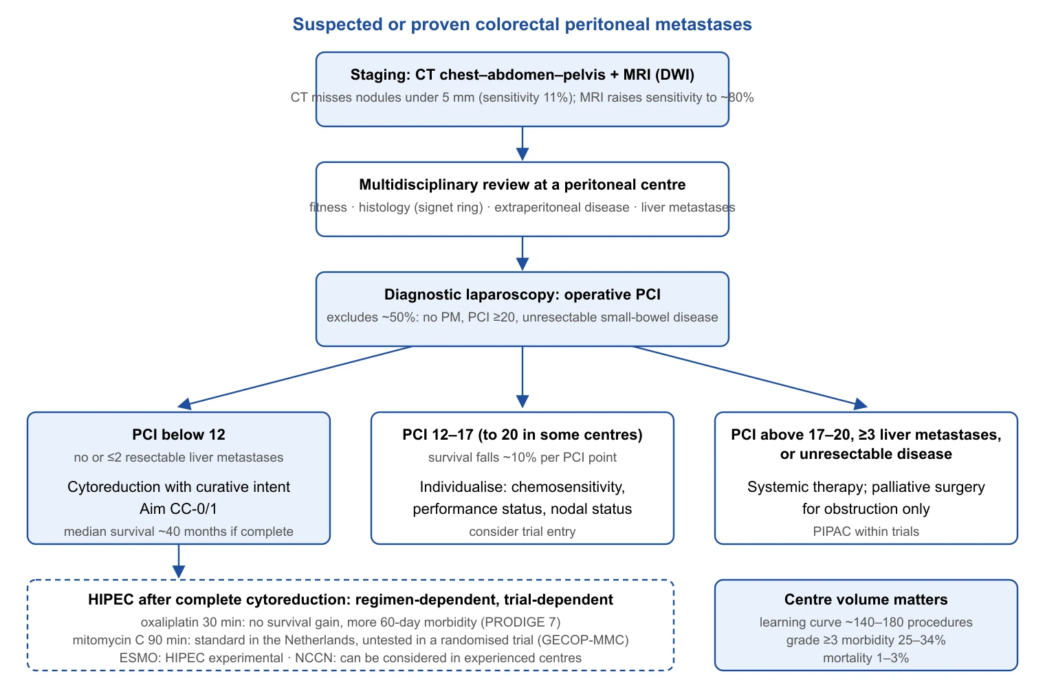
Selecting a patient for cytoreduction: what the evidence supports
- Operative PCI and completeness of cytoreduction determine survival; imaging underestimates both, so decisions rest on laparoscopy or laparotomy findings [216,220].
- Below a PCI of 12 cytoreduction is generally indicated, above 17 rarely justified; one or two resectable liver metastases with a low PCI do not exclude it [221,235].
- The modern benefit of surgery over systemic therapy rests on observational data of moderate certainty; routine perioperative chemotherapy added toxicity without survival in CAIRO6 [229,234].
HIPEC: The Evidence Appraised
Pharmacological rationale and the two regimens
HIPEC assumes that the peritoneal–plasma barrier permits intraperitoneal concentrations far above systemic exposure, that hyperthermia (42 °C in the trials below) enhances cytotoxicity and penetration, and that residual disease after CC-0/1 cytoreduction lies within the millimetre the drug reaches. In a 20-patient dose-escalation study of oxaliplatin at 460 mg/m² and 42–44 °C for 30 minutes, half the dose was absorbed during the perfusion, peritoneal concentration was 25 times plasma, and intratumoral penetration 17.8 times that of unexposed tissue [238]. The short perfusion reflects rapid absorption; the price is an exposure time a fraction of that of mitomycin C, which the Dutch protocol gives at 35 mg/m² over 90 minutes, the regimen the 2022 PSOGI consensus named as preferred [44]; HIPECT4 used 30 mg/m² over 60 minutes [27]. The best experimental evidence that HIPEC adds to cytoreduction is a rat model in which cytoreduction alone gave a median survival of 43 days against 97 days with mitomycin C at 35 mg/m² [239]; the gap between that model and the clinic is the subject of this section.
PRODIGE 7: the trial that isolated HIPEC
PRODIGE 7, run by the UNICANCER-GI and BIG-RENAPE groups, is the only trial to have tested HIPEC as the sole variable. At 17 French centres between 2008 and 2014, 265 patients aged 18–70 with a PCI of 25 or less and complete or near-complete cytoreduction (residual under 1 mm) were randomised in theatre to oxaliplatin HIPEC (360 mg/m² closed or 460 mg/m² open, 30 minutes) or no HIPEC; all received 6 months of systemic chemotherapy [24]. After a median follow-up of 63.8 months, median overall survival was 41.7 months (36.2–53.8) with HIPEC and 41.2 months (35.1–49.7) without (HR 1.00, 95.37% CI 0.63–1.58; p=0.99). Treatment-related mortality at 30 days was 2% in each arm. Grade 3 or worse events were similar at 30 days (42% versus 32%, p=0.083) but more frequent with HIPEC at 60 days (26% versus 15%, p=0.035)[24]. The 41-month median in both arms is the trial's second message: complete cytoreduction with modern systemic therapy produces the survival previously attributed to the combination.
A post-hoc analysis found that patients with a PCI of 11–15 had a median survival of 41.6 months with HIPEC against 32.7 months without (p=0.0209), and GECOP-MMC stratifies by this band for that reason [223]. The signal is hypothesis-generating: the subgroup was not pre-specified or powered, several strata were examined without correction, and no mechanism explains a drug effect at PCI 11–15 but not 1–10. Critics add that prior oxaliplatin-based systemic therapy may have selected resistant disease, that the 30-minute regimen differs from 90-minute mitomycin C, and that overall survival is a blunt endpoint for a locoregional intervention [223,240]. These limit the trial's external validity for mitomycin C; they do not rescue oxaliplatin HIPEC, which the 2022 PSOGI panel rejected by strong negative consensus [44].
Prophylactic and adjuvant HIPEC: PROPHYLOCHIP, COLOPEC and HIPECT4
PROPHYLOCHIP-PRODIGE 15 randomised 150 patients at 23 French hospitals, all with resected synchronous localised PM, ovarian metastases or a perforated primary and no recurrence after 6 months of adjuvant chemotherapy, to surveillance or systematic second-look laparotomy with oxaliplatin HIPEC. Three-year disease-free survival was 53% (41–64) with surveillance and 44% (33–56) with second-look surgery (HR 0.97, 0.61–1.56); 41% of operated patients had grade 3–4 complications [25]. Surveillance with treatment of recurrence when it appears is as effective and far less morbid.
COLOPEC randomised 204 patients with cT4 or pT4 N0–2 M0 or perforated colon cancer at nine Dutch centres to adjuvant oxaliplatin HIPEC (460 mg/m², 30 minutes, given 5–8 weeks after resection in 91%) plus systemic chemotherapy, or chemotherapy alone, with laparoscopy at 18 months. Peritoneal metastasis-free survival at 18 months was 80.9% (73.3–88.5) versus 76.2% (68.0–84.4) (p=0.28); PM was diagnosed in 19% and 23%, and 9 of the 19 HIPEC-arm cases were found at the exploration preceding the planned HIPEC, so nearly half of the "metachronous" disease was already present at 5–8 weeks [241]. At 5 years, overall survival was 69.6% versus 70.9% (p=0.69), disease-free survival 55.7% versus 52.3% (p=0.88) and the peritoneal metastasis endpoint 63.9% versus 63.2% (p=0.91, as reported in the published abstract, to which an erratum followed in 2026); the investigators concluded that adjuvant HIPEC belongs in trials only [26].
HIPECT4 reached the opposite conclusion with a different drug and endpoint. At 17 Spanish centres, 184 patients with cT4 N0–2 M0 colon cancer were randomised to resection with intraoperative mitomycin C HIPEC (30 mg/m², 60 minutes) or resection alone, both with adjuvant chemotherapy. Three-year locoregional control, the primary endpoint, was 97.6% with HIPEC versus 87.6% without (p=0.03; HR 0.21, 0.05–0.95), larger in the pT4-confirmed subgroup (98.3% versus 82.1%; HR 0.09, 0.01–0.70); disease-free survival (81.2% versus 78.0%; HR 0.71, 0.41–1.22) and overall survival (91.7% versus 92.9%; HR 0.79, 0.26–2.37) did not differ, nor did morbidity [27]. An individual-patient-data meta-analysis pooling COLOPEC and HIPECT4 (386 patients) found 36-month locoregional recurrence of 16.0% with HIPEC and 21.2% without (p=0.295), a significant reduction only in the pre-specified right-sided subgroup (HR 0.56, 0.48–0.67), and no survival difference [242]. One-off intraoperative mitomycin C may therefore reduce peritoneal recurrence after a T4 primary, but this has not translated into survival, and the two trials differ in drug, timing and endpoint.
How guidelines responded
The ESMO guideline, as set out in its Pan-Asian adaptation, states that where metastasis is confined to the peritoneum complete cytoreductive surgery should be carried out [II, A] and that HIPEC "can be considered as an experimental procedure, still to be validated in clinical trials"[47,243]. The NCCN guideline, as quoted in a 2025 debate, states that cytoreductive surgery and/or intraperitoneal chemotherapy "can be considered in experienced centers for selected patients with limited peritoneal metastases for whom R0 resection can be achieved"[244]. The Dutch guideline module (2022) takes the opposite view: it recommends considering CRS/HIPEC for isolated PM with a PCI below 20, directs mitomycin C after oxaliplatin-containing induction, and states that primary CRS/HIPEC with mitomycin C remains standard in the Netherlands because PRODIGE 7 makes no statement about upfront surgery with 90-minute mitomycin C [222]. A PSOGI survey of experts from 18 countries recorded a switch to mitomycin C, longer perfusions, fewer referrals and removal of HIPEC from some national guidelines after PRODIGE 7 [237], and the 2022 PSOGI consensus, grading the evidence for 9 of 10 questions as weak or very weak, concluded that HIPEC "could be conditionally recommended" with the Dutch mitomycin C protocol as preferred regimen [44]. The disagreement is explicit: ESMO calls HIPEC experimental, the Dutch guideline and PSOGI call mitomycin C HIPEC standard or conditionally recommended, and NCCN sits between.
Mitomycin C versus oxaliplatin: observational comparisons
| Study | Design | n | Median OS, mitomycin C | Median OS, oxaliplatin | Comment |
|---|---|---|---|---|---|
| ASPSM registry [245] | Retrospective, complete CRS | 539 | 32.7 mo | 31.4 mo (p=0.925) | PSDSS I/II: 54.3 vs 28.2 mo (p=0.012); III/IV: 19.4 vs 30.4 mo (p=0.427) |
| St George, Sydney [246] | Retrospective, single centre | 201 | 29.0 mo | 56.0 mo | HR 0.59 (0.37–0.91) favouring oxaliplatin |
| International, 39 centres [228] | Retrospective, propensity-matched | 2,093 | 39 mo (matched 40) | 47 mo (matched 48) | aHR 0.78 (0.65–0.94) favouring oxaliplatin; high-dose mitomycin C equivalent; 90-day mortality worse with mitomycin C |
| Systematic review, 46 studies [247] | Narrative synthesis | — | — | — | Cohorts not comparable; severe complications higher with oxaliplatin; no valid OS comparison |
The observational literature points both ways. The ASPSM analysis found no overall difference but a large advantage for mitomycin C in low-severity disease; the Sydney series and the 39-centre registry found the reverse, with oxaliplatin associated with 8–27 months longer median survival after adjustment and mitomycin C with higher 90-day mortality [228,245,246]. The systematic review explains why none of this is interpretable: oxaliplatin cohorts more often received oxaliplatin-based neoadjuvant therapy and shorter perfusions, and regimen followed institutional habit [247]. The authors' judgement is that no observational comparison can establish that mitomycin C would have succeeded where oxaliplatin failed; the current preference rests on pharmacology and tradition, not evidence of efficacy.
Trials under way, PIPAC and the overall verdict
| Trial | Design, n | Regimen | Primary endpoint | Result | Main criticism |
|---|---|---|---|---|---|
| Dutch trial 2003 [232] | CRS + HIPEC vs 5-FU; 105 | Mitomycin C | OS | 22.3 vs 12.6 mo (p=0.032) | Surgery and HIPEC bundled; 5-FU-only comparator |
| PRODIGE 7, 2021 [24] | CRS ± HIPEC, PCI ≤25; 265 | Oxaliplatin 30 min | OS | 41.7 vs 41.2 mo, HR 1.00; 60-day grade ≥3 26% vs 15% | Short high-dose regimen; prior oxaliplatin |
| PROPHYLOCHIP, 2020 [25] | Second-look + HIPEC vs surveillance; 150 | Oxaliplatin | 3-yr DFS | 44% vs 53%, HR 0.97; grade 3–4 41% | Oxaliplatin regimen |
| COLOPEC, 2019/2024 [26,241] | Adjuvant HIPEC vs none, T4/perforated; 204 | Oxaliplatin 30 min, delayed | 18-mo PM-free survival | 80.9% vs 76.2% (p=0.28); 5-yr OS 69.6% vs 70.9% | Delayed HIPEC; PM already present in 9 of 19 |
| HIPECT4, 2023 [27] | Intraoperative HIPEC vs none, cT4; 184 | Mitomycin C 60 min | 3-yr locoregional control | 97.6% vs 87.6%, HR 0.21; DFS, OS unchanged | Surrogate endpoint; few events |
| CAIRO6, 2026 [229] | Perioperative chemotherapy + CRS-HIPEC vs CRS-HIPEC; 358 | HIPEC in both arms | OS | 44 vs 39 mo, HR 0.85 (0.62–1.15) | Does not test HIPEC |
| ICARuS, 2023 abstract [248] | HIPEC vs EPIC after CRS; 75 | Mitomycin C vs floxuridine | 3-yr PFS | 7.7 vs 8.8 mo, HR 0.69 (0.42–1.14) | Closed early; abstract only |
| GECOP-MMC, recruiting [223] | CRS ± HIPEC, CC-0, PCI ≤20; 216 planned | Mitomycin C 35 mg/m² | 3-yr peritoneal RFS | Awaited | Surrogate endpoint |
GECOP-MMC will answer the question PRODIGE 7 left open: 31 Spanish centres randomise intraoperatively, after complete cytoreduction with a PCI of 20 or less, to high-dose mitomycin C HIPEC or none, stratified by PCI 1–10, 11–15 and 16–20, with 3-year peritoneal recurrence-free survival as primary endpoint [223]. ICARuS closed its colorectal cohort once PRODIGE 7 was presented and has reported only in abstract [248]; this review found no peer-reviewed long-term update of PRODIGE 7.
PIPAC (pressurised intraperitoneal aerosol chemotherapy) delivers low-dose aerosolised oxaliplatin laparoscopically and repeatedly to patients with unresectable PM. A review of 45 studies (1,810 procedures, various primaries) found adverse events above grade 2 after 12–15% of procedures and, in colorectal cancer, objective responses of 71–86% with a median survival of 16 months, almost entirely from single-arm series [45]. In the first prospective monotherapy trial in colorectal cancer (20 patients, 59 procedures), major treatment-related events occurred in 15% including one possibly related death, radiological response was 0%, and median progression-free and overall survival were 3.5 and 8.0 months [249]; a phase II trial alternating PIPAC with first-line systemic therapy preserved most patient-reported outcomes [250]. No randomised trial of PIPAC in colorectal PM has reported; the evidence is phase I–II and the indication palliative, within trials.
HIPEC in colorectal cancer: the authors' bottom line
- Cytoreductive surgery is the active component: both arms of PRODIGE 7 and of CAIRO6 reached a median survival of about 40 months after complete cytoreduction [24,229].
- Oxaliplatin HIPEC for 30 minutes adds morbidity without survival (HR 1.00; 60-day grade ≥3 complications 26% versus 15%) and should not be given outside a trial, a position shared by ESMO and the 2022 PSOGI consensus [24,44,243].
- Prophylactic HIPEC fails: second-look surgery with HIPEC did not improve disease-free survival in PROPHYLOCHIP, and adjuvant oxaliplatin HIPEC did not improve any 5-year endpoint in COLOPEC [25,26].
- Mitomycin C HIPEC is unproven in a randomised trial for established peritoneal metastases; the only randomised signal is HIPECT4's locoregional control endpoint after a T4 primary (HR 0.21), without a survival difference [27,242].
- Where mitomycin C HIPEC is standard, as in the Netherlands, it is a policy choice awaiting GECOP-MMC; where it is not, patients lose nothing established by complete cytoreduction alone at a high-volume centre [222,223].
Systemic Therapy and the Surgeon
Systemic therapy for metastatic colon cancer is chosen by molecular profile and sidedness, and the surgeon needs enough command of that framework to time surgery, to recognise conversion to resectability and to understand what drugs do to the liver that will be resected. Metastasectomy itself is covered elsewhere in this review.
First-line framework for metastatic disease
For RAS and BRAF wild-type tumours the choice between an anti-EGFR antibody and bevacizumab is governed by sidedness. In PARADIGM (802 RAS wild-type patients), panitumumab with mFOLFOX6 improved median overall survival over bevacizumab in left-sided tumours (37.9 vs 34.3 months; HR 0.82, 95% CI 0.68–0.99) and overall (36.2 vs 31.3 months; HR 0.84, 0.72–0.98), and raised the curative resection rate in left-sided disease from 11.6% to 18.3%[81]. The earlier trials pointed the same way without being decisive: FIRE-3 found longer overall survival with FOLFIRI plus cetuximab (28.7 vs 25.0 months; HR 0.77, 0.62–0.96)[251], whereas CALGB/SWOG 80405 found no significant difference (30.0 vs 29.0 months; HR 0.88, 0.77–1.01)[252]. For RAS-mutant or right-sided disease bevacizumab is the partner, and the triplet is used in fit patients when depth of response matters: in TRIBE2, upfront FOLFOXIRI plus bevacizumab with reintroduction at progression lengthened progression-free survival 2 over sequential doublets (19.2 vs 16.4 months; HR 0.74, 0.63–0.88) at the cost of more grade 3–4 diarrhoea (17% vs 5%) and neutropenia (50% vs 21%)[253].
| Subgroup | Preferred first line | Pivotal evidence | Key result |
|---|---|---|---|
| dMMR/MSI-H | Pembrolizumab, or nivolumab + ipilimumab | KEYNOTE-177; CheckMate 8HW | PFS 16.5 vs 8.2 mo [60]; 24-mo PFS 72% vs 14%[254] |
| RAS/BRAF wt, left-sided | Doublet + anti-EGFR | PARADIGM | OS 37.9 vs 34.3 mo, HR 0.82 [81] |
| RAS/BRAF wt, right-sided | Doublet or triplet + bevacizumab | PARADIGM subgroup; TRIBE2 | No OS gain from anti-EGFR on the right [81]; PFS2 HR 0.74 for triplet [253] |
| RAS mutant | Doublet or triplet + bevacizumab | TRIBE2 | PFS2 19.2 vs 16.4 mo [253] |
| BRAF V600E | Encorafenib + cetuximab + mFOLFOX6 | BREAKWATER | OS 30.3 vs 15.1 mo, HR 0.49 [70] |
| HER2-amplified, RAS wt (later line) | Tucatinib + trastuzumab; trastuzumab deruxtecan | MOUNTAINEER; DESTINY-CRC02 | ORR 38.1%[74]; 37.8%[255] |
| KRAS G12C (later line) | Sotorasib 960 mg + panitumumab | CodeBreaK 300 | PFS 5.6 vs 2.2 mo, HR 0.49 [78] |
Immunotherapy in dMMR/MSI-H disease and surgery after immunotherapy
KEYNOTE-177 randomised 307 patients with untreated dMMR/MSI-H metastatic colorectal cancer to pembrolizumab or chemotherapy: progression-free survival doubled (16.5 vs 8.2 months; HR 0.60, 95% CI 0.45–0.80)[60]. At the final analysis (median follow-up 44.5 months) median overall survival was not reached versus 36.7 months (HR 0.74, 0.53–1.03); formal superiority was not demonstrated, which the investigators attributed to the 60% of chemotherapy patients who crossed over to anti-PD-1 therapy, and grade 3 or worse treatment-related events were 22% versus 66%[61]. In CheckMate 8HW, nivolumab plus ipilimumab gave a 24-month progression-free survival of 72% versus 14% with chemotherapy [254] and outperformed nivolumab alone (HR 0.62, 0.48–0.81) with grade 3–4 events of 22% versus 14%[256]; the 2026 final analysis reported overall survival HRs of 0.61 (0.46–0.82) versus nivolumab and 0.37 (0.24–0.57) versus chemotherapy [257].
These results raise surgical questions the trials did not address. Radiological residual disease after checkpoint blockade often contains no viable tumour: of 121 patients with advanced dMMR colorectal cancer, 14 underwent resection after immunotherapy and 13 had a pathological complete response, although 12 of them had residual tumour on preoperative imaging; the authors concluded that residual radiographic tumour may not require systematic resection [258]. Early radiological enlargement may be pseudo-progression, so a decision to abandon immunotherapy and operate should rest on confirmed MMR status, repeat imaging and, where possible, biopsy [259]. The non-metastatic data sharpen the dilemma. In NICHE-2, 111 patients with locally advanced dMMR colon cancer received one dose of ipilimumab and two of nivolumab before colectomy: major pathological response occurred in 95% and pathological complete response in 68%[6], and at 58.7 months 3-year disease-free and 5-year overall survival were both 100%[158]. Across six neoadjuvant series, pathological complete response ranged from 44% to 78%[260], and in a phase II study of neoadjuvant pembrolizumab 10 patients chose a year of pembrolizumab and surveillance instead of surgery [261]. For colon cancer there is no validated definition of clinical complete response and no trial of organ preservation with survival endpoints, so resection remains the standard after neoadjuvant immunotherapy (authors' judgement). Immune-related adverse events (colitis, hypophysitis, adrenal insufficiency) must be excluded before anaesthesia; the operation itself is no more morbid after short-course immunotherapy [6,260].
BRAF V600E, HER2 and KRAS G12C
BRAF V600E mutation, concentrated in right-sided, older and dMMR-associated disease, carries the worst prognosis of the common molecular subgroups. In BEACON, previously treated patients received encorafenib plus cetuximab with or without binimetinib versus irinotecan-based chemotherapy with cetuximab: median overall survival was 9.0 months with the triplet versus 5.4 months with control (HR 0.52, 95% CI 0.39–0.70)[68], and in the updated analysis the doublet matched the triplet (9.3 months in both; doublet HR 0.61, 0.48–0.77, versus 5.9 months with control) with a response rate of 19.5% versus 1.8%[69]. BREAKWATER moved the combination to first line: encorafenib, cetuximab and mFOLFOX6 improved progression-free survival (12.8 vs 7.1 months; HR 0.53, 0.41–0.68) and overall survival (30.3 vs 15.1 months; HR 0.49, 0.38–0.63) over standard care, with serious adverse events in 46.1% versus 38.9%[70]. A responding BRAF-mutant patient is a new category of conversion candidate with a high risk of early recurrence.
HER2 amplification predicts resistance to anti-EGFR antibodies. Tucatinib plus trastuzumab gave a confirmed response rate of 38.1% (95% CI 27.7–49.3) in 84 chemotherapy-refractory patients in MOUNTAINEER [74], and trastuzumab deruxtecan 5.4 mg/kg achieved 37.8% in DESTINY-CRC02, with interstitial lung disease in 8%[255]. KRAS G12C, present in 3–4% of metastatic disease, is targetable with sotorasib plus panitumumab, which lengthened progression-free survival over trifluridine/tipiracil or regorafenib in CodeBreaK 300 (5.6 vs 2.2 months; HR 0.49, 0.30–0.80)[78]; the underpowered overall survival analysis gave an HR of 0.70 (0.41–1.18)[262]. None of these later-line agents has a defined perioperative role, but each can re-open metastasectomy in individual patients.
Conversion therapy and the surgeon's role in planning
The purpose of conversion therapy is resection, so the surgeon must own the timetable. In the Paul Brousse series, 138 (12.5%) of patients with initially unresectable liver metastases were resected after chemotherapy, with 0.7% operative mortality and 5- and 10-year survival of 33% and 23%, lower than after primary resection (48% and 30%) [46]. In CELIM, cetuximab-based chemotherapy raised resectability from 32% to 60% on blinded review and produced R0 resection in 30–38%[190]. Response is time-limited and so is the opportunity: ESMO recommends regular re-evaluation of resectability, in practice approximately every 2 months, so that surgery follows as soon as the disease becomes resectable rather than at the end of a fixed course [47].
The second reason for urgency is chemotherapy-associated liver injury. Oxaliplatin causes sinusoidal obstruction, seen in 78% of oxaliplatin-treated livers in the original Geneva series [263]. In 406 hepatectomies, oxaliplatin was associated with sinusoidal dilatation (18.9% vs 1.9%; OR 8.3, 95% CI 2.9–23.6) and irinotecan with steatohepatitis (20.2% vs 4.4%; OR 5.4, 2.2–13.5), and steatohepatitis raised 90-day mortality from 1.6% to 14.7% (OR 10.5, 2.0–36.4)[264]. Injury is dose-dependent: morbidity after major hepatectomy was 38% in chemotherapy-treated versus 13.5% in untreated patients and correlated with the number of cycles rather than the regimen [265], and nine or more cycles doubled postoperative liver insufficiency (11% vs 4%; OR 3.90) without improving pathological response (57% vs 55%)[266]. The rules that follow are that resectable disease should not be treated beyond about six cycles before hepatectomy, that bevacizumab should be stopped some 5–6 weeks before surgery, and that metastases that become resectable after two or three cycles should be resected then, not after a further course to consolidate the response (authors' judgement).
Resection of the asymptomatic primary in unresectable metastatic disease
Whether to remove an asymptomatic primary before chemotherapy was debated for two decades on retrospective series that favoured resection. Three randomised trials have answered it, all negative (Table). JCOG1007 (iPACS) was stopped for futility: median overall survival was 25.9 months with resection plus chemotherapy versus 26.7 months with chemotherapy alone (HR 1.10, 95% CI 0.76–1.59), with three postoperative deaths [28]. CAIRO4 first reported 60-day mortality of 11% after upfront resection versus 3% with systemic treatment, concentrated in patients with raised lactate dehydrogenase, transaminases or neutrophils [29], and then median overall survival of 20.1 versus 18.3 months (P = 0.32)[267]. The combined SYNCHRONOUS and CCRe-IV trials found 16.7 versus 18.6 months (HR 0.94, 0.74–1.21); 24.1% of resected patients never received chemotherapy compared with 6.4% of the unresected [30]. ESMO and NCCN now recommend systemic therapy first in the asymptomatic patient [47,56]. The residual indications are the complications chemotherapy cannot treat: transfusion-dependent bleeding, obstruction (managed by the pathways in Part 2 of this series, with stent or diversion where possible) and perforation. A primary that remains quiet is resected only if the metastases become resectable (Figure).
| Trial | n | Median OS, PTR vs no PTR | HR (95% CI) | Note |
|---|---|---|---|---|
| JCOG1007 (iPACS)[28] | 165 | 25.9 vs 26.7 mo | 1.10 (0.76–1.59) | Stopped for futility; 3 postoperative deaths |
| CAIRO4 [29,267] | 204 | 20.1 vs 18.3 mo | P = 0.32 | 60-day mortality 11% vs 3% |
| SYNCHRONOUS + CCRe-IV [30] | 393 | 16.7 vs 18.6 mo | 0.94 (0.74–1.21) | 24.1% of PTR arm never received chemotherapy |
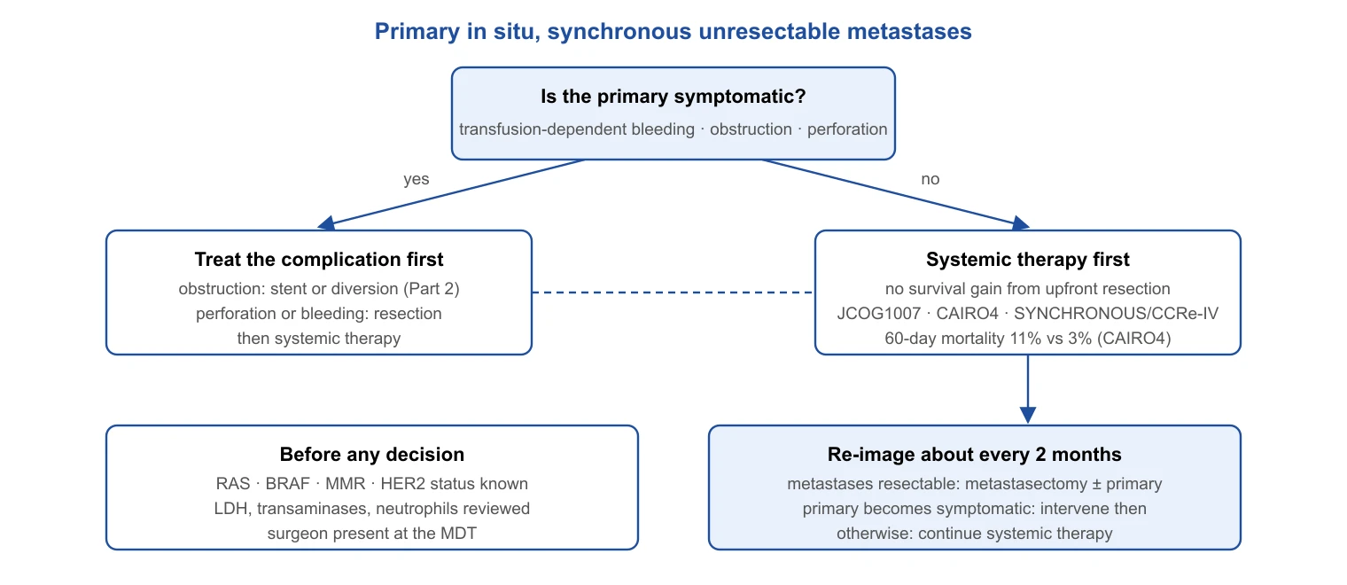
Systemic therapy: what the surgeon must carry into the multidisciplinary meeting
- Molecular status (RAS, BRAF, MMR, HER2) before treatment is planned; it decides the regimen, the likelihood of conversion and, for dMMR tumours, whether surgery should follow immunotherapy at all.
- Immunotherapy in dMMR disease produces pathological complete response in two-thirds of resected colon primaries and most resected metastases; radiological residual disease and early enlargement are not reliable signs of viable tumour.
- Conversion therapy is a race against liver injury: reassess at about 2-month intervals, operate when resectable, avoid more than about six cycles before hepatectomy.
- The asymptomatic primary stays in place: three randomised trials found no survival benefit from upfront resection and one found a four-fold rise in 60-day mortality.
Pathology, Surgical Quality Metrics and ctDNA in Surgical Decision-Making
Structured pathology reporting
The pathology report is the surgeon's audit as well as the oncologist's staging document. The International Collaboration on Cancer Reporting dataset, developed with the College of American Pathologists and the Royal College of Pathologists, defines 18 core (essential) and 7 non-core (recommended) elements; the sum of the core elements is the minimum reporting standard for every colorectal resection, covering site, type and grade, depth of invasion, margins, lymphovascular and perineural invasion, tumour deposits, nodes examined and involved and MMR status, with tumour budding and the plane of dissection also specified [268]. Staging follows the eighth edition of TNM [269]. Two elements changed. Tumour deposits are classified as N1c only when no nodal metastasis is present; a meta-analysis of 10,106 patients found them in 22%, invariably associated with worse outcome and with a stronger effect than nodal metastasis or extramural venous invasion, and argued that their number should be added to the node count [270]. Tumour budding, defined by the International Tumor Budding Consensus Conference as a single cell or cluster of up to four cells at the invasive front, counted in a 0.785 mm² hotspot and reported in three tiers, independently predicts nodal metastasis in pT1 cancer and survival in stage II disease [124].
Surgical quality metrics
Quality metrics convert the report and the audit database into feedback on the operation (Table). Lymph node yield of at least 12 is discussed with the evidence on lymphadenectomy in Part 1 of this series; the ratio of involved to examined nodes adds prognostic information once at least 10 nodes have been examined [271]. Plane of surgery is graded on the specimen: in the Leeds series of 399 specimens, 32% were mesocolic, 44% intramesocolic and 24% muscularis propria plane, mesocolic surgery removed more tissue (44 vs 21 mm from muscularis propria to margin) and was associated with a 15% absolute 5-year survival advantage over muscularis propria plane surgery (HR 0.57, 95% CI 0.38–0.85 univariate), significant on multivariate analysis in stage III (HR 0.45, 0.24–0.85)[272]. Thirty- and 90-day mortality and failure to rescue are hospital-level metrics; the evidence that variation in mortality between hospitals is driven more by rescue than by complication rate is set out in Part 2 of this series.
| Metric | Measure | Target | Evidence |
|---|---|---|---|
| Lymph node yield | Nodes examined per specimen | ≥12 | See Part 1 of this series |
| Lymph node ratio | Involved / examined nodes | Report; prognostic when ≥10 examined | [271] |
| Plane of surgery | Mesocolic / intramesocolic / muscularis propria | Mesocolic plane | [272] |
| Margin status | R0 / R1 / R2; circumferential margin | R0 | [268] |
| Tumour deposits | Present / number | Report number | [270] |
| Tumour budding | ITBCC three-tier count | Report | [124] |
| Mortality | 30-day and 90-day | Risk-adjusted, benchmarked | See Part 2 of this series |
| Failure to rescue | Deaths among patients with a serious complication | Hospital-level benchmark | See Part 2 of this series |
Circulating tumour DNA as a surgical tool
Circulating tumour DNA (ctDNA) detects molecular residual disease after resection with a precision no other test matches. In 125 Danish patients with stage I–III disease, a tumour-informed assay positive at postoperative day 30 carried an HR of 7.2 (95% CI 2.7–19.0) for relapse, surveillance positivity an HR of 43.5, 14 of 16 relapses (87.5%) were detected, and ctDNA preceded radiological recurrence by a mean of 8.7 months (up to 16.5)[273]. In 96 stage III patients, post-chemotherapy ctDNA separated 3-year recurrence-free intervals of 30% and 77%[274]. GALAXY scaled this to 1,039 patients with stage II–IV resectable disease: ctDNA positivity at 4 weeks carried an HR of 10.0 for recurrence and identified the patients who benefited from adjuvant chemotherapy (HR 6.59)[142]; the 2,240-patient update confirmed the effect on disease-free (HR 11.99) and overall survival (HR 9.68), and sustained rather than transient clearance on chemotherapy predicted 24-month disease-free survival of 89.0% versus 3.3%[143]. Serial testing every 3 months gave a median lead time of 9.8 months over CT, and the rate of ctDNA rise distinguished fast from slow recurrences [275].
Three applications matter to the surgeon. After metastasectomy, ctDNA was detectable in 24% of 54 patients resected for liver metastases, recurrence-free survival was worse when it was (HR 6.3, 95% CI 2.58–15.2), and end-of-treatment detection predicted a 5-year recurrence-free survival of 0% versus 75.6%[276]. Before metastasectomy, persistently rising ctDNA identifies disease that is biologically systemic and unlikely to be cured by major hepatectomy, although this use rests on cohort data only. In surveillance, the 8–10-month lead time may find oligometastatic recurrence while still resectable, but whether this converts to survival is untested, and the first randomised test of acting on a positive result was negative: in ALTAIR, trifluridine/tipiracil versus placebo in 243 ctDNA-positive patients without radiological disease gave disease-free survival of 9.30 versus 5.55 months (HR 0.79, 0.60–1.05)[16]. The DYNAMIC trial of ctDNA-guided adjuvant therapy is discussed in the stage-specific sections of this review.
Two limitations govern interpretation. Tumour-informed assays, which sequence the resected tumour and track a personalised mutation panel in plasma, are more sensitive than tumour-naive panels but need tissue and weeks of turnaround; both have a false-negative floor, so an undetectable result does not exclude residual disease [36]. On decision-making, ESMO states that molecular residual disease detection has high clinical validity for anticipating relapse but cannot be recommended in routine practice because there is no evidence of utility in directing treatment [36], and the NCCN panel likewise regards the data as insufficient to recommend ctDNA outside a clinical trial [56]. The surgical corollary is that ctDNA should inform the discussion of adjuvant therapy, surveillance intensity and the timing of metastasectomy, but should not on its own deny a patient a potentially curative operation (authors' judgement).
Recurrence Patterns, Salvage Surgery and Surveillance
Patterns and timing of recurrence
Recurrence after curative colon resection is less frequent than historical series suggested. In the entire Swedish population operated on in 2007–2012, 5-year recurrence was 5% in stage I, 12% in stage II and 33% in stage III; vascular and perineural invasion, emergency surgery, absence of central ligation, a short longitudinal margin and postoperative complications shortened time to recurrence [3]. Timing is front-loaded: in a Dutch population cohort, 19% developed metachronous metastases, 83% of them within 3 years, the liver being the most frequent site [277], and in 20,898 patients from 18 adjuvant trials recurrence fell below 1.5% per year after 5 years and 0.5% per year after 8 [278]. Metachronous peritoneal carcinomatosis developed in 4.2% of patients in Stockholm, with T4 (HR 9.98, 95% CI 3.10–32.11), N2, emergency surgery (HR 2.11) and R2 resection (HR 2.75) as independent predictors [2]. After resection of liver metastases, 56.7% of 1,669 patients recurred, first in the liver only in 43.2%, extrahepatic only in 35.8% and both in 21.0%, with a median recurrence-free survival of 16.3 months [279].
Survival after recurrence depends on when it occurs. In the ACCENT dataset of 17,381 stage II–III trial patients, 32.9% recurred and median survival after recurrence was 13.3 months; a longer disease-free interval, initial stage II and no prior adjuvant therapy predicted longer survival [280]. These figures predate routine metastasectomy; the roughly 10% of patients with metachronous metastases treated with curative intent now reach 5-year survival of about 60%[277].
Surveillance intensity and guideline schedules
The randomised evidence on surveillance intensity, including FACS and COLOFOL, was summarised in Part 2 of this series (Section 15.4); only the pooled conclusion is restated. The 2019 Cochrane review of 19 trials (13,216 participants) found that intensive follow-up made little or no difference to overall survival (HR 0.91, 95% CI 0.80–1.04; high-quality evidence), cancer-specific survival (HR 0.93, 0.81–1.07) or relapse-free survival (HR 1.05, 0.92–1.21), but doubled salvage surgery with curative intent (RR 1.98, 1.53–2.56; about 60 more operations per 1,000 patients)[31]. GILDA (1,228 patients) found that intensive surveillance detected recurrence 5.9 months earlier without a survival or quality-of-life difference [281], and in 8,529 National Cancer Database patients, high-intensity testing neither shortened time to detection (15.1 vs 16.0 months) nor increased resection of recurrence [282]. Surveillance should therefore be targeted at candidates for salvage surgery, in whom CT should not be omitted (Table).
| Guideline | History, examination, CEA | CT chest and abdomen | Colonoscopy |
|---|---|---|---|
| ASCO/CCO 2013 [283] | Every 3–6 months for 5 years | Annually for 3 years | 1 year after surgery, then every 5 years |
| NCCN 2024 [56] | Every 3–6 months for 2 years, then every 6 months to 5 years | Every 6–12 months for up to 5 years | 1 year, then 3 years, then every 5 years |
| ESMO 2020 [57] | Every 3–6 months for 3 years, then every 6–12 months to 5 years | Every 6–12 months for 3 years | 1 year, then every 3–5 years |
| ASCRS 2021 [284] | Risk-adapted intervals for 5 years | Risk-adapted, for 5 years | 1 year after surgery |
Salvage surgery and survival after recurrence
Isolated locoregional recurrence after colon resection is rare and is resected far less often than hepatic recurrence. In the Memorial Sloan Kettering series, 100 of 744 patients with recurrent colon cancer (13.4%) were explored with curative intent for locoregional recurrence; median survival was 30 months, R0 resection was achieved in 56 and was the strongest predictor of outcome, with a median survival of 66 months, whereas mesenteric or nodal recurrence and more than one site predicted poor survival [285]. Hepatic recurrence after hepatectomy is the commonest salvage scenario. In 1,036 patients, repeat hepatectomy carried morbidity similar to the first operation (27.1%, 34.4% and 33.3% after first, second and third hepatectomy) with mortality of 3.1% versus 1.6%, and patients who underwent repeat resection had 5-year survival of 54% from the first hepatectomy compared with 45% in those resected once [286]. The original Paul Brousse series reported 5-year survival of 41% after a second hepatectomy, with a curative second resection and an interval over 1 year as independent predictors [287], and a third hepatectomy was achieved in 88% of those attempted, with 25% morbidity, no mortality and 32% 5-year survival [288]. Across all sites, salvage surgery requires that complete resection be technically achievable, that the patient be fit for it, that the disease-free interval and ctDNA trajectory suggest indolent rather than explosive biology, and that systemic therapy be integrated rather than omitted (authors' judgement).
Special Populations
Older and frail patients
Chronological age is a poor guide to surgical risk; frailty is a better one. In 9,397 Dutch patients with stage I–III colorectal cancer, elderly patients who survived the first postoperative year had the same cancer-related survival as younger patients; the excess mortality of age was concentrated in the first year, and 30-day mortality greatly underestimated it [289]. A 2024 meta-analysis of 16 studies (245,747 patients) found a pooled frailty prevalence of 31% and nearly twice the odds of postoperative complications in frail patients (OR 1.94, 95% CI 1.47–2.56)[99]; over 65, frailty also predicted longer stay, readmission and worse long-term survival [100]. Fit older patients therefore receive standard oncological surgery; the effort goes into identifying and optimising the frail. Geriatric assessment changes outcomes when it changes management: in the cluster-randomised GAP70+ trial (718 patients, mean age 77), an assessment-guided intervention reduced grade 3–5 treatment toxicity from 71% to 51% (RR 0.74, 0.64–0.86)[290].
Adjuvant chemotherapy after 75 is a separate decision. In ACCENT, patients aged 70 and over derived little or no benefit from adding oxaliplatin to a fluoropyrimidine (disease-free survival HR 0.94, 95% CI 0.78–1.13; overall survival HR 1.04, 0.85–1.27), whereas fluoropyrimidines retained their efficacy [150]. In Medicare patients with stage III disease diagnosed after 75, adjuvant chemotherapy was associated with a survival benefit of trial magnitude (HR 0.60, 0.53–0.68), but oxaliplatin added at most a small increment (HR 0.84, 0.69–1.04)[291]. A fluoropyrimidine alone is therefore reasonable beyond 75, with oxaliplatin reserved for fit patients.
Early-onset colorectal cancer
Colorectal cancer before 50 is rising in most high-income countries. In the United States, colon cancer incidence has increased by 1.0–2.4% annually since the mid-1980s at ages 20–39 and by 0.5–1.3% since the mid-1990s at 40–54, while falling over 55 [48]; the proportion of cases under 55 rose from 11% in 1995 to 20% in 2019, with about 19,550 cases and 3,750 deaths under 50 expected in 2023 [292]. Across 50 countries, early-onset incidence rose in 27 over the latest decade, most steeply in New Zealand (3.97% per year) and England (3.59%)[293]. The disease is left-sided more often (distal colon 41% vs 34%), presents at a more advanced stage (72% vs 63% stage III–IV) and is diagnosed later (median 128 vs 79 days from first symptom), although the delay did not explain the stage difference [294]. Stage for stage, outcomes equal those of older patients despite more treatment: in the National Cancer Database, patients under 50 were two to four times more likely to receive systemic chemotherapy at every stage, with no survival gain in stage II (RR 0.90, 95% CI 0.69–1.17) and a marginal one in stage III (RR 0.89, 0.81–0.97)[295]. Germline testing should be offered to every patient diagnosed under 50: pathogenic variants were found in 16% of 450 unselected patients, a third of whom did not meet conventional testing criteria [116], and in about one in five of 430 in Michigan [296]. Fertility preservation and the implications of a hereditary syndrome for the extent of resection are discussed before surgery.
Hereditary syndromes
Lynch syndrome. In the Colon Cancer Family Registry, none of 50 MMR variant carriers treated by extended colectomy developed a metachronous cancer, compared with 22% of 332 treated by segmental resection; cumulative risk after segmental colectomy was 16% at 10 years, 41% at 20 and 62% at 30, and each 10 cm of bowel removed reduced it by 31%[297]. A meta-analysis of six studies (871 patients) gave metachronous rates of 22.8% after segmental and 6% after extended colectomy (OR 4.02, 95% CI 2.01–8.04), despite 1–2-yearly colonoscopy [298]. Risk is gene-specific: MLH1 and MSH2 variants carry high colorectal risk, MSH6 a modest one and PMS2 no demonstrable increase, and 10-year survival after colon cancer exceeds 80%[299]. The EHTG/ESCP guideline therefore recommends that extended colectomy be discussed with younger carriers, particularly of MLH1 and MSH2 variants, with segmental resection plus intensive colonoscopy as an acceptable alternative after shared decision-making [300]; ASCRS and ESMO take the same position [114,301]. Aspirin is the proven chemopreventive: in CAPP2, 600 mg daily reduced colorectal cancer over 10 years (9% vs 13%; HR 0.65, 0.43–0.97)[302].
Familial adenomatous polyposis. Prophylactic colectomy is timed by polyp burden, usually in the late teens or early twenties, and the choice between ileorectal anastomosis and ileal pouch-anal anastomosis depends on rectal polyp density, genotype and desmoid risk [303]. In a meta-analysis of 12 studies (1,002 patients), ileorectal anastomosis gave fewer bowel movements and less night-time defaecation, the pouch had more 30-day reoperations (23.4% vs 11.6%; OR 2.11), and rectal cancer occurred only in the ileorectal group (5.5%)[304]. Desmoids developed in 9% of 735 Dutch patients, 95% after abdominal surgery, with a family history raising the risk from 6.7% to 30%, and often made later conversion to a pouch impossible, which argues for primary pouch surgery in desmoid-prone families [305].
MUTYH-associated polyposis. Biallelic carriers are managed as attenuated polyposis; monoallelic carriers have a modest lifetime risk (7.2% in men and 5.6% in women to age 70), justifying enhanced screening but not prophylactic surgery [114,306]. NCCN provides gene-specific surveillance recommendations for these syndromes [307].
Pregnancy
Colorectal cancer in pregnancy is rare (pooled risk about 0.002%). In a systematic review of 119 cases, median age was 32 years, 12%, 41% and 47% were diagnosed in the first, second and third trimesters, 53% were colonic, bleeding (47%) and abdominal pain (38%) were the usual presentations and were easily attributed to pregnancy, and 9.8% received chemotherapy during pregnancy without permanent neonatal disability [308]. Resection is timed by gestational age and stage, usually in the second trimester or after delivery, with obstetric, oncological and neonatal teams planning together (authors' judgement).
Special populations: the decisions that differ from the standard pathway
- In older patients, assess frailty rather than age; fit patients receive standard surgery, frail patients receive optimisation first, and oxaliplatin is of little value after 70.
- Under 50, offer germline testing to all, discuss fertility before surgery, and do not escalate treatment beyond the stage-matched standard.
- In Lynch syndrome, discuss extended colectomy with young MLH1 and MSH2 carriers; the metachronous risk after segmental resection is 16% at 10 years and 41% at 20.
- In FAP, the pouch prevents rectal cancer at the cost of function and reoperation; desmoid-prone families should have it first.
Clinical Guidelines
The positions below are those cited in the body of this review; wording is paraphrased from the source and the strength or level is given only where the source states it. Where a body has no position extracted for a decision point, the cell says so rather than inferring one; access limitations are listed in Appendix A.
| Decision point | NCCN | ESMO or other European body | ASCO, ASCRS or other body | Sources |
|---|---|---|---|---|
| MMR/MSI testing | All newly diagnosed colon cancers; extended RAS, BRAF V600E, HER2 and NTRK in metastatic disease | MMR status required in all stage II tumours; RAS, BRAF V600E and MMR at diagnosis of metastatic disease | US Multi-Society Task Force: universal tumour testing for Lynch syndrome | [47,55–57] |
| Malignant polyp (pT1) | Not extracted | ESGE: low-risk resection is en bloc R0, sm1, well or moderately differentiated, no lymphovascular invasion, no high-grade budding; high-risk features prompt staging and MDT discussion, not automatic colectomy | JSCCR: surgery for any single adverse feature; USMSTF and ASCRS: a single adverse feature indicates oncological resection in a fit patient | [125–128] |
| Stage II adjuvant therapy | Not extracted | Same high-risk feature list as ASCO; no adjuvant chemotherapy for stage I; no fluoropyrimidine monotherapy in dMMR stage II | ASCO: against routine chemotherapy without risk features; offer for pT4; may offer for other features; oxaliplatin allowed in high-risk disease, 3 months CAPOX or 6 months FOLFOX | [57,134] |
| Stage III duration | Not extracted | 6 months for T4 or N2; 3 months of CAPOX for T1–3 N1 after shared decision-making | ASCO: same | [57,146] |
| ctDNA in adjuvant decisions | Data insufficient to recommend outside a clinical trial | ESMO Precision Medicine Working Group: high clinical validity for predicting relapse; not recommended in routine practice | ASCO stage II guideline predates DYNAMIC; JSCO: supported as a prognostic tool, with cost, assay standardisation and lack of interventional evidence as barriers | [36,56,134,145] |
| Immunotherapy for localised dMMR disease | Not extracted | Not extracted | No guideline statement extracted; the evidence is ATOMIC (adjuvant atezolizumab, phase III) and NICHE-2 (neoadjuvant ipilimumab–nivolumab, single arm); AZUR-2 is recruiting | [6,7,158] |
| Resectability of liver metastases | Not extracted | EGOSLIM consensus: resection not denied for stable disease after optimal chemotherapy if an adequate remnant can be preserved; ESMO: local treatment chosen from an oligometastatic "toolbox" | IHPBA consensus: standardised selection for transplantation | [47,164,201] |
| Synchronous disease and the asymptomatic primary | Systemic therapy first when the primary is asymptomatic and metastases unresectable | ESMO: same; EGOSLIM: chemotherapy before surgery in most scenarios and liver-first resection when the primary is asymptomatic | Not extracted | [47,56,164] |
| Lung metastasectomy | Not extracted | ESMO: one option within the oligometastatic toolbox, not a default | STS consensus: endorsed for selected patients with controlled primary disease, resectable pulmonary disease and adequate reserve, within MDT review | [47,209] |
| Cytoreduction and HIPEC | Cytoreductive surgery and/or intraperitoneal chemotherapy "can be considered in experienced centers for selected patients with limited peritoneal metastases for whom R0 resection can be achieved" (as quoted) | ESMO: complete cytoreduction [II, A]; HIPEC experimental, to be validated in trials. Dutch guideline: CRS/HIPEC with mitomycin C for isolated PM with PCI below 20 | PSOGI 2022: HIPEC conditionally recommended; Dutch mitomycin C protocol preferred; 30-minute oxaliplatin rejected | [44,47,222,243,244] |
| Surveillance after stage II–III resection | CEA every 3–6 months for 2 years then 6-monthly to 5 years; CT every 6–12 months for up to 5 years; colonoscopy at 1 year, 3 years, then every 5 years | CEA every 3–6 months for 3 years then every 6–12 months to 5 years; CT every 6–12 months for 3 years; colonoscopy at 1 year then every 3–5 years | ASCO/CCO: CEA every 3–6 months for 5 years, CT annually for 3 years, colonoscopy at 1 year then every 5 years; ASCRS: risk-adapted intervals for 5 years | [56,57,283,284] |
| Extent of colectomy in Lynch syndrome | Genetic/familial guideline: gene-specific surveillance | EHTG/ESCP: discuss extended colectomy with younger carriers, particularly MLH1 and MSH2; segmental resection with intensive colonoscopy acceptable after shared decision-making; ESMO hereditary guideline: same | ASCRS: same | [114,300,301,307] |
| Germline referral | Not extracted | ESMO: dMMR tumour without BRAF mutation or MLH1 methylation, family history criteria, diagnosis before 50, polyposis, known familial variant | USMSTF: same triggers | [55,114] |
Where guidelines disagree, the disagreement is about HIPEC, the malignant polyp and the frequency of imaging. On HIPEC the positions are irreconcilable: ESMO calls it experimental [47,243], the Dutch guideline retains mitomycin C HIPEC as standard for a PCI below 20 because PRODIGE 7 tested a different regimen [222], PSOGI conditionally recommends it while grading nine of its ten questions as weak or very weak evidence [44], and NCCN sits between [244]. On the malignant polyp the features are agreed and the arithmetic is not: JSCCR acts on any single adverse feature [126], whereas ESGE weighs cumulative risk [125]. On surveillance, NCCN permits CT every 6–12 months for up to five years, ESMO for three years and ASCO/CCO annually for three years [56,57,283], although randomised evidence finds no survival difference between intensities [31]. No guideline consulted directs treatment on ctDNA, and none had incorporated the ATOMIC result at the time of search.
Evidence Gaps
- Whether treating ctDNA-positive patients changes outcome. Trifluridine–tipiracil in ALTAIR was negative (HR 0.79, 95% CI 0.60–1.05)[16] and the escalation arm of DYNAMIC-III did not reach its endpoint [15]; CIRCULATE-US (NRG-GI008) continues to recruit [144].
- Whether ctDNA negativity can safely shorten chemotherapy in stage III. DYNAMIC-III failed its non-inferiority margin for de-escalation (three-year recurrence-free survival 85.3% vs 88.1%)[15]; VEGA has not reported, and COBRA (NRG-GI005) closed for futility on a clearance endpoint reported only at a congress.
- Perioperative versus adjuvant immunotherapy in localised dMMR disease. ATOMIC improved three-year disease-free survival with adjuvant atezolizumab [7]; NICHE-2 reported 100% three-year disease-free survival after neoadjuvant ipilimumab–nivolumab in a single arm [158]. The comparison is indirect and ATOMIC has not reported overall survival; AZUR-2 (NCT05855200) is the randomised test.
- Which prehabilitation, for whom. The positive trials selected unfit patients or used supervised high-intensity programmes [9,103], the negative trial enrolled frail octogenarians [10], and the network meta-analysis rates every component comparison as very low certainty [104].
- Long-term survival after ablation versus resection. COLLISION stopped at a pre-planned interim analysis with a median follow-up of 28.9 months and an immature survival endpoint (HR 1.05, 95% CI 0.69–1.58)[19].
- The sequencing of synchronous liver metastases. METASYNC analysed 85 patients [175], the LiverMetSurvey comparison is confounded by indication [176] and the network meta-analysis found no significant differences [177].
- Remnant augmentation. Liver venous deprivation outperformed portal vein embolisation in cohorts [180,181] and the randomised HYPER-LIV01 trial was in progress when they were reported [179]; ALPPS rests on a single 97-patient trial with 90-day mortality of 6–8% in both arms [38].
- Transplantation beyond the TransMet criteria. TransMet randomised 94 patients aged 18–65 with BRAF wild-type, liver-only, chemotherapy-responsive disease [20]; the ten-year SECA-II data come from 25 patients [200]; access depends on allocation policy [201].
- The effect of pulmonary metastasectomy. PulMiCC closed after 65 randomised patients, 93 in the updated analysis, with confidence intervals that include both benefit and harm [21,22].
- Mitomycin C HIPEC for established peritoneal metastases. No randomised trial has isolated it; HIPECT4 reported locoregional control without a survival difference after a T4 primary [27,242]; GECOP-MMC (216 planned) will answer the question [223].
- Perioperative systemic therapy before cytoreduction, and PIPAC. CAIRO6 found no overall survival benefit (HR 0.85, 95% CI 0.62–1.15) with more morbidity [229]; PIPAC rests on phase I–II data, with a 0% radiological response in the first prospective monotherapy trial in colorectal cancer [249].
- Surgery and surveillance in the era of immunotherapy and ctDNA. There is no validated definition of clinical complete response after neoadjuvant immunotherapy and no organ-preservation trial with survival endpoints [258,261]; ctDNA surveillance gives a lead time of 8–10 months over imaging [273,275], but whether earlier detection converts to survival is untested.
Scenario-Based Decision Framework
The scenarios below condense Sections 6 to 16 into eight presentations that recur in multidisciplinary meetings. Each assumes complete staging, MMR status on the biopsy, a frailty assessment and a multidisciplinary discussion.
Scenario 1. The malignant polyp with deep invasion alone. A 64-year-old has a 2 cm sigmoid polyp removed en bloc: adenocarcinoma invading 1,500 µm into the submucosa (sm2), well differentiated, no lymphovascular invasion, no high-grade budding, clear vertical margin.
Evidence. Depth of invasion was not an independent predictor of nodal metastasis after adjustment (OR 1.73, 95% CI 0.96–3.12), and as the only adverse feature it carried an absolute risk of 2.6%[122]; lymphovascular invasion (OR 7.42) and budding (OR 4.00) carry far more weight [121].
Approach. Under ESGE this justifies discussion, not automatic colectomy [125]; under JSCCR and ASCRS it is an indication for resection [126,128]. A fit patient should hear both the 2.6% nodal risk and the risks of sigmoid colectomy, and decide.
Caveat. The staged approach costs nothing oncologically only when the specimen is en bloc and fully assessable [131].
Scenario 2. Stage II, mismatch repair-deficient. A 58-year-old woman undergoes right hemicolectomy for a pT3 N0 caecal tumour, 18 nodes negative, no obstruction or perforation; immunohistochemistry shows loss of MLH1 and PMS2.
Evidence. Fluorouracil gave no disease-free survival benefit in dMMR tumours (HR 1.10) and was associated with worse overall survival in stage II (HR 2.95)[5]. MLH1 loss with BRAF V600E mutation or MLH1 methylation indicates sporadic disease [55].
Approach. Observation and no fluoropyrimidine monotherapy, as ASCO and ESMO recommend [57,134]. Reflex BRAF or methylation testing; if neither is present, genetic counselling and germline testing.
Caveat. pT4 dMMR stage II has no proven regimen; whether oxaliplatin restores a benefit is unknown [139].
Scenario 3. Stage III, ctDNA-negative at four weeks. A 61-year-old man has a pT3 N1a pMMR descending colon cancer resected; a tumour-informed ctDNA assay at four weeks is undetectable and he asks whether chemotherapy can be omitted.
Evidence. In DYNAMIC-III, de-escalation in ctDNA-negative stage III patients did not meet non-inferiority (three-year recurrence-free survival 85.3% vs 88.1%)[15]. For T1–3 N1 disease, 3 months of CAPOX matched 6 months (83.1% vs 83.3%) with grade 2 or worse neuropathy in 25% rather than 58%[11,12].
Approach. Three months of CAPOX, started within eight weeks of surgery [146,148]. The ctDNA result belongs in the prognostic discussion, but a negative assay has a false-negative floor and no guideline recommends treating on ctDNA outside a trial [36,56].
Caveat. For T4 or N2 disease the course is 6 months [11].
Scenario 4. Synchronous liver metastases, right colon. A 55-year-old with an asymptomatic pT3 right colon cancer has three unilobar liver metastases, the largest 4 cm, no extrahepatic disease, a normal liver and an adequate remnant; the tumour is KRAS-mutant.
Evidence. Benchmark five-year survival after resection of two to three metastases is at least 48%[17]; RAS mutation worsens it but does not contraindicate resection [169]. In unilobar disease the three sequencing strategies gave equal survival in LiverMetSurvey [176], and METASYNC found no difference in major complications between simultaneous and delayed resection [175].
Approach. Liver MRI before any chemotherapy; hepatobiliary MDT with RAS and BRAF status. Parenchymal-sparing resection of all three lesions, simultaneous with the colectomy if the hepatectomy is minor and the patient fit, otherwise staged; ablation is an alternative for lesions of 3 cm or less [19].
Caveat. Perioperative chemotherapy delays recurrence without improving survival [187]; if given, it should not exceed about six cycles before hepatectomy [266].
Scenario 5. Disappearing liver metastases. After six cycles of FOLFOX and bevacizumab for four bilobar metastases, two lesions under 2 cm are no longer visible on CT.
Evidence. Residual disease or early in situ recurrence was found at 83% of sites of vanished metastases [192]; contrast-enhanced MRI detects residual disease best, and resecting the sites reduces local recurrence without a demonstrated survival benefit [193].
Approach. Gadoxetic-acid MRI, then operate rather than consolidate: resect or ablate the sites of the vanished lesions using the baseline imaging as the map, and the visible lesions with parenchymal sparing; stop bevacizumab some five to six weeks before surgery.
Caveat. The scenario is avoidable by baseline liver MRI and surgery planned before the target disappears; chemotherapy-associated liver injury is dose-dependent [264,265].
Scenario 6. Isolated peritoneal disease, PCI 12. A fit 49-year-old has peritoneal metastases from a sigmoid primary with an operative PCI of 12 at laparoscopy, no small-bowel involvement, no liver or extraperitoneal disease; the tumour is pMMR and RAS wild-type.
Evidence. Survival after cytoreduction falls linearly with PCI (HR 1.1 per point); surgery is generally indicated below 12, contraindicated above 17 and individualised between [220,221]. Both arms of PRODIGE 7 reached about 41 months after complete cytoreduction, and oxaliplatin HIPEC added 60-day morbidity (26% vs 15%) without survival [24].
Approach. Referral to a peritoneal centre, as PSOGI recommends whether or not HIPEC is offered [237]. Cytoreduction aiming at CC-0; mitomycin C HIPEC according to local policy or within GECOP-MMC [223]; no oxaliplatin HIPEC outside a trial; upfront surgery, since routine perioperative chemotherapy added toxicity without survival in CAIRO6 [229].
Caveat. A PCI of 12 lies at the edge of the favourable band, and imaging underestimates the operative PCI [216]. The expectation to share is a median survival of about 40 months.
Scenario 7. Asymptomatic primary, unresectable liver disease, RAS-mutant. A 67-year-old presents with a non-obstructing ascending colon cancer and more than ten bilobar liver metastases; KRAS is mutated and the tumour is pMMR.
Evidence. Upfront resection of the primary did not improve survival in JCOG1007 (HR 1.10), CAIRO4 (20.1 vs 18.3 months) or SYNCHRONOUS/CCRe-IV (HR 0.94), carried 60-day mortality of 11% versus 3% in CAIRO4, and left 24.1% of resected patients without any chemotherapy [28–30,267]. For right-sided or RAS-mutant disease, FOLFOXIRI plus bevacizumab improved progression-free survival as induction in CAIRO5 [191].
Approach. Systemic therapy first, as ESMO and NCCN recommend [47,56], with the surgeon at every multidisciplinary review and resectability reassessed about every two months. Operate on the liver as soon as it becomes resectable; operate on the primary earlier only for bleeding, obstruction or perforation (Part 2 of this series).
Caveat. Raised lactate dehydrogenase, transaminases or neutrophils marked the patients who died after upfront resection in CAIRO4 [29]; if the primary obstructs, a stent or diversion is preferable to resection where feasible.
Scenario 8. Lynch syndrome at 32. A 32-year-old woman with a known germline MLH1 variant presents with a non-obstructing pT3 N1 caecal cancer, dMMR on immunohistochemistry.
Evidence. After segmental colectomy in MMR variant carriers, metachronous cancer occurred in 22%, with a cumulative risk of 16% at 10 years, 41% at 20 and 62% at 30, against none of 50 after extended colectomy [297]; pooled rates were 22.8% versus 6% (OR 4.02)[298]. Aspirin 600 mg daily reduced colorectal cancer over ten years in CAPP2 (HR 0.65)[302].
Approach. Genetic counselling before, not after, the operation, as EHTG/ESCP, ASCRS and ESMO advise [114,300,301]. Discuss extended colectomy with ileorectal anastomosis against right hemicolectomy with intensive colonoscopy, with the functional cost and the 10- and 20-year metachronous risks on the table; fertility counselling before surgery. For stage III dMMR disease, mFOLFOX6 with atezolizumab is the trial-supported adjuvant regimen [7]; aspirin chemoprevention afterwards.
Caveat. The choice of extent is hers after shared decision-making; segmental resection with surveillance is an accepted alternative in every guideline cited. Neoadjuvant immunotherapy is an option within trials or expert centres [6].
Conclusions
The molecular result that most changes a surgical plan is also the simplest to obtain. MMR status from the diagnostic biopsy identifies Lynch syndrome in one patient in 35 [4], removes fluoropyrimidine monotherapy from stage II [5], and opens checkpoint inhibition before surgery in locally advanced disease [6] and after it in stage III [7]. RAS, BRAF and HER2 status and the side of the primary then decide the first-line regimen in metastatic disease and with it the likelihood that liver or lung surgery will follow [80,81].
Adjuvant therapy has become shorter where risk is low and more targeted where it is high. Three months of CAPOX suffices for T1–3 N1 stage III disease at a fraction of the neuropathy of six [11,12], and the largest effects belong to interventions targeted by biology, immunotherapy in dMMR disease and aspirin in PI3K-altered tumours [7,153]. ctDNA has so far improved only the decision not to treat [13,15].
Metastasectomy is defined by benchmarks and bounded by biology. Liver resection yields five-year survival of 45–58% for solitary disease and at least 28% for more than three metastases [17]; parenchymal sparing preserves the repeat resection that accounts for many long-term survivors [39,163]; thermal ablation has become an equal option for lesions of 3 cm or less [19]; and transplantation offers five-year survival of 56.6% to a selected few, constrained by organ supply [20,201]; the lung evidence is weaker [21]. In peritoneal disease, complete cytoreduction in an expert centre gives a median survival of about 40 months [24,229]; oxaliplatin HIPEC should not be given outside a trial, prophylactic HIPEC fails, and mitomycin C HIPEC awaits GECOP-MMC [25,26,223].
The surgeon's contribution to systemic therapy is timing. Conversion is a race against chemotherapy-associated liver injury, so resectability is reassessed about every two months and metastases are resected when they become resectable, not after a further course [47,266]; the asymptomatic primary stays in place because three randomised trials found no survival benefit from removing it [28,30,267]. After resection, surveillance should be targeted at candidates for salvage surgery, since intensity doubles salvage operations without improving survival [31].
Finally, the patient who does not fit the pathway needs the pathway adjusted, not abandoned. In older patients, frailty is optimised and the fluoropyrimidine retained while oxaliplatin adds little after 70 [150]; under 50, germline testing is universal and treatment is not escalated beyond the stage-matched standard [116,295]; in Lynch syndrome, the extent of colectomy is decided with the patient before the operation, because the metachronous risk after segmental resection reaches 41% at 20 years [297]; and in pregnancy, timing is set by gestation and stage with the obstetric, oncological and neonatal teams planning together [308].
Appendix A. Verification Notes and Limitations of This Review
- Engstrand J, Nilsson H, Strömberg C, Jonas E, Freedman J. Colorectal cancer liver metastases - a population-based study on incidence, management and survival. BMC Cancer. 2018;18(1):78. doi:10.1186/s12885-017-3925-x. PMID: 29334918.
- Segelman J, Granath F, Holm T, Machado M, Mahteme H, Martling A. Incidence, prevalence and risk factors for peritoneal carcinomatosis from colorectal cancer. Br J Surg. 2012;99(5):699-705. doi:10.1002/bjs.8679. PMID: 22287157.
- Osterman E, Glimelius B. Recurrence risk after up-to-date colon cancer staging, surgery, and pathology: analysis of the entire Swedish population. Dis Colon Rectum. 2018;61(9):1016-1025. doi:10.1097/DCR.0000000000001158. PMID: 30086050.
- Hampel H, Frankel WL, Martin E, Arnold M, Khanduja K, Kuebler P, et al. Feasibility of screening for Lynch syndrome among patients with colorectal cancer. J Clin Oncol. 2008;26(35):5783-8. doi:10.1200/JCO.2008.17.5950. PMID: 18809606.
- Sargent DJ, Marsoni S, Monges G, Thibodeau SN, Labianca R, Hamilton SR, et al. Defective mismatch repair as a predictive marker for lack of efficacy of fluorouracil-based adjuvant therapy in colon cancer. J Clin Oncol. 2010;28(20):3219-26. doi:10.1200/JCO.2009.27.1825. PMID: 20498393.
- Chalabi M, Verschoor YL, Tan PB, Balduzzi S, Van Lent AU, Grootscholten C, et al. Neoadjuvant immunotherapy in locally advanced mismatch repair-deficient colon cancer. N Engl J Med. 2024;390(21):1949-1958. doi:10.1056/NEJMoa2400634. PMID: 38838311.
- Sinicrope FA, Ou FS, Arnold D, Peters WR, Behrens RJ, Lieu CH, et al. Atezolizumab plus FOLFOX for stage III mismatch repair-deficient colon cancer. N Engl J Med. 2026;394(12):1155-66. doi:10.1056/NEJMoa2507874. PMID: 41880612.
- Molenaar CJ, van Rooijen SJ, Fokkenrood HJ, Roumen RM, Janssen L, Slooter GD. Prehabilitation versus no prehabilitation to improve functional capacity, reduce postoperative complications and improve quality of life in colorectal cancer surgery. Cochrane Database Syst Rev. 2022;5:CD013259. doi:10.1002/14651858.CD013259.pub2. PMID: 35588252.
- Molenaar CJL, Minnella EM, Coca-Martinez M, Ten Cate DWG, Regis M, Awasthi R, et al. Effect of multimodal prehabilitation on reducing postoperative complications and enhancing functional capacity following colorectal cancer surgery: the PREHAB randomized clinical trial. JAMA Surg. 2023;158(6):572-81. doi:10.1001/jamasurg.2023.0198. PMID: 36988937.
- Carli F, Bousquet-Dion G, Awasthi R, Elsherbini N, Liberman S, Boutros M, et al. Effect of multimodal prehabilitation vs postoperative rehabilitation on 30-day postoperative complications for frail patients undergoing resection of colorectal cancer: a randomized clinical trial. JAMA Surg. 2020;155(3):233-42. doi:10.1001/jamasurg.2019.5474. PMID: 31968063.
- Grothey A, Sobrero AF, Shields AF, Yoshino T, Paul J, Taieb J, et al. Duration of adjuvant chemotherapy for stage III colon cancer. N Engl J Med. 2018;378(13):1177-88. doi:10.1056/NEJMoa1713709. PMID: 29590544.
- Iveson TJ, Kerr RS, Saunders MP, Cassidy J, Hollander NH, Tabernero J, et al. 3 versus 6 months of adjuvant oxaliplatin-fluoropyrimidine combination therapy for colorectal cancer (SCOT): an international, randomised, phase 3, non-inferiority trial. Lancet Oncol. 2018;19(4):562-78. doi:10.1016/S1470-2045(18)30093-7. PMID: 29611518.
- Tie J, Cohen JD, Lahouel K, Lo SN, Wang Y, Kosmider S, et al. Circulating tumor DNA analysis guiding adjuvant therapy in stage II colon cancer. N Engl J Med. 2022;386(24):2261-72. doi:10.1056/NEJMoa2200075. PMID: 35657320.
- Tie J, Wang Y, Lo SN, Lahouel K, Cohen JD, Wong R, et al. Circulating tumor DNA analysis guiding adjuvant therapy in stage II colon cancer: 5-year outcomes of the randomized DYNAMIC trial. Nat Med. 2025;31(5):1509-18. doi:10.1038/s41591-025-03579-w. PMID: 40055522.
- Tie J, Wang Y, Loree JM, Cohen JD, Wong R, Price T, et al. Circulating tumor DNA-guided adjuvant therapy in locally advanced colon cancer: the randomized phase 2/3 DYNAMIC-III trial. Nat Med. 2025;31(12):4291-300. doi:10.1038/s41591-025-04030-w. PMID: 41115959.
- Bando H, Watanabe J, Takahashi Y, Kotaka M, Matsuhashi N, Oki E, et al. Post-adjuvant chemotherapy in ctDNA-positive patients with resected colorectal cancer: a randomized phase 3 trial. Nat Med. 2026;32(7):2473-2480. doi:10.1038/s41591-026-04428-0. PMID: 42260101.
- Viganò L, Risi L, Dasari BVM, Marques HP, Giuliante F, Nordin A, et al. Benchmarking oncologic outcomes of liver resection for colorectal metastases: LiverMetSurvey-based reference values for evaluating alternative treatments. Ann Surg. 2025;282(5):876-87. doi:10.1097/SLA.0000000000006884. PMID: 40772629.
- Deng G, Li H, Jia GQ, Fang D, Tang YY, Xie J, et al. Parenchymal-sparing versus extended hepatectomy for colorectal liver metastases: a systematic review and meta-analysis. Cancer Med. 2019;8(14):6165-75. doi:10.1002/cam4.2515. PMID: 31464101.
- van der Lei S, Puijk RS, Dijkstra M, Schulz HH, Vos DJW, De Vries JJJ, et al. Thermal ablation versus surgical resection of small-size colorectal liver metastases (COLLISION): an international, randomised, controlled, phase 3 non-inferiority trial. Lancet Oncol. 2025;26(2):187-99. doi:10.1016/S1470-2045(24)00660-0. PMID: 39848272.
- Adam R, Piedvache C, Chiche L, Adam JP, Salamé E, Bucur P, et al. Liver transplantation plus chemotherapy versus chemotherapy alone in patients with permanently unresectable colorectal liver metastases (TransMet): results from a multicentre, open-label, prospective, randomised controlled trial. Lancet. 2024;404(10458):1107-18. doi:10.1016/S0140-6736(24)01595-2. PMID: 39306468.
- Treasure T, Farewell V, Macbeth F, Monson K, Williams NR, Brew-Graves C, et al. Pulmonary Metastasectomy versus Continued Active Monitoring in Colorectal Cancer (PulMiCC): a multicentre randomised clinical trial. Trials. 2019;20(1):718. doi:10.1186/s13063-019-3837-y. PMID: 31831062.
- Milosevic M, Edwards J, Tsang D, Dunning J, Shackcloth M, Batchelor T, et al. Pulmonary Metastasectomy in Colorectal Cancer: updated analysis of 93 randomized patients - control survival is much better than previously assumed. Colorectal Dis. 2020;22(10):1314-24. doi:10.1111/codi.15113. PMID: 32388895.
- Treasure T, Farewell V, Macbeth F, Batchelor T, Milošević M, King J, et al. The Pulmonary Metastasectomy in Colorectal Cancer cohort study: analysis of case selection, risk factors and survival in a prospective observational study of 512 patients. Colorectal Dis. 2021;23(7):1793-803. doi:10.1111/codi.15651. PMID: 33783109.
- Quénet F, Elias D, Roca L, Goéré D, Ghouti L, Pocard M, et al. Cytoreductive surgery plus hyperthermic intraperitoneal chemotherapy versus cytoreductive surgery alone for colorectal peritoneal metastases (PRODIGE 7): a multicentre, randomised, open-label, phase 3 trial. Lancet Oncol. 2021;22(2):256-266. doi:10.1016/S1470-2045(20)30599-4. PMID: 33476595.
- Goéré D, Glehen O, Quenet F, Guilloit JM, Bereder JM, Lorimier G, et al. Second-look surgery plus hyperthermic intraperitoneal chemotherapy versus surveillance in patients at high risk of developing colorectal peritoneal metastases (PROPHYLOCHIP-PRODIGE 15): a randomised, phase 3 study. Lancet Oncol. 2020;21(9):1147-1154. doi:10.1016/S1470-2045(20)30322-3. PMID: 32717180.
- Zwanenburg ES, El Klaver C, Wisselink DD, Punt CJA, Snaebjornsson P, Crezee J, et al. Adjuvant hyperthermic intraperitoneal chemotherapy in patients with locally advanced colon cancer (COLOPEC): 5-year results of a randomized multicenter trial. J Clin Oncol. 2024;42(2):140-145. doi:10.1200/JCO.22.02644. PMID: 37922442.
- Arjona-Sánchez A, Espinosa-Redondo E, Gutiérrez-Calvo A, Segura-Sampedro JJ, Pérez-Viejo E, Concepción-Martín V, et al. Efficacy and safety of intraoperative hyperthermic intraperitoneal chemotherapy for locally advanced colon cancer: a phase 3 randomized clinical trial. JAMA Surg. 2023;158(7):683-691. doi:10.1001/jamasurg.2023.0662. PMID: 37099280.
- Kanemitsu Y, Shitara K, Mizusawa J, Hamaguchi T, Shida D, Komori K, et al. Primary tumor resection plus chemotherapy versus chemotherapy alone for colorectal cancer patients with asymptomatic, synchronous unresectable metastases (JCOG1007; iPACS): a randomized clinical trial. J Clin Oncol. 2021;39(10):1098-1107. doi:10.1200/JCO.20.02447. PMID: 33560877.
- van der Kruijssen DEW, Elias SG, Vink GR, van Rooijen KL, 't Lam-Boer J, Mol L, et al. Sixty-day mortality of patients with metastatic colorectal cancer randomized to systemic treatment vs primary tumor resection followed by systemic treatment: the CAIRO4 phase 3 randomized clinical trial. JAMA Surg. 2021;156(12):1093-1101. doi:10.1001/jamasurg.2021.4992. PMID: 34613339.
- Rahbari NN, Biondo S, Frago R, Feißt M, Kreisler E, Rossion I, et al. Primary tumor resection before systemic therapy in patients with colon cancer and unresectable metastases: combined results of the SYNCHRONOUS and CCRe-IV trials. J Clin Oncol. 2024;42(13):1531-1541. doi:10.1200/JCO.23.01540. PMID: 38412408.
- Jeffery M, Hickey BE, Hider PN. Follow-up strategies for patients treated for non-metastatic colorectal cancer. Cochrane Database Syst Rev. 2019;9(9):CD002200. doi:10.1002/14651858.CD002200.pub4. PMID: 31483854.
- Pino MS, Chung DC. The chromosomal instability pathway in colon cancer. Gastroenterology. 2010;138(6):2059-72. doi:10.1053/j.gastro.2009.12.065. PMID: 20420946.
- Luchini C, Bibeau F, Ligtenberg MJL, Singh N, Nottegar A, Bosse T, et al. ESMO recommendations on microsatellite instability testing for immunotherapy in cancer, and its relationship with PD-1/PD-L1 expression and tumour mutational burden: a systematic review-based approach. Ann Oncol. 2019;30(8):1232-43. doi:10.1093/annonc/mdz116. PMID: 31056702.
- Weisenberger DJ, Siegmund KD, Campan M, Young J, Long TI, Faasse MA, et al. CpG island methylator phenotype underlies sporadic microsatellite instability and is tightly associated with BRAF mutation in colorectal cancer. Nat Genet. 2006;38(7):787-93. doi:10.1038/ng1834. PMID: 16804544.
- Guinney J, Dienstmann R, Wang X, de Reyniès A, Schlicker A, Soneson C, et al. The consensus molecular subtypes of colorectal cancer. Nat Med. 2015;21(11):1350-6. doi:10.1038/nm.3967. PMID: 26457759.
- Pascual J, Attard G, Bidard FC, Curigliano G, De Mattos-Arruda L, Diehn M, et al. ESMO recommendations on the use of circulating tumour DNA assays for patients with cancer: a report from the ESMO Precision Medicine Working Group. Ann Oncol. 2022;33(8):750-768. doi:10.1016/j.annonc.2022.05.520. PMID: 35809752.
- Kishi Y, Abdalla EK, Chun YS, Zorzi D, Madoff DC, Wallace MJ, et al. Three hundred and one consecutive extended right hepatectomies: evaluation of outcome based on systematic liver volumetry. Ann Surg. 2009;250(4):540-8. doi:10.1097/SLA.0b013e3181b674df. PMID: 19730239.
- Sandström P, Røsok BI, Sparrelid E, Larsen PN, Larsson AL, Lindell G, et al. ALPPS improves resectability compared with conventional two-stage hepatectomy in patients with advanced colorectal liver metastasis: results from a Scandinavian multicenter randomized controlled trial (LIGRO trial). Ann Surg. 2018;267(5):833-40. doi:10.1097/SLA.0000000000002511. PMID: 28902669.
- Mise Y, Aloia TA, Brudvik KW, Schwarz L, Vauthey JN, Conrad C. Parenchymal-sparing hepatectomy in colorectal liver metastasis improves salvageability and survival. Ann Surg. 2016;263(1):146-52. doi:10.1097/SLA.0000000000001194. PMID: 25775068.
- Viganò L, Procopio F, Cimino MM, Donadon M, Gatti A, Costa G, et al. Is tumor detachment from vascular structures equivalent to R0 resection in surgery for colorectal liver metastases? An observational cohort. Ann Surg Oncol. 2016;23(4):1352-60. doi:10.1245/s10434-015-5009-y. PMID: 26714946.
- Lam VW, Laurence JM, Johnston E, Hollands MJ, Pleass HC, Richardson AJ. A systematic review of two-stage hepatectomy in patients with initially unresectable colorectal liver metastases. HPB (Oxford). 2013;15(7):483-91. doi:10.1111/j.1477-2574.2012.00607.x. PMID: 23750490.
- Schnitzbauer AA, Lang SA, Goessmann H, Nadalin S, Baumgart J, Farkas SA, et al. Right portal vein ligation combined with in situ splitting induces rapid left lateral liver lobe hypertrophy enabling 2-staged extended right hepatic resection in small-for-size settings. Ann Surg. 2012;255(3):405-14. doi:10.1097/SLA.0b013e31824856f5. PMID: 22330038.
- Jacquet P, Sugarbaker PH. Clinical research methodologies in diagnosis and staging of patients with peritoneal carcinomatosis. Cancer Treat Res. 1996;82:359-374. doi:10.1007/978-1-4613-1247-5_23. PMID: 8849962.
- Hübner M, van Der Speeten K, Govaerts K, de Hingh I, Villeneuve L, Kusamura S, et al. 2022 Peritoneal Surface Oncology Group International consensus on HIPEC regimens for peritoneal malignancies: colorectal cancer. Ann Surg Oncol. 2024;31(1):567-576. doi:10.1245/s10434-023-14368-5. PMID: 37940803.
- Alyami M, Hübner M, Grass F, Bakrin N, Villeneuve L, Laplace N, et al. Pressurised intraperitoneal aerosol chemotherapy: rationale, evidence, and potential indications. Lancet Oncol. 2019;20(7):e368-e377. doi:10.1016/S1470-2045(19)30318-3. PMID: 31267971.
- Adam R, Delvart V, Pascal G, Valeanu A, Castaing D, Azoulay D, et al. Rescue surgery for unresectable colorectal liver metastases downstaged by chemotherapy: a model to predict long-term survival. Ann Surg. 2004;240(4):644-657. doi:10.1097/01.sla.0000141198.92114.f6. PMID: 15383792.
- Cervantes A, Adam R, Roselló S, Arnold D, Normanno N, Taïeb J, et al. Metastatic colorectal cancer: ESMO Clinical Practice Guideline for diagnosis, treatment and follow-up. Ann Oncol. 2023;34(1):10-32. doi:10.1016/j.annonc.2022.10.003. PMID: 36307056.
- Siegel RL, Fedewa SA, Anderson WF, Miller KD, Ma J, Rosenberg PS, et al. Colorectal cancer incidence patterns in the United States, 1974-2013. J Natl Cancer Inst. 2017;109(8):djw322. doi:10.1093/jnci/djw322. PMID: 28376186.
- Fearon ER, Vogelstein B. A genetic model for colorectal tumorigenesis. Cell. 1990;61(5):759-67. doi:10.1016/0092-8674(90)90186-i. PMID: 2188735.
- East JE, Atkin WS, Bateman AC, Clark SK, Dolwani S, Ket SN, et al. British Society of Gastroenterology position statement on serrated polyps in the colon and rectum. Gut. 2017;66(7):1181-96. doi:10.1136/gutjnl-2017-314005. PMID: 28450390.
- Crockett SD, Nagtegaal ID. Terminology, molecular features, epidemiology, and management of serrated colorectal neoplasia. Gastroenterology. 2019;157(4):949-66.e4. doi:10.1053/j.gastro.2019.06.041. PMID: 31323292.
- Cancer Genome Atlas Network. Comprehensive molecular characterization of human colon and rectal cancer. Nature. 2012;487(7407):330-7. doi:10.1038/nature11252. PMID: 22810696.
- Juo YY, Johnston FM, Zhang DY, Juo HH, Wang H, Pappou EP, et al. Prognostic value of CpG island methylator phenotype among colorectal cancer patients: a systematic review and meta-analysis. Ann Oncol. 2014;25(12):2314-27. doi:10.1093/annonc/mdu149. PMID: 24718889.
- Dienstmann R, Vermeulen L, Guinney J, Kopetz S, Tejpar S, Tabernero J. Consensus molecular subtypes and the evolution of precision medicine in colorectal cancer. Nat Rev Cancer. 2017;17(2):79-92. doi:10.1038/nrc.2016.126. PMID: 28050011.
- Giardiello FM, Allen JI, Axilbund JE, Boland CR, Burke CA, Burt RW, et al. Guidelines on genetic evaluation and management of Lynch syndrome: a consensus statement by the US Multi-Society Task Force on colorectal cancer. Am J Gastroenterol. 2014;109(8):1159-79. doi:10.1038/ajg.2014.186. PMID: 25070057.
- Benson AB, Venook AP, Adam M, Chang G, Chen YJ, Ciombor KK, et al. Colon Cancer, Version 3.2024, NCCN Clinical Practice Guidelines in Oncology. J Natl Compr Canc Netw. 2024;22(2D):e240029. doi:10.6004/jnccn.2024.0029. PMID: 38862008.
- Argilés G, Tabernero J, Labianca R, Hochhauser D, Salazar R, Iveson T, et al. Localised colon cancer: ESMO Clinical Practice Guidelines for diagnosis, treatment and follow-up. Ann Oncol. 2020;31(10):1291-1305. doi:10.1016/j.annonc.2020.06.022. PMID: 32702383.
- Popat S, Hubner R, Houlston RS. Systematic review of microsatellite instability and colorectal cancer prognosis. J Clin Oncol. 2005;23(3):609-18. doi:10.1200/JCO.2005.01.086. PMID: 15659508.
- Ribic CM, Sargent DJ, Moore MJ, Thibodeau SN, French AJ, Goldberg RM, et al. Tumor microsatellite-instability status as a predictor of benefit from fluorouracil-based adjuvant chemotherapy for colon cancer. N Engl J Med. 2003;349(3):247-57. doi:10.1056/NEJMoa022289. PMID: 12867608.
- André T, Shiu KK, Kim TW, Jensen BV, Jensen LH, Punt C, et al. Pembrolizumab in microsatellite-instability-high advanced colorectal cancer. N Engl J Med. 2020;383(23):2207-2218. doi:10.1056/NEJMoa2017699. PMID: 33264544.
- Diaz LA Jr, Shiu KK, Kim TW, Jensen BV, Jensen LH, Punt C, et al. Pembrolizumab versus chemotherapy for microsatellite instability-high or mismatch repair-deficient metastatic colorectal cancer (KEYNOTE-177): final analysis of a randomised, open-label, phase 3 study. Lancet Oncol. 2022;23(5):659-670. doi:10.1016/S1470-2045(22)00197-8. PMID: 35427471.
- Morton D, Seymour M, Magill L, Handley K, Glasbey J, Glimelius B, et al. Preoperative chemotherapy for operable colon cancer: mature results of an international randomized controlled trial. J Clin Oncol. 2023;41(8):1541-52. doi:10.1200/JCO.22.00046. PMID: 36657089.
- Strickler JH, Yoshino T, Stevinson K, Eichinger CS, Giannopoulou C, Rehn M, et al. Prevalence of KRAS G12C mutation and co-mutations and associated clinical outcomes in patients with colorectal cancer: a systematic literature review. Oncologist. 2023;28(11):e981-e994. doi:10.1093/oncolo/oyad138. PMID: 37432264.
- Douillard JY, Oliner KS, Siena S, Tabernero J, Burkes R, Barugel M, et al. Panitumumab-FOLFOX4 treatment and RAS mutations in colorectal cancer. N Engl J Med. 2013;369(11):1023-34. doi:10.1056/NEJMoa1305275. PMID: 24024839.
- Sorich MJ, Wiese MD, Rowland A, Kichenadasse G, McKinnon RA, Karapetis CS. Extended RAS mutations and anti-EGFR monoclonal antibody survival benefit in metastatic colorectal cancer: a meta-analysis of randomized, controlled trials. Ann Oncol. 2015;26(1):13-21. doi:10.1093/annonc/mdu378. PMID: 25115304.
- Innocenti F, Ou FS, Qu X, Zemla TJ, Niedzwiecki D, Tam R, et al. Mutational analysis of patients with colorectal cancer in CALGB/SWOG 80405 identifies new roles of microsatellite instability and tumor mutational burden for patient outcome. J Clin Oncol. 2019;37(14):1217-27. doi:10.1200/JCO.18.01798. PMID: 30865548.
- Venderbosch S, Nagtegaal ID, Maughan TS, Smith CG, Cheadle JP, Fisher D, et al. Mismatch repair status and BRAF mutation status in metastatic colorectal cancer patients: a pooled analysis of the CAIRO, CAIRO2, COIN, and FOCUS studies. Clin Cancer Res. 2014;20(20):5322-30. doi:10.1158/1078-0432.CCR-14-0332. PMID: 25139339.
- Kopetz S, Grothey A, Yaeger R, Van Cutsem E, Desai J, Yoshino T, et al. Encorafenib, binimetinib, and cetuximab in BRAF V600E-mutated colorectal cancer. N Engl J Med. 2019;381(17):1632-1643. doi:10.1056/NEJMoa1908075. PMID: 31566309.
- Tabernero J, Grothey A, Van Cutsem E, Yaeger R, Wasan H, Yoshino T, et al. Encorafenib plus cetuximab as a new standard of care for previously treated BRAF V600E-mutant metastatic colorectal cancer: updated survival results and subgroup analyses from the BEACON study. J Clin Oncol. 2021;39(4):273-284. doi:10.1200/JCO.20.02088. PMID: 33503393.
- Elez E, Yoshino T, Shen L, Lonardi S, Van Cutsem E, Eng C, et al. Encorafenib, cetuximab, and mFOLFOX6 in BRAF-mutated colorectal cancer. N Engl J Med. 2025;392(24):2425-2437. doi:10.1056/NEJMoa2501912. PMID: 40444708.
- Siena S, Di Bartolomeo M, Raghav K, Masuishi T, Loupakis F, Kawakami H, et al. Trastuzumab deruxtecan (DS-8201) in patients with HER2-expressing metastatic colorectal cancer (DESTINY-CRC01): a multicentre, open-label, phase 2 trial. Lancet Oncol. 2021;22(6):779-89. doi:10.1016/S1470-2045(21)00086-3. PMID: 33961795.
- Missiaglia E, Jacobs B, D'Ario G, Di Narzo AF, Soneson C, Budinska E, et al. Distal and proximal colon cancers differ in terms of molecular, pathological, and clinical features. Ann Oncol. 2014;25(10):1995-2001. doi:10.1093/annonc/mdu275. PMID: 25057166.
- Ross JS, Fakih M, Ali SM, Elvin JA, Schrock AB, Suh J, et al. Targeting HER2 in colorectal cancer: the landscape of amplification and short variant mutations in ERBB2 and ERBB3. Cancer. 2018;124(7):1358-73. doi:10.1002/cncr.31125. PMID: 29338072.
- Strickler JH, Cercek A, Siena S, André T, Ng K, Van Cutsem E, et al. Tucatinib plus trastuzumab for chemotherapy-refractory, HER2-positive, RAS wild-type unresectable or metastatic colorectal cancer (MOUNTAINEER): a multicentre, open-label, phase 2 study. Lancet Oncol. 2023;24(5):496-508. doi:10.1016/S1470-2045(23)00150-X. PMID: 37142372.
- Cocco E, Benhamida J, Middha S, Zehir A, Mullaney K, Shia J, et al. Colorectal carcinomas containing hypermethylated MLH1 promoter and wild-type BRAF/KRAS are enriched for targetable kinase fusions. Cancer Res. 2019;79(6):1047-53. doi:10.1158/0008-5472.CAN-18-3126. PMID: 30643016.
- Drilon A, Laetsch TW, Kummar S, DuBois SG, Lassen UN, Demetri GD, et al. Efficacy of larotrectinib in TRK fusion-positive cancers in adults and children. N Engl J Med. 2018;378(8):731-9. doi:10.1056/NEJMoa1714448. PMID: 29466156.
- Yaeger R, Weiss J, Pelster MS, Spira AI, Barve M, Ou SI, et al. Adagrasib with or without cetuximab in colorectal cancer with mutated KRAS G12C. N Engl J Med. 2023;388(1):44-54. doi:10.1056/NEJMoa2212419. PMID: 36546659.
- Fakih MG, Salvatore L, Esaki T, Modest DP, Lopez-Bravo DP, Taieb J, et al. Sotorasib plus panitumumab in refractory colorectal cancer with mutated KRAS G12C. N Engl J Med. 2023;389(23):2125-2139. doi:10.1056/NEJMoa2308795. PMID: 37870968.
- Domingo E, Freeman-Mills L, Rayner E, Glaire M, Briggs S, Vermeulen L, et al. Somatic POLE proofreading domain mutation, immune response, and prognosis in colorectal cancer: a retrospective, pooled biomarker study. Lancet Gastroenterol Hepatol. 2016;1(3):207-16. doi:10.1016/S2468-1253(16)30014-0. PMID: 28404093.
- Arnold D, Lueza B, Douillard JY, Peeters M, Lenz HJ, Venook A, et al. Prognostic and predictive value of primary tumour side in patients with RAS wild-type metastatic colorectal cancer treated with chemotherapy and EGFR directed antibodies in six randomized trials. Ann Oncol. 2017;28(8):1713-29. doi:10.1093/annonc/mdx175. PMID: 28407110.
- Watanabe J, Muro K, Shitara K, Yamazaki K, Shiozawa M, Ohori H, et al. Panitumumab vs bevacizumab added to standard first-line chemotherapy and overall survival among patients with RAS wild-type, left-sided metastatic colorectal cancer: a randomized clinical trial. JAMA. 2023;329(15):1271-1282. doi:10.1001/jama.2023.4428. PMID: 37071094.
- Petrelli F, Tomasello G, Borgonovo K, Ghidini M, Turati L, Dallera P, et al. Prognostic survival associated with left-sided vs right-sided colon cancer: a systematic review and meta-analysis. JAMA Oncol. 2017;3(2):211-9. doi:10.1001/jamaoncol.2016.4227. PMID: 27787550.
- Mulder SA, Kranse R, Damhuis RA, de Wilt JH, Ouwendijk RJ, Kuipers EJ, et al. Prevalence and prognosis of synchronous colorectal cancer: a Dutch population-based study. Cancer Epidemiol. 2011;35(5):442-7. doi:10.1016/j.canep.2010.12.007. PMID: 21470938.
- Pickhardt PJ, Hassan C, Halligan S, Marmo R. Colorectal cancer: CT colonography and colonoscopy for detection--systematic review and meta-analysis. Radiology. 2011;259(2):393-405. doi:10.1148/radiol.11101887. PMID: 21415247.
- Nerad E, Lahaye MJ, Maas M, Nelemans P, Bakers FC, Beets GL, et al. Diagnostic accuracy of CT for local staging of colon cancer: a systematic review and meta-analysis. AJR Am J Roentgenol. 2016;207(5):984-95. doi:10.2214/AJR.15.15785. PMID: 27490941.
- Foxtrot Collaborative Group. Feasibility of preoperative chemotherapy for locally advanced, operable colon cancer: the pilot phase of a randomised controlled trial. Lancet Oncol. 2012;13(11):1152-60. doi:10.1016/S1470-2045(12)70348-0. PMID: 23017669.
- Thirunavukarasu P, Sukumar S, Sathaiah M, Mahan M, Pragatheeshwar KD, Pingpank JF, et al. C-stage in colon cancer: implications of carcinoembryonic antigen biomarker in staging, prognosis, and management. J Natl Cancer Inst. 2011;103(8):689-97. doi:10.1093/jnci/djr078. PMID: 21421861.
- Konishi T, Shimada Y, Hsu M, Tufts L, Jimenez-Rodriguez R, Cercek A, et al. Association of preoperative and postoperative serum carcinoembryonic antigen and colon cancer outcome. JAMA Oncol. 2018;4(3):309-15. doi:10.1001/jamaoncol.2017.4420. PMID: 29270608.
- Niekel MC, Bipat S, Stoker J. Diagnostic imaging of colorectal liver metastases with CT, MR imaging, FDG PET, and/or FDG PET/CT: a meta-analysis of prospective studies including patients who have not previously undergone treatment. Radiology. 2010;257(3):674-84. doi:10.1148/radiol.10100729. PMID: 20829538.
- Vreugdenburg TD, Ma N, Duncan JK, Riitano D, Cameron AL, Maddern GJ. Comparative diagnostic accuracy of hepatocyte-specific gadoxetic acid (Gd-EOB-DTPA) enhanced MR imaging and contrast enhanced CT for the detection of liver metastases: a systematic review and meta-analysis. Int J Colorectal Dis. 2016;31(11):1739-49. doi:10.1007/s00384-016-2664-9. PMID: 27682648.
- Moulton CA, Gu CS, Law CH, Tandan VR, Hart R, Quan D, et al. Effect of PET before liver resection on surgical management for colorectal adenocarcinoma metastases: a randomized clinical trial. JAMA. 2014;311(18):1863-9. doi:10.1001/jama.2014.3740. PMID: 24825641.
- Hackett NJ, De Oliveira GS, Jain UK, Kim JY. ASA class is a reliable independent predictor of medical complications and mortality following surgery. Int J Surg. 2015;18:184-90. doi:10.1016/j.ijsu.2015.04.079. PMID: 25937154.
- Bilimoria KY, Liu Y, Paruch JL, Zhou L, Kmiecik TE, Ko CY, et al. Development and evaluation of the universal ACS NSQIP surgical risk calculator: a decision aid and informed consent tool for patients and surgeons. J Am Coll Surg. 2013;217(5):833-42.e1-3. doi:10.1016/j.jamcollsurg.2013.07.385. PMID: 24055383.
- West MA, Asher R, Browning M, Minto G, Swart M, Richardson K, et al. Validation of preoperative cardiopulmonary exercise testing-derived variables to predict in-hospital morbidity after major colorectal surgery. Br J Surg. 2016;103(6):744-52. doi:10.1002/bjs.10112. PMID: 26914526.
- Moran J, Wilson F, Guinan E, McCormick P, Hussey J, Moriarty J. Role of cardiopulmonary exercise testing as a risk-assessment method in patients undergoing intra-abdominal surgery: a systematic review. Br J Anaesth. 2016;116(2):177-91. doi:10.1093/bja/aev454. PMID: 26787788.
- Rockwood K, Song X, MacKnight C, Bergman H, Hogan DB, McDowell I, et al. A global clinical measure of fitness and frailty in elderly people. CMAJ. 2005;173(5):489-95. doi:10.1503/cmaj.050051. PMID: 16129869.
- Subramaniam S, Aalberg JJ, Soriano RP, Divino CM. New 5-factor modified frailty index using American College of Surgeons NSQIP data. J Am Coll Surg. 2018;226(2):173-81.e8. doi:10.1016/j.jamcollsurg.2017.11.005. PMID: 29155268.
- Al-Khamis A, Warner C, Park J, Marecik S, Davis N, Mellgren A, et al. Modified frailty index predicts early outcomes after colorectal surgery: an ACS-NSQIP study. Colorectal Dis. 2019;21(10):1192-205. doi:10.1111/codi.14725. PMID: 31162882.
- Xia L, Yin R, Mao L, Shi X. Prevalence and impact of frailty in patients undergoing colorectal cancer surgery: a systematic review and meta-analysis based on modified frailty index. J Surg Oncol. 2024;130(3):604-612. doi:10.1002/jso.27778. PMID: 39016206.
- Fagard K, Leonard S, Deschodt M, Devriendt E, Wolthuis A, Prenen H, et al. The impact of frailty on postoperative outcomes in individuals aged 65 and over undergoing elective surgery for colorectal cancer: a systematic review. J Geriatr Oncol. 2016;7(6):479-491. doi:10.1016/j.jgo.2016.06.001. PMID: 27338516.
- Montroni I, Ugolini G, Saur NM, Rostoft S, Spinelli A, Van Leeuwen BL, et al. Quality of life in older adults after major cancer surgery: the GOSAFE international study. J Natl Cancer Inst. 2022;114(7):969-78. doi:10.1093/jnci/djac071. PMID: 35394037.
- Gillis C, Li C, Lee L, Awasthi R, Augustin B, Gamsa A, et al. Prehabilitation versus rehabilitation: a randomized control trial in patients undergoing colorectal resection for cancer. Anesthesiology. 2014;121(5):937-47. doi:10.1097/ALN.0000000000000393. PMID: 25076007.
- Berkel AEM, Bongers BC, Kotte H, Weltevreden P, de Jongh FHC, Eijsvogel MMM, et al. Effects of community-based exercise prehabilitation for patients scheduled for colorectal surgery with high risk for postoperative complications: results of a randomized clinical trial. Ann Surg. 2022;275(2):e299-e306. doi:10.1097/SLA.0000000000004702. PMID: 33443905.
- McIsaac DI, Kidd G, Gillis C, Branje K, Al-Bayati M, Baxi A, et al. Relative efficacy of prehabilitation interventions and their components: systematic review with network and component network meta-analyses of randomised controlled trials. BMJ. 2025;388:e081164. doi:10.1136/bmj-2024-081164. PMID: 39843215.
- Weimann A, Braga M, Carli F, Higashiguchi T, Hübner M, Klek S, et al. ESPEN practical guideline: clinical nutrition in surgery. Clin Nutr. 2021;40(7):4745-61. doi:10.1016/j.clnu.2021.03.031. PMID: 34242915.
- Talboom K, Borstlap WAA, Roodbeen SX, Bruns ERJ, Buskens CJ, Hompes R, et al. Ferric carboxymaltose infusion versus oral iron supplementation for preoperative iron deficiency anaemia in patients with colorectal cancer (FIT): a multicentre, open-label, randomised, controlled trial. Lancet Haematol. 2023;10(4):e250-e260. doi:10.1016/S2352-3026(22)00402-1. PMID: 36863386.
- Munoz M, Acheson AG, Auerbach M, Besser M, Habler O, Kehlet H, et al. International consensus statement on the peri-operative management of anaemia and iron deficiency. Anaesthesia. 2017;72(2):233-47. doi:10.1111/anae.13773. PMID: 27996086.
- Keeler BD, Simpson JA, Ng O, Padmanabhan H, Brookes MJ, Acheson AG; IVICA Trial Group. Randomized clinical trial of preoperative oral versus intravenous iron in anaemic patients with colorectal cancer. Br J Surg. 2017;104(3):214-21. doi:10.1002/bjs.10328. PMID: 28092401.
- Richards T, Baikady RR, Clevenger B, Butcher A, Abeysiri S, Chau M, et al. Preoperative intravenous iron to treat anaemia before major abdominal surgery (PREVENTT): a randomised, double-blind, controlled trial. Lancet. 2020;396(10259):1353-61. doi:10.1016/S0140-6736(20)31539-7. PMID: 32896294.
- Mills E, Eyawo O, Lockhart I, Kelly S, Wu P, Ebbert JO. Smoking cessation reduces postoperative complications: a systematic review and meta-analysis. Am J Med. 2011;124(2):144-54.e8. doi:10.1016/j.amjmed.2010.09.013. PMID: 21295194.
- Thomsen T, Villebro N, Møller AM. Interventions for preoperative smoking cessation. Cochrane Database Syst Rev. 2014;(3):CD002294. doi:10.1002/14651858.CD002294.pub4. PMID: 24671929.
- Eliasen M, Grønkjær M, Skov-Ettrup LS, Mikkelsen SS, Becker U, Tolstrup JS, et al. Preoperative alcohol consumption and postoperative complications: a systematic review and meta-analysis. Ann Surg. 2013;258(6):930-42. doi:10.1097/SLA.0b013e3182988d59. PMID: 23732268.
- Oppedal K, Møller AM, Pedersen B, Tønnesen H. Preoperative alcohol cessation prior to elective surgery. Cochrane Database Syst Rev. 2012;(7):CD008343. doi:10.1002/14651858.CD008343.pub2. PMID: 22786514.
- Stjepanovic N, Moreira L, Carneiro F, Balaguer F, Cervantes A, Balmaña J, et al. Hereditary gastrointestinal cancers: ESMO Clinical Practice Guidelines for diagnosis, treatment and follow-up. Ann Oncol. 2019;30(10):1558-1571. doi:10.1093/annonc/mdz233. PMID: 31378807.
- Yurgelun MB, Kulke MH, Fuchs CS, Allen BA, Uno H, Hornick JL, et al. Cancer susceptibility gene mutations in individuals with colorectal cancer. J Clin Oncol. 2017;35(10):1086-95. doi:10.1200/JCO.2016.71.0012. PMID: 28135145.
- Pearlman R, Frankel WL, Swanson B, Zhao W, Yilmaz A, Miller K, et al. Prevalence and spectrum of germline cancer susceptibility gene mutations among patients with early-onset colorectal cancer. JAMA Oncol. 2017;3(4):464-471. doi:10.1001/jamaoncol.2016.5194. PMID: 27978560.
- Uson PLS Jr, Riegert-Johnson D, Boardman L, Kisiel J, Mountjoy L, Patel N, et al. Germline cancer susceptibility gene testing in unselected patients with colorectal adenocarcinoma: a multicenter prospective study. Clin Gastroenterol Hepatol. 2022;20(3):e508-e528. doi:10.1016/j.cgh.2021.04.013. PMID: 33857637.
- Haggitt RC, Glotzbach RE, Soffer EE, Wruble LD. Prognostic factors in colorectal carcinomas arising in adenomas: implications for lesions removed by endoscopic polypectomy. Gastroenterology. 1985;89(2):328-36. doi:10.1016/0016-5085(85)90333-6. PMID: 4007423.
- Kikuchi R, Takano M, Takagi K, Fujimoto N, Nozaki R, Fujiyoshi T, et al. Management of early invasive colorectal cancer. Risk of recurrence and clinical guidelines. Dis Colon Rectum. 1995;38(12):1286-95. doi:10.1007/BF02049154. PMID: 7497841.
- Bosch SL, Teerenstra S, de Wilt JH, Cunningham C, Nagtegaal ID. Predicting lymph node metastasis in pT1 colorectal cancer: a systematic review of risk factors providing rationale for therapy decisions. Endoscopy. 2013;45(10):827-34. doi:10.1055/s-0033-1344238. PMID: 23884793.
- Ebbehøj AL, Jørgensen LN, Krarup PM, Smith HG. Histopathological risk factors for lymph node metastases in T1 colorectal cancer: meta-analysis. Br J Surg. 2021;108(7):769-76. doi:10.1093/bjs/znab168. PMID: 34244752.
- Zwager LW, Bastiaansen BAJ, Montazeri NSM, Hompes R, Barresi V, Ichimasa K, et al. Deep submucosal invasion is not an independent risk factor for lymph node metastasis in T1 colorectal cancer: a meta-analysis. Gastroenterology. 2022;163(1):174-89. doi:10.1053/j.gastro.2022.04.010. PMID: 35436498.
- Dykstra MA, Gimon TI, Ronksley PE, Buie WD, MacLean AR. Classic and novel histopathologic risk factors for lymph node metastasis in T1 colorectal cancer: a systematic review and meta-analysis. Dis Colon Rectum. 2021;64(9):1139-50. doi:10.1097/DCR.0000000000002164. PMID: 34397562.
- Lugli A, Kirsch R, Ajioka Y, Bosman F, Cathomas G, Dawson H, et al. Recommendations for reporting tumor budding in colorectal cancer based on the International Tumor Budding Consensus Conference (ITBCC) 2016. Mod Pathol. 2017;30(9):1299-1311. doi:10.1038/modpathol.2017.46. PMID: 28548122.
- Pimentel-Nunes P, Libânio D, Bastiaansen BAJ, Bhandari P, Bisschops R, Bourke MJ, et al. Endoscopic submucosal dissection for superficial gastrointestinal lesions: European Society of Gastrointestinal Endoscopy (ESGE) Guideline - Update 2022. Endoscopy. 2022;54(6):591-622. doi:10.1055/a-1811-7025. PMID: 35523224.
- Hashiguchi Y, Muro K, Saito Y, Ito Y, Ajioka Y, Hamaguchi T, et al. Japanese Society for Cancer of the Colon and Rectum (JSCCR) guidelines 2019 for the treatment of colorectal cancer. Int J Clin Oncol. 2020;25(1):1-42. doi:10.1007/s10147-019-01485-z. PMID: 31203527.
- Shaukat A, Kaltenbach T, Dominitz JA, Robertson DJ, Anderson JC, Cruise M, et al. Endoscopic recognition and management strategies for malignant colorectal polyps: recommendations of the US Multi-Society Task Force on Colorectal Cancer. Gastroenterology. 2020;159(5):1916-34.e2. doi:10.1053/j.gastro.2020.08.050. PMID: 33159840.
- Vogel JD, Felder SI, Bhama AR, Hawkins AT, Langenfeld SJ, Shaffer VO, et al. The American Society of Colon and Rectal Surgeons clinical practice guidelines for the management of colon cancer. Dis Colon Rectum. 2022;65(2):148-77. doi:10.1097/DCR.0000000000002323. PMID: 34775402.
- Lim HT, Sohn DK. Pathological risk stratification after endoscopic resection of T1 colorectal cancer: a comparative analysis of international guidelines. J Minim Invasive Surg. 2026;29(1):31-9. doi:10.7602/jmis.2026.29.1.31. PMID: 41847788.
- Ichimasa K, Kudo SE, Miyachi H, Kouyama Y, Mochizuki K, Takashina Y, et al. Current problems and perspectives of pathological risk factors for lymph node metastasis in T1 colorectal cancer: systematic review. Dig Endosc. 2022;34(5):901-12. doi:10.1111/den.14220. PMID: 34942683.
- Overwater A, Kessels K, Elias SG, Backes Y, Spanier BWM, Seerden TCJ, et al. Endoscopic resection of high-risk T1 colorectal carcinoma prior to surgical resection has no adverse effect on long-term outcomes. Gut. 2018;67(2):284-90. doi:10.1136/gutjnl-2015-310961. PMID: 27811313.
- Kajiwara Y, Oka S, Tanaka S, Nakamura T, Saito S, Fukunaga Y, et al. Nomogram as a novel predictive tool for lymph node metastasis in T1 colorectal cancer treated with endoscopic resection: a nationwide, multicenter study. Gastrointest Endosc. 2023;97(6):1119-28.e5. doi:10.1016/j.gie.2023.01.022. PMID: 36669574.
- Kudo SE, Ichimasa K, Villard B, Mori Y, Misawa M, Saito S, et al. Artificial intelligence system to determine risk of T1 colorectal cancer metastasis to lymph node. Gastroenterology. 2021;160(4):1075-84.e2. doi:10.1053/j.gastro.2020.09.027. PMID: 32979355.
- Baxter NN, Kennedy EB, Bergsland E, Berlin J, George TJ, Gill S, et al. Adjuvant therapy for stage II colon cancer: ASCO guideline. J Clin Oncol. 2022;40(8):892-910. doi:10.1200/JCO.21.02538. PMID: 34936379.
- QUASAR Collaborative Group, Gray R, Barnwell J, McConkey C, Hills RK, Williams NS, et al. Adjuvant chemotherapy versus observation in patients with colorectal cancer: a randomised study. Lancet. 2007;370(9604):2020-9. doi:10.1016/S0140-6736(07)61866-2. PMID: 18083404.
- Figueredo A, Coombes ME, Mukherjee S. Adjuvant therapy for completely resected stage II colon cancer. Cochrane Database Syst Rev. 2008;(3):CD005390. doi:10.1002/14651858.CD005390.pub2. PMID: 18646127.
- André T, Boni C, Navarro M, Tabernero J, Hickish T, Topham C, et al. Improved overall survival with oxaliplatin, fluorouracil, and leucovorin as adjuvant treatment in stage II or III colon cancer in the MOSAIC trial. J Clin Oncol. 2009;27(19):3109-16. doi:10.1200/JCO.2008.20.6771. PMID: 19451431.
- Tournigand C, André T, Bonnetain F, Chibaudel B, Lledo G, Hickish T, et al. Adjuvant therapy with fluorouracil and oxaliplatin in stage II and elderly patients (between ages 70 and 75 years) with colon cancer: subgroup analyses of the MOSAIC trial. J Clin Oncol. 2012;30(27):3353-60. doi:10.1200/JCO.2012.42.5645. PMID: 22915656.
- André T, de Gramont A, Vernerey D, Chibaudel B, Bonnetain F, Tijeras-Raballand A, et al. Adjuvant fluorouracil, leucovorin, and oxaliplatin in stage II to III colon cancer: updated 10-year survival and outcomes according to BRAF mutation and mismatch repair status of the MOSAIC study. J Clin Oncol. 2015;33(35):4176-87. doi:10.1200/JCO.2015.63.4238. PMID: 26527776.
- Iveson TJ, Sobrero AF, Yoshino T, Souglakos I, Ou FS, Meyers JP, et al. Duration of adjuvant doublet chemotherapy (3 or 6 months) in patients with high-risk stage II colorectal cancer. J Clin Oncol. 2021;39(6):631-41. doi:10.1200/JCO.20.01330. PMID: 33439695.
- Tie J, Wang Y, Tomasetti C, Li L, Springer S, Kinde I, et al. Circulating tumor DNA analysis detects minimal residual disease and predicts recurrence in patients with stage II colon cancer. Sci Transl Med. 2016;8(346):346ra92. doi:10.1126/scitranslmed.aaf6219. PMID: 27384348.
- Kotani D, Oki E, Nakamura Y, Yukami H, Mishima S, Bando H, et al. Molecular residual disease and efficacy of adjuvant chemotherapy in patients with colorectal cancer. Nat Med. 2023;29(1):127-134. doi:10.1038/s41591-022-02115-4. PMID: 36646802.
- Nakamura Y, Watanabe J, Akazawa N, Hirata K, Kataoka K, Yokota M, et al. ctDNA-based molecular residual disease and survival in resectable colorectal cancer. Nat Med. 2024;30(11):3272-3283. doi:10.1038/s41591-024-03254-6. PMID: 39284954.
- Sahin IH, Lin Y, Yothers G, Lucas PC, Deming D, George TJ, et al. Minimal residual disease-directed adjuvant therapy for patients with early-stage colon cancer: CIRCULATE-US. Oncology (Williston Park). 2022;36(10):604-8. doi:10.46883/2022.25920976. PMID: 36260786.
- Kobayashi S, Nakamura Y, Hashimoto T, Bando H, Oki E, Karasaki T, et al. Japan Society of Clinical Oncology position paper on appropriate clinical use of molecular residual disease (MRD) testing. Int J Clin Oncol. 2025;30(4):605-54. doi:10.1007/s10147-024-02683-0. PMID: 39920551.
- Lieu C, Kennedy EB, Bergsland E, Berlin J, George TJ, Gill S, et al. Duration of oxaliplatin-containing adjuvant therapy for stage III colon cancer: ASCO clinical practice guideline. J Clin Oncol. 2019;37(16):1436-47. doi:10.1200/JCO.19.00281. PMID: 30986117.
- André T, Meyerhardt J, Iveson T, Sobrero A, Yoshino T, Souglakos I, et al. Effect of duration of adjuvant chemotherapy for patients with stage III colon cancer (IDEA collaboration): final results from a prospective, pooled analysis of six randomised, phase 3 trials. Lancet Oncol. 2020;21(12):1620-9. doi:10.1016/S1470-2045(20)30527-1. PMID: 33271092.
- Biagi JJ, Raphael MJ, Mackillop WJ, Kong W, King WD, Booth CM. Association between time to initiation of adjuvant chemotherapy and survival in colorectal cancer: a systematic review and meta-analysis. JAMA. 2011;305(22):2335-42. doi:10.1001/jama.2011.749. PMID: 21642686.
- Haller DG, O'Connell MJ, Cartwright TH, Twelves CJ, McKenna EF, Sun W, et al. Impact of age and medical comorbidity on adjuvant treatment outcomes for stage III colon cancer: a pooled analysis of individual patient data from four randomized, controlled trials. Ann Oncol. 2015;26(4):715-24. doi:10.1093/annonc/mdv003. PMID: 25595934.
- McCleary NJ, Meyerhardt JA, Green E, Yothers G, de Gramont A, Van Cutsem E, et al. Impact of age on the efficacy of newer adjuvant therapies in patients with stage II/III colon cancer: findings from the ACCENT database. J Clin Oncol. 2013;31(20):2600-2606. doi:10.1200/JCO.2013.49.6638. PMID: 23733765.
- Meyerhardt JA, Shi Q, Fuchs CS, Meyer J, Niedzwiecki D, Zemla T, et al. Effect of celecoxib vs placebo added to standard adjuvant therapy on disease-free survival among patients with stage III colon cancer: the CALGB/SWOG 80702 (Alliance) randomized clinical trial. JAMA. 2021;325(13):1277-86. doi:10.1001/jama.2021.2454. PMID: 33821899.
- Chia JWK, Segelov E, Deng Y, Ho GF, Wang W, Han S, et al. Aspirin after completion of standard adjuvant therapy for colorectal cancer (ASCOLT): an international, multicentre, phase 3, randomised, double-blind, placebo-controlled trial. Lancet Gastroenterol Hepatol. 2025;10(3):198-209. doi:10.1016/S2468-1253(24)00387-X. PMID: 39824200.
- Martling A, Hed Myrberg I, Nilbert M, Grönberg H, Granath F, Eklund M, et al. Low-dose aspirin for colorectal cancer with PI3K pathway alterations. N Engl J Med. 2025;393(11):1051-64. doi:10.1056/NEJMoa2504650. PMID: 40961426.
- Ng K, Nimeiri HS, McCleary NJ, Abrams TA, Yurgelun MB, Cleary JM, et al. Effect of high-dose vs standard-dose vitamin D3 supplementation on progression-free survival among patients with advanced or metastatic colorectal cancer: the SUNSHINE randomized clinical trial. JAMA. 2019;321(14):1370-9. doi:10.1001/jama.2019.2402. PMID: 30964527.
- Ng K, Ou FS, Zemla T, Jackson NA, Kalyan A, Devoe C, et al. Addition of high-dose vitamin D3 to standard treatment in patients with metastatic colorectal cancer: the SOLARIS randomized clinical trial (Alliance A021703). JAMA. 2026;336(9):746-54. doi:10.1001/jama.2026.9350. PMID: 42545685.
- Urashima M, Ohdaira H, Akutsu T, Okada S, Yoshida M, Kitajima M, et al. Effect of vitamin D supplementation on relapse-free survival among patients with digestive tract cancers: the AMATERASU randomized clinical trial. JAMA. 2019;321(14):1361-9. doi:10.1001/jama.2019.2210. PMID: 30964526.
- Courneya KS, Vardy JL, O'Callaghan CJ, Gill S, Friedenreich CM, Wong RKS, et al. Structured exercise after adjuvant chemotherapy for colon cancer. N Engl J Med. 2025;393(1):13-25. doi:10.1056/NEJMoa2502760. PMID: 40450658.
- Chalabi M, van den Dungen LDW, de Gooyer PGM, Balduzzi S, Aalbers AGJ, Kerver ED, et al. Disease-free survival and overall survival after neoadjuvant immunotherapy in mismatch repair-deficient colon cancer in the Netherlands: results of the single-arm, multicentre, phase 2, NICHE-2 trial. Lancet. 2026; published online (S0140-6736(26)01602-8). doi:10.1016/S0140-6736(26)01602-8. PMID: 42906564.
- Manfredi S, Lepage C, Hatem C, Coatmeur O, Faivre J, Bouvier AM. Epidemiology and management of liver metastases from colorectal cancer. Ann Surg. 2006;244(2):254-9. doi:10.1097/01.sla.0000217629.94941.cf. PMID: 16858188.
- van der Geest LG, Lam-Boer J, Koopman M, Verhoef C, Elferink MA, de Wilt JH. Nationwide trends in incidence, treatment and survival of colorectal cancer patients with synchronous metastases. Clin Exp Metastasis. 2015;32(5):457-65. doi:10.1007/s10585-015-9719-0. PMID: 25899064.
- Kanas GP, Taylor A, Primrose JN, Langeberg WJ, Kelsh MA, Mowat FS, et al. Survival after liver resection in metastatic colorectal cancer: review and meta-analysis of prognostic factors. Clin Epidemiol. 2012;4:283-301. doi:10.2147/CLEP.S34285. PMID: 23152705.
- Tomlinson JS, Jarnagin WR, DeMatteo RP, Fong Y, Kornprat P, Gonen M, et al. Actual 10-year survival after resection of colorectal liver metastases defines cure. J Clin Oncol. 2007;25(29):4575-80. doi:10.1200/JCO.2007.11.0833. PMID: 17925551.
- Creasy JM, Sadot E, Koerkamp BG, Chou JF, Gonen M, Kemeny NE, et al. Actual 10-year survival after hepatic resection of colorectal liver metastases: what factors preclude cure? Surgery. 2018;163(6):1238-44. doi:10.1016/j.surg.2018.01.004. PMID: 29455841.
- Adam R, de Gramont A, Figueras J, Kokudo N, Kunstlinger F, Loyer E, et al. Managing synchronous liver metastases from colorectal cancer: a multidisciplinary international consensus. Cancer Treat Rev. 2015;41(9):729-41. doi:10.1016/j.ctrv.2015.06.006. PMID: 26417845.
- Shindoh J, Truty MJ, Aloia TA, Curley SA, Zimmitti G, Huang SY, et al. Kinetic growth rate after portal vein embolization predicts posthepatectomy outcomes: toward zero liver-related mortality in patients with colorectal liver metastases and small future liver remnant. J Am Coll Surg. 2013;216(2):201-9. doi:10.1016/j.jamcollsurg.2012.10.018. PMID: 23219349.
- Fong Y, Fortner J, Sun RL, Brennan MF, Blumgart LH. Clinical score for predicting recurrence after hepatic resection for metastatic colorectal cancer: analysis of 1001 consecutive cases. Ann Surg. 1999;230(3):309-18. doi:10.1097/00000658-199909000-00004. PMID: 10493478.
- Margonis GA, Sasaki K, Gholami S, Kim Y, Andreatos N, Rezaee N, et al. Genetic And Morphological Evaluation (GAME) score for patients with colorectal liver metastases. Br J Surg. 2018;105(9):1210-20. doi:10.1002/bjs.10838. PMID: 29691844.
- Brudvik KW, Jones RP, Giuliante F, Shindoh J, Passot G, Chung MH, et al. RAS mutation clinical risk score to predict survival after resection of colorectal liver metastases. Ann Surg. 2019;269(1):120-6. doi:10.1097/SLA.0000000000002319. PMID: 28549012.
- Vauthey JN, Zimmitti G, Kopetz SE, Shindoh J, Chen SS, Andreou A, et al. RAS mutation status predicts survival and patterns of recurrence in patients undergoing hepatectomy for colorectal liver metastases. Ann Surg. 2013;258(4):619-26. doi:10.1097/SLA.0b013e3182a5025a. PMID: 24018645.
- Gagnière J, Dupré A, Gholami SS, Pezet D, Boerner T, Gönen M, et al. Is hepatectomy justified for BRAF mutant colorectal liver metastases? A multi-institutional analysis of 1497 patients. Ann Surg. 2020;271(1):147-54. doi:10.1097/SLA.0000000000002968. PMID: 29995686.
- Moris D, Ronnekleiv-Kelly S, Rahnemai-Azar AA, Felekouras E, Dillhoff M, Schmidt C, et al. Parenchymal-sparing versus anatomic liver resection for colorectal liver metastases: a systematic review. J Gastrointest Surg. 2017;21(6):1076-85. doi:10.1007/s11605-017-3397-y. PMID: 28364212.
- Sadot E, Groot Koerkamp B, Leal JN, Shia J, Gonen M, Allen PJ, et al. Resection margin and survival in 2368 patients undergoing hepatic resection for metastatic colorectal cancer: surgical technique or biologic surrogate? Ann Surg. 2015;262(3):476-85. doi:10.1097/SLA.0000000000001427. PMID: 26258316.
- Procopio F, Viganò L, Cimino M, Donadon M, Del Fabbro D, Torzilli G. Does KRAS mutation status impact the risk of local recurrence after R1 vascular resection for colorectal liver metastasis? An observational cohort study. Eur J Surg Oncol. 2020;46(5):818-24. doi:10.1016/j.ejso.2019.12.004. PMID: 31839435.
- Mentha G, Majno PE, Andres A, Rubbia-Brandt L, Morel P, Roth AD. Neoadjuvant chemotherapy and resection of advanced synchronous liver metastases before treatment of the colorectal primary. Br J Surg. 2006;93(7):872-8. doi:10.1002/bjs.5346. PMID: 16671066.
- Boudjema K, Locher C, Sabbagh C, Ortega-Deballon P, Heyd B, Bachellier P, et al. Simultaneous versus delayed resection for initially resectable synchronous colorectal cancer liver metastases: a prospective, open-label, randomized, controlled trial. Ann Surg. 2021;273(1):49-56. doi:10.1097/SLA.0000000000003848. PMID: 32209911.
- Giuliante F, Viganò L, De Rose AM, Mirza DF, Lapointe R, Kaiser G, et al. Liver-first approach for synchronous colorectal metastases: analysis of 7360 patients from the LiverMetSurvey registry. Ann Surg Oncol. 2021;28(13):8198-208. doi:10.1245/s10434-021-10220-w. PMID: 34212254.
- Gavriilidis P, Katsanos K, Sutcliffe RP, Simopoulos C, Azoulay D, Roberts KJ. Simultaneous, delayed and liver-first hepatic resections for synchronous colorectal liver metastases: a systematic review and network meta-analysis. J Clin Med Res. 2019;11(8):572-82. doi:10.14740/jocmr3887. PMID: 31413769.
- van Lienden KP, van den Esschert JW, de Graaf W, Bipat S, Lameris JS, van Gulik TM, et al. Portal vein embolization before liver resection: a systematic review. Cardiovasc Intervent Radiol. 2013;36(1):25-34. doi:10.1007/s00270-012-0440-y. PMID: 22806245.
- Guiu B, Quenet F, Panaro F, Piron L, Cassinotto C, Herrerro A, et al. Liver venous deprivation versus portal vein embolization before major hepatectomy: future liver remnant volumetric and functional changes. Hepatobiliary Surg Nutr. 2020;9(5):564-76. doi:10.21037/hbsn.2020.02.06. PMID: 33163507.
- Heil J, Korenblik R, Heid F, Bechstein WO, Bemelmans M, Binkert C, et al. Preoperative portal vein or portal and hepatic vein embolization: DRAGON collaborative group analysis. Br J Surg. 2021;108(7):834-42. doi:10.1093/bjs/znaa149. PMID: 33661306.
- Korenblik R, Heil J, Smits J, James S, Olij B, Bechstein WO, et al. Liver regeneration after portal and hepatic vein embolization improves overall survival compared with portal vein embolization alone: mid-term survival analysis of the multicentre DRAGON 0 cohort. Br J Surg. 2024;111(4):znae087. doi:10.1093/bjs/znae087. PMID: 38662462.
- Schadde E, Raptis DA, Schnitzbauer AA, Ardiles V, Tschuor C, Lesurtel M, et al. Prediction of mortality after ALPPS stage-1: an analysis of 320 patients from the International ALPPS Registry. Ann Surg. 2015;262(5):780-5. doi:10.1097/SLA.0000000000001450. PMID: 26583666.
- Hasselgren K, Røsok BI, Larsen PN, Sparrelid E, Lindell G, Schultz NA, et al. ALPPS improves survival compared with TSH in patients affected of CRLM: survival analysis from the randomized controlled trial LIGRO. Ann Surg. 2021;273(3):442-8. doi:10.1097/SLA.0000000000003701. PMID: 32049675.
- Ruers T, Van Coevorden F, Punt CJ, Pierie JE, Borel-Rinkes I, Ledermann JA, et al. Local treatment of unresectable colorectal liver metastases: results of a randomized phase II trial. J Natl Cancer Inst. 2017;109(9):djx015. doi:10.1093/jnci/djx015. PMID: 28376151.
- Meijerink MR, Puijk RS, van Tilborg AAJM, Henningsen KH, Fernandez LG, Neyt M, et al. Radiofrequency and microwave ablation compared to systemic chemotherapy and to partial hepatectomy in the treatment of colorectal liver metastases: a systematic review and meta-analysis. Cardiovasc Intervent Radiol. 2018;41(8):1189-204. doi:10.1007/s00270-018-1959-3. PMID: 29666906.
- Petrelli F, Comito T, Barni S, Pancera G, Scorsetti M, Ghidini A. Stereotactic body radiotherapy for colorectal cancer liver metastases: a systematic review. Radiother Oncol. 2018;129(3):427-34. doi:10.1016/j.radonc.2018.06.035. PMID: 29997034.
- Nordlinger B, Sorbye H, Glimelius B, Poston GJ, Schlag PM, Rougier P, et al. Perioperative FOLFOX4 chemotherapy and surgery versus surgery alone for resectable liver metastases from colorectal cancer (EORTC 40983): long-term results of a randomised, controlled, phase 3 trial. Lancet Oncol. 2013;14(12):1208-15. doi:10.1016/S1470-2045(13)70447-9. PMID: 24120480.
- Kanemitsu Y, Shimizu Y, Mizusawa J, Inaba Y, Hamaguchi T, Shida D, et al. Hepatectomy followed by mFOLFOX6 versus hepatectomy alone for liver-only metastatic colorectal cancer (JCOG0603): a phase II or III randomized controlled trial. J Clin Oncol. 2021;39(34):3789-99. doi:10.1200/JCO.21.01032. PMID: 34520230.
- Kanemitsu Y, Shimizu Y, Mizusawa J, Inaba Y, Tsukamoto S, Takashima A, et al. Randomized phase II/III trial comparing hepatectomy, followed by mFOLFOX6 with hepatectomy alone for liver metastasis from colorectal cancer: long-term results of JCOG0603. J Clin Oncol. 2026;44(15):1395-400. doi:10.1200/JCO-25-01231. PMID: 41564372.
- Folprecht G, Gruenberger T, Bechstein WO, Raab HR, Lordick F, Hartmann JT, et al. Tumour response and secondary resectability of colorectal liver metastases following neoadjuvant chemotherapy with cetuximab: the CELIM randomised phase 2 trial. Lancet Oncol. 2010;11(1):38-47. doi:10.1016/S1470-2045(09)70330-4. PMID: 19942479.
- Bond MJG, Bolhuis K, Loosveld OJL, de Groot JWB, Droogendijk H, Helgason HH, et al. First-line systemic treatment strategies in patients with initially unresectable colorectal cancer liver metastases (CAIRO5): an open-label, multicentre, randomised, controlled, phase 3 study from the Dutch Colorectal Cancer Group. Lancet Oncol. 2023;24(7):757-71. doi:10.1016/S1470-2045(23)00219-X. PMID: 37329889.
- Benoist S, Brouquet A, Penna C, Julié C, El Hajjam M, Chagnon S, et al. Complete response of colorectal liver metastases after chemotherapy: does it mean cure? J Clin Oncol. 2006;24(24):3939-45. doi:10.1200/JCO.2006.05.8727. PMID: 16921046.
- Tsilimigras DI, Ntanasis-Stathopoulos I, Paredes AZ, Moris D, Gavriatopoulou M, Cloyd JM, et al. Disappearing liver metastases: a systematic review of the current evidence. Surg Oncol. 2019;29:7-13. doi:10.1016/j.suronc.2019.02.005. PMID: 31196496.
- Fretland ÅA, Dagenborg VJ, Bjørnelv GMW, Kazaryan AM, Kristiansen R, Fagerland MW, et al. Laparoscopic versus open resection for colorectal liver metastases: the OSLO-COMET randomized controlled trial. Ann Surg. 2018;267(2):199-207. doi:10.1097/SLA.0000000000002353. PMID: 28657937.
- Aghayan DL, Kazaryan AM, Dagenborg VJ, Røsok BI, Fagerland MW, Waaler Bjørnelv GM, et al. Long-term oncologic outcomes after laparoscopic versus open resection for colorectal liver metastases: a randomized trial. Ann Intern Med. 2021;174(2):175-82. doi:10.7326/M20-4011. PMID: 33197213.
- Robles-Campos R, Lopez-Lopez V, Brusadin R, Lopez-Conesa A, Gil-Vazquez PJ, Navarro-Barrios Á, et al. Open versus minimally invasive liver surgery for colorectal liver metastases (LapOpHuva): a prospective randomized controlled trial. Surg Endosc. 2019;33(12):3926-36. doi:10.1007/s00464-019-06679-0. PMID: 30701365.
- Fichtinger RS, Aldrighetti LA, Abu Hilal M, Troisi RI, Sutcliffe RP, Besselink MG, et al. Laparoscopic versus open hemihepatectomy: the ORANGE II PLUS multicenter randomized controlled trial. J Clin Oncol. 2024;42(15):1799-809. doi:10.1200/JCO.23.01019. PMID: 38640453.
- Hagness M, Foss A, Line PD, Scholz T, Jørgensen PF, Fosby B, et al. Liver transplantation for nonresectable liver metastases from colorectal cancer. Ann Surg. 2013;257(5):800-6. doi:10.1097/SLA.0b013e3182823957. PMID: 23360920.
- Dueland S, Syversveen T, Solheim JM, Solberg S, Grut H, Bjørnbeth BA, et al. Survival following liver transplantation for patients with nonresectable liver-only colorectal metastases. Ann Surg. 2020;271(2):212-8. doi:10.1097/SLA.0000000000003404. PMID: 31188200.
- Dueland S, Smedman TM, Syversveen T, Khan A, Grut H, Line PD. Liver transplantation for nonresectable colorectal liver metastases: ten-year follow-up and assessment of curative rate in the SECA-II study. Ann Surg. 2026; published online ahead of print. doi:10.1097/SLA.0000000000007200. PMID: 42572136.
- Bonney GK, Chew CA, Lodge P, Hubbard J, Halazun KJ, Trunecka P, et al. Liver transplantation for non-resectable colorectal liver metastases: the International Hepato-Pancreato-Biliary Association consensus guidelines. Lancet Gastroenterol Hepatol. 2021;6(11):933-46. doi:10.1016/S2468-1253(21)00219-3. PMID: 34506756.
- Hellingman T, de Swart ME, Heymans MW, Jansma EP, van der Vliet HJ, Kazemier G. Repeat hepatectomy justified in patients with early recurrence of colorectal cancer liver metastases: a systematic review and meta-analysis. Cancer Epidemiol. 2021;74:101977. doi:10.1016/j.canep.2021.101977. PMID: 34303642.
- Mitry E, Guiu B, Cosconea S, Jooste V, Faivre J, Bouvier AM. Epidemiology, management and prognosis of colorectal cancer with lung metastases: a 30-year population-based study. Gut. 2010;59(10):1383-8. doi:10.1136/gut.2010.211557. PMID: 20732912.
- Pfannschmidt J, Dienemann H, Hoffmann H. Surgical resection of pulmonary metastases from colorectal cancer: a systematic review of published series. Ann Thorac Surg. 2007;84(1):324-38. doi:10.1016/j.athoracsur.2007.02.093. PMID: 17588454.
- Gonzalez M, Poncet A, Combescure C, Robert J, Ris HB, Gervaz P. Risk factors for survival after lung metastasectomy in colorectal cancer patients: a systematic review and meta-analysis. Ann Surg Oncol. 2013;20(2):572-9. doi:10.1245/s10434-012-2726-3. PMID: 23104709.
- Lee BM, Kim HE, Yang YH, Yang SY, Kim HS, Choi SH, et al. Stereotactic ablative radiotherapy versus surgery in patients with pulmonary metastases from colorectal cancer. Cancer Res Treat. 2025;57(4):1135-43. doi:10.4143/crt.2024.1040. PMID: 39930854.
- van Dorp M, Trimbos C, Schreurs WH, Dickhoff C, Heineman DJ, Torensma B, et al. Colorectal pulmonary metastases: pulmonary metastasectomy or stereotactic radiotherapy? Cancers (Basel). 2023;15(21):5186. doi:10.3390/cancers15215186. PMID: 37958360.
- Andres A, Mentha G, Adam R, Gerstel E, Skipenko OG, Barroso E, et al. Surgical management of patients with colorectal cancer and simultaneous liver and lung metastases. Br J Surg. 2015;102(6):691-9. doi:10.1002/bjs.9783. PMID: 25789941.
- Handy JR, Bremner RM, Crocenzi TS, Detterbeck FC, Fernando HC, Fidias PM, et al. Expert consensus document on pulmonary metastasectomy. Ann Thorac Surg. 2019;107(2):631-49. doi:10.1016/j.athoracsur.2018.10.028. PMID: 30476477.
- Lurvink RJ, Bakkers C, Rijken A, van Erning FN, Nienhuijs SW, Burger JW, et al. Increase in the incidence of synchronous and metachronous peritoneal metastases in patients with colorectal cancer: a nationwide study. Eur J Surg Oncol. 2021;47(5):1026-1033. doi:10.1016/j.ejso.2020.11.135. PMID: 33272737.
- Hugen N, van de Velde CJH, de Wilt JHW, Nagtegaal ID. Metastatic pattern in colorectal cancer is strongly influenced by histological subtype. Ann Oncol. 2014;25(3):651-657. doi:10.1093/annonc/mdt591. PMID: 24504447.
- van Santvoort HC, Braam HJ, Spekreijse KR, Koning NR, de Bruin PC, de Vries Reilingh TS, et al. Peritoneal carcinomatosis in T4 colorectal cancer: occurrence and risk factors. Ann Surg Oncol. 2014;21(5):1686-1691. doi:10.1245/s10434-013-3461-0. PMID: 24398543.
- Honoré C, Goéré D, Souadka A, Dumont F, Elias D. Definition of patients presenting a high risk of developing peritoneal carcinomatosis after curative surgery for colorectal cancer: a systematic review. Ann Surg Oncol. 2013;20(1):183-192. doi:10.1245/s10434-012-2473-5. PMID: 23090572.
- Franko J, Shi Q, Meyers JP, Maughan TS, Adams RA, Seymour MT, et al. Prognosis of patients with peritoneal metastatic colorectal cancer given systemic therapy: an analysis of individual patient data from prospective randomised trials from the Analysis and Research in Cancers of the Digestive System (ARCAD) database. Lancet Oncol. 2016;17(12):1709-1719. doi:10.1016/S1470-2045(16)30500-9. PMID: 27743922.
- Hentzen JEKR, van der Plas WY, Constansia RDN, Been LB, Hoogwater FJH, van Ginkel RJ, et al. Role of diagnostic laparoscopy in patients with suspicion of colorectal peritoneal metastases to evaluate suitability for cytoreductive surgery with hyperthermic intraperitoneal chemotherapy. BJS Open. 2019;3(6):812-821. doi:10.1002/bjs5.50193. PMID: 31832588.
- Koh JL, Yan TD, Glenn D, Morris DL. Evaluation of preoperative computed tomography in estimating peritoneal cancer index in colorectal peritoneal carcinomatosis. Ann Surg Oncol. 2009;16(2):327-333. doi:10.1245/s10434-008-0234-2. PMID: 19050972.
- Dohan A, Hoeffel C, Soyer P, Jannot AS, Valette PJ, Thivolet A, et al. Evaluation of the peritoneal carcinomatosis index with CT and MRI. Br J Surg. 2017;104(9):1244-1249. doi:10.1002/bjs.10527. PMID: 28376270.
- Furtado FS, Wu MZ, Esfahani SA, Ferrone CR, Blaszkowsky LS, Clark JW, et al. Positron emission tomography/magnetic resonance imaging (PET/MRI) versus the standard of care imaging in the diagnosis of peritoneal carcinomatosis. Ann Surg. 2023;277(4):e893-e899. doi:10.1097/SLA.0000000000005418. PMID: 35185121.
- Elias D, Goéré D, Di Pietrantonio D, Boige V, Malka D, Kohneh-Shahri N, et al. Results of systematic second-look surgery in patients at high risk of developing colorectal peritoneal carcinomatosis. Ann Surg. 2008;247(3):445-450. doi:10.1097/SLA.0b013e31815f0113. PMID: 18376188.
- Goéré D, Souadka A, Faron M, Cloutier AS, Viana B, Honoré C, et al. Extent of colorectal peritoneal carcinomatosis: attempt to define a threshold above which HIPEC does not offer survival benefit: a comparative study. Ann Surg Oncol. 2015;22(9):2958-2964. doi:10.1245/s10434-015-4387-5. PMID: 25631064.
- Faron M, Macovei R, Goéré D, Honoré C, Benhaim L, Elias D. Linear relationship of peritoneal cancer index and survival in patients with peritoneal metastases from colorectal cancer. Ann Surg Oncol. 2016;23(1):114-119. doi:10.1245/s10434-015-4627-8. PMID: 26014158.
- Nederlandse Vereniging voor Heelkunde. Richtlijn Colorectaal carcinoom (CRC): Lokale therapie peritoneale metastasen. Utrecht: Federatie Medisch Specialisten, Richtlijnendatabase; 2022. https://richtlijnendatabase.nl/richtlijn/colorectaal_carcinoom_crc/gemetastaseerd_colorectaalcarcinoom_crc/lokale_therapie_peritoneale_metastasen.html
- Pereira F, Serrano A, Manzanedo I, Pérez-Viejo E, González-Moreno S, González-Bayón L, et al. GECOP-MMC: phase IV randomized clinical trial to evaluate the efficacy of hyperthermic intraperitoneal chemotherapy (HIPEC) with mytomicin-C after complete surgical cytoreduction in patients with colon cancer peritoneal metastases. BMC Cancer. 2022;22(1):536. doi:10.1186/s12885-022-09572-7. PMID: 35549912.
- Pelz JO, Stojadinovic A, Nissan A, Hohenberger W, Esquivel J. Evaluation of a peritoneal surface disease severity score in patients with colon cancer with peritoneal carcinomatosis. J Surg Oncol. 2009;99(1):9-15. doi:10.1002/jso.21169. PMID: 18937291.
- Esquivel J, Lowy AM, Markman M, Chua T, Pelz J, Baratti D, et al. The American Society of Peritoneal Surface Malignancies (ASPSM) multiinstitution evaluation of the Peritoneal Surface Disease Severity Score (PSDSS) in 1,013 patients with colorectal cancer with peritoneal carcinomatosis. Ann Surg Oncol. 2014;21(13):4195-4201. doi:10.1245/s10434-014-3798-z. PMID: 24854493.
- Elias D, Gilly F, Boutitie F, Quenet F, Bereder JM, Mansvelt B, et al. Peritoneal colorectal carcinomatosis treated with surgery and perioperative intraperitoneal chemotherapy: retrospective analysis of 523 patients from a multicentric French study. J Clin Oncol. 2010;28(1):63-68. doi:10.1200/JCO.2009.23.9285. PMID: 19917863.
- Kuijpers AM, Mirck B, Aalbers AG, Nienhuijs SW, de Hingh IH, Wiezer MJ, et al. Cytoreduction and HIPEC in the Netherlands: nationwide long-term outcome following the Dutch protocol. Ann Surg Oncol. 2013;20(13):4224-4230. doi:10.1245/s10434-013-3145-9. PMID: 23897008.
- Fisher OM, Brown C, Esquivel J, Larsen SG, Liauw W, Alzahrani NA, et al. Hyperthermic intraperitoneal chemotherapy in colorectal cancer. BJS Open. 2024;8(3):zrae017. doi:10.1093/bjsopen/zrae017. PMID: 38722737.
- Rovers KP, Bakkers C, van den Heuvel TBM, van de Vlasakker VCJ, Kerkhoff TME, Nienhuijs SW, et al. Perioperative systemic therapy versus surgery alone for resectable colorectal peritoneal-only metastases (CAIRO6): a randomised, open-label, phase 3 trial. Lancet Oncol. 2026;27(7):849-863. doi:10.1016/S1470-2045(26)00085-9. PMID: 42372745.
- Simkens GA, van Oudheusden TR, Luyer MD, Nienhuijs SW, Nieuwenhuijzen GA, Rutten HJ, et al. Predictors of severe morbidity after cytoreductive surgery and hyperthermic intraperitoneal chemotherapy for patients with colorectal peritoneal carcinomatosis. Ann Surg Oncol. 2016;23(3):833-841. doi:10.1245/s10434-015-4892-6. PMID: 26442921.
- Foster JM, Sleightholm R, Patel A, Shostrom V, Hall B, Neilsen B, et al. Morbidity and mortality rates following cytoreductive surgery combined with hyperthermic intraperitoneal chemotherapy compared with other high-risk surgical oncology procedures. JAMA Netw Open. 2019;2(1):e186847. doi:10.1001/jamanetworkopen.2018.6847. PMID: 30646202.
- Verwaal VJ, van Ruth S, de Bree E, van Sloothen GW, van Tinteren H, Boot H, et al. Randomized trial of cytoreduction and hyperthermic intraperitoneal chemotherapy versus systemic chemotherapy and palliative surgery in patients with peritoneal carcinomatosis of colorectal cancer. J Clin Oncol. 2003;21(20):3737-3743. doi:10.1200/JCO.2003.04.187. PMID: 14551293.
- Verwaal VJ, Bruin S, Boot H, van Slooten G, van Tinteren H. 8-year follow-up of randomized trial: cytoreduction and hyperthermic intraperitoneal chemotherapy versus systemic chemotherapy in patients with peritoneal carcinomatosis of colorectal cancer. Ann Surg Oncol. 2008;15(9):2426-2432. doi:10.1245/s10434-008-9966-2. PMID: 18521686.
- Kazi M, Pawar A, Saklani A, Bhatt A. Cytoreductive surgery for colorectal peritoneal metastasis in the era of modern systemic therapies: a systematic review and meta-analysis of survival outcomes. Int J Colorectal Dis. 2025;40(1):178. doi:10.1007/s00384-025-04978-8. PMID: 40813511.
- Maggiori L, Goéré D, Viana B, Tzanis D, Dumont F, Honoré C, et al. Should patients with peritoneal carcinomatosis of colorectal origin with synchronous liver metastases be treated with a curative intent? A case-control study. Ann Surg. 2013;258(1):116-121. doi:10.1097/SLA.0b013e3182778089. PMID: 23207243.
- Kusamura S, Baratti D, Deraco M. Multidimensional analysis of the learning curve for cytoreductive surgery and hyperthermic intraperitoneal chemotherapy in peritoneal surface malignancies. Ann Surg. 2012;255(2):348-356. doi:10.1097/SLA.0b013e3182436c28. PMID: 22202584.
- van de Vlasakker VCJ, Lurvink RJ, Cashin PH, Ceelen W, Deraco M, Goéré D, et al. The impact of PRODIGE 7 on the current worldwide practice of CRS-HIPEC for colorectal peritoneal metastases: a web-based survey and 2021 statement by Peritoneal Surface Oncology Group International (PSOGI). Eur J Surg Oncol. 2021;47(11):2888-2892. doi:10.1016/j.ejso.2021.05.023. PMID: 34020808.
- Elias D, Bonnay M, Puizillou JM, Antoun S, Demirdjian S, et al. Heated intra-operative intraperitoneal oxaliplatin after complete resection of peritoneal carcinomatosis: pharmacokinetics and tissue distribution. Ann Oncol. 2002;13(2):267-272. doi:10.1093/annonc/mdf019. PMID: 11886004.
- Klaver YL, Hendriks T, Lomme RM, Rutten HJ, Bleichrodt RP, de Hingh IH. Intraoperative hyperthermic intraperitoneal chemotherapy after cytoreductive surgery for peritoneal carcinomatosis in an experimental model. Br J Surg. 2010;97(12):1874-1880. doi:10.1002/bjs.7249. PMID: 20806291.
- Rovers KP, Kok NFM, Punt CJA, Tanis PJ, de Hingh IHJT. Limitations of the PRODIGE 7 trial. Lancet Oncol. 2021;22(5):e174. doi:10.1016/S1470-2045(21)00063-2. PMID: 33932372.
- Klaver CEL, Wisselink DD, Punt CJA, Snaebjornsson P, Crezee J, Aalbers AGJ, et al. Adjuvant hyperthermic intraperitoneal chemotherapy in patients with locally advanced colon cancer (COLOPEC): a multicentre, open-label, randomised trial. Lancet Gastroenterol Hepatol. 2019;4(10):761-770. doi:10.1016/S2468-1253(19)30239-0. PMID: 31371228.
- Hamm JJM, van den Berg R, Andrinopoulou ER, Zwanenburg ES, Musters GD, Tanis PJ, et al. Adjuvant hyperthermic intraperitoneal chemotherapy in patients with colon cancer at high risk of peritoneal metastases: individual patient data meta-analysis. Br J Surg. 2025;112(4):znaf076. doi:10.1093/bjs/znaf076. PMID: 40296220.
- Yoshino T, Cervantes A, Bando H, Martinelli E, et al. Pan-Asian adapted ESMO Clinical Practice Guidelines for the diagnosis, treatment and follow-up of patients with metastatic colorectal cancer. ESMO Open. 2023;8(3):101558. doi:10.1016/j.esmoop.2023.101558. PMID: 37236086.
- Foster JM, Nash GM, Möller MG. Great Debate: hyperthermic intraperitoneal chemotherapy for colorectal peritoneal metastases: should it be offered? Ann Surg Oncol. 2025;32(9):6215-6222. doi:10.1245/s10434-025-17651-9. PMID: 40593448.
- Prada-Villaverde A, Esquivel J, Lowy AM, Markman M, Chua T, Pelz J, et al. The American Society of Peritoneal Surface Malignancies evaluation of HIPEC with mitomycin C versus oxaliplatin in 539 patients with colon cancer undergoing a complete cytoreductive surgery. J Surg Oncol. 2014;110(7):779-785. doi:10.1002/jso.23728. PMID: 25088304.
- Leung V, Huo YR, Liauw W, Morris DL. Oxaliplatin versus mitomycin C for HIPEC in colorectal cancer peritoneal carcinomatosis. Eur J Surg Oncol. 2017;43(1):144-149. doi:10.1016/j.ejso.2016.09.015. PMID: 27780675.
- Wisselink DD, Braakhuis LLF, Gallo G, van Grevenstein WMU, van Dieren S, Kok NFM, et al. Systematic review of published literature on oxaliplatin and mitomycin C as chemotherapeutic agents for hyperthermic intraperitoneal chemotherapy in patients with peritoneal metastases from colorectal cancer. Crit Rev Oncol Hematol. 2019;142:119-129. doi:10.1016/j.critrevonc.2019.06.014. PMID: 31400583.
- Nash GM, Garcia-Aguilar J, Paty P, Gonen M, Foote MB, Chung S, et al. Colorectal cohort analysis from the Intraperitoneal Chemotherapy After Cytoreductive Surgery for Peritoneal Metastasis (ICARuS) clinical trial. J Clin Oncol. 2023;41(4 Suppl):160. doi:10.1200/JCO.2023.41.4_suppl.160.
- Rovers KP, Wassenaar ECE, Lurvink RJ, Creemers GM, Burger JWA, Los M, et al. Pressurized intraperitoneal aerosol chemotherapy (oxaliplatin) for unresectable colorectal peritoneal metastases: a multicenter, single-arm, phase II trial (CRC-PIPAC). Ann Surg Oncol. 2021;28(9):5311-5326. doi:10.1245/s10434-020-09558-4. PMID: 33544279.
- van de Vlasakker VCJ, Rauwerdink P, Rovers KPB, Wassenaar EC, Creemers GJ, Los M, et al. Patient-reported outcomes during first-line palliative systemic therapy alternated with pressurized intraperitoneal aerosol chemotherapy for unresectable colorectal peritoneal metastases: a single-arm phase II trial (CRC-PIPAC-II). Surg Endosc. 2024;38(11):6566-6576. doi:10.1007/s00464-024-11185-z. PMID: 39285036.
- Heinemann V, von Weikersthal LF, Decker T, Kiani A, Vehling-Kaiser U, Al-Batran SE, et al. FOLFIRI plus cetuximab versus FOLFIRI plus bevacizumab as first-line treatment for patients with metastatic colorectal cancer (FIRE-3): a randomised, open-label, phase 3 trial. Lancet Oncol. 2014;15(10):1065-1075. doi:10.1016/S1470-2045(14)70330-4. PMID: 25088940.
- Venook AP, Niedzwiecki D, Lenz HJ, Innocenti F, Fruth B, Meyerhardt JA, et al. Effect of first-line chemotherapy combined with cetuximab or bevacizumab on overall survival in patients with KRAS wild-type advanced or metastatic colorectal cancer: a randomized clinical trial. JAMA. 2017;317(23):2392-2401. doi:10.1001/jama.2017.7105. PMID: 28632865.
- Cremolini C, Antoniotti C, Rossini D, Lonardi S, Loupakis F, Pietrantonio F, et al. Upfront FOLFOXIRI plus bevacizumab and reintroduction after progression versus mFOLFOX6 plus bevacizumab followed by FOLFIRI plus bevacizumab in the treatment of patients with metastatic colorectal cancer (TRIBE2): a multicentre, open-label, phase 3, randomised, controlled trial. Lancet Oncol. 2020;21(4):497-507. doi:10.1016/S1470-2045(19)30862-9. PMID: 32164906.
- Andre T, Elez E, Van Cutsem E, Jensen LH, Bennouna J, Mendez G, et al. Nivolumab plus ipilimumab in microsatellite-instability-high metastatic colorectal cancer. N Engl J Med. 2024;391(21):2014-2026. doi:10.1056/NEJMoa2402141. PMID: 39602630.
- Raghav K, Siena S, Takashima A, Kato T, Van den Eynde M, Pietrantonio F, et al. Trastuzumab deruxtecan in patients with HER2-positive advanced colorectal cancer (DESTINY-CRC02): primary results from a multicentre, randomised, phase 2 trial. Lancet Oncol. 2024;25(9):1147-1162. doi:10.1016/S1470-2045(24)00380-2. PMID: 39116902.
- André T, Elez E, Lenz HJ, Jensen LH, Touchefeu Y, Van Cutsem E, et al. Nivolumab plus ipilimumab versus nivolumab in microsatellite instability-high metastatic colorectal cancer (CheckMate 8HW): a randomised, open-label, phase 3 trial. Lancet. 2025;405(10476):383-395. doi:10.1016/S0140-6736(24)02848-4. PMID: 39874977.
- Lonardi S, Lenz HJ, Elez E, Jensen LH, Van Cutsem E, Touchefeu Y, et al. Final results of nivolumab plus ipilimumab versus nivolumab or chemotherapy in microsatellite instability-high/mismatch repair-deficient metastatic colorectal cancer from CheckMate 8HW. Ann Oncol. 2026; published online (S0923-7534(26)06652-4). doi:10.1016/j.annonc.2026.09.009. PMID: 42926106.
- Ludford K, Cohen R, Svrcek M, Foo WC, Colle R, Parc Y, et al. Pathological tumor response following immune checkpoint blockade for deficient mismatch repair advanced colorectal cancer. J Natl Cancer Inst. 2021;113(2):208-211. doi:10.1093/jnci/djaa052. PMID: 32294214.
- André T, Lonardi S, Zaanan A, Cohen R, Taieb J. Checkpoint inhibitors in microsatellite instability-high metastatic colorectal cancer: treatment strategies, specificities, and care management questions. ESMO Gastrointest Oncol. 2026;12:100341. doi:10.1016/j.esmogo.2026.100341. PMID: 42282106.
- Gosavi R, Smith B, Chouhan H, Nguyen TC, Teoh W, McMurrick P, et al. Neoadjuvant immunotherapy in localized dMMR/MSI-H colon cancer: a systematic review of pathological response and surgical outcomes. Ann Surg Oncol. 2026;33(12):10641-10650. doi:10.1245/s10434-026-20430-9. PMID: 42606655.
- Ludford K, Ho WJ, Thomas JV, Raghav KPS, Murphy MB, Fleming ND, et al. Neoadjuvant pembrolizumab in localized microsatellite instability high/deficient mismatch repair solid tumors. J Clin Oncol. 2023;41(12):2181-2190. doi:10.1200/JCO.22.01351. PMID: 36623241.
- Pietrantonio F, Salvatore L, Esaki T, Modest DP, Lopez-Bravo DP, Taieb J, et al. Overall survival analysis of the phase III CodeBreaK 300 study of sotorasib plus panitumumab versus investigator's choice in chemorefractory KRAS G12C colorectal cancer. J Clin Oncol. 2025;43(19):2147-2154. doi:10.1200/JCO-24-02026. PMID: 40215429.
- Rubbia-Brandt L, Audard V, Sartoretti P, Roth AD, Brezault C, Le Charpentier M, et al. Severe hepatic sinusoidal obstruction associated with oxaliplatin-based chemotherapy in patients with metastatic colorectal cancer. Ann Oncol. 2004;15(3):460-466. doi:10.1093/annonc/mdh095. PMID: 14998849.
- Vauthey JN, Pawlik TM, Ribero D, Wu TT, Zorzi D, Hoff PM, et al. Chemotherapy regimen predicts steatohepatitis and an increase in 90-day mortality after surgery for hepatic colorectal metastases. J Clin Oncol. 2006;24(13):2065-2072. doi:10.1200/JCO.2005.05.3074. PMID: 16648507.
- Karoui M, Penna C, Amin-Hashem M, Mitry E, Benoist S, Franc B, et al. Influence of preoperative chemotherapy on the risk of major hepatectomy for colorectal liver metastases. Ann Surg. 2006;243(1):1-7. doi:10.1097/01.sla.0000193603.26265.c3. PMID: 16371728.
- Kishi Y, Zorzi D, Contreras CM, Maru DM, Kopetz S, Ribero D, et al. Extended preoperative chemotherapy does not improve pathologic response and increases postoperative liver insufficiency after hepatic resection for colorectal liver metastases. Ann Surg Oncol. 2010;17(11):2870-2876. doi:10.1245/s10434-010-1166-1. PMID: 20567921.
- van der Kruijssen DEW, Elias SG, van de Ven PM, van Rooijen KL, 't Lam-Boer J, Mol L, et al. Upfront resection versus no resection of the primary tumor in patients with synchronous metastatic colorectal cancer: the randomized phase III CAIRO4 study conducted by the Dutch Colorectal Cancer Group and the Danish Colorectal Cancer Group. Ann Oncol. 2024;35(9):769-779. doi:10.1016/j.annonc.2024.06.001. PMID: 38852675.
- Loughrey MB, Webster F, Arends MJ, Brown I, Burgart LJ, Cunningham C, et al. Dataset for pathology reporting of colorectal cancer: recommendations from the International Collaboration on Cancer Reporting (ICCR). Ann Surg. 2022;275(3):e549-e561. doi:10.1097/SLA.0000000000005051. PMID: 34238814.
- Weiser MR. AJCC 8th edition: colorectal cancer. Ann Surg Oncol. 2018;25(6):1454-1455. doi:10.1245/s10434-018-6462-1. PMID: 29616422.
- Nagtegaal ID, Knijn N, Hugen N, Marshall HC, Sugihara K, Tot T, et al. Tumor deposits in colorectal cancer: improving the value of modern staging-a systematic review and meta-analysis. J Clin Oncol. 2017;35(10):1119-1127. doi:10.1200/JCO.2016.68.9091. PMID: 28029327.
- Berger AC, Sigurdson ER, LeVoyer T, Hanlon A, Mayer RJ, Macdonald JS, et al. Colon cancer survival is associated with decreasing ratio of metastatic to examined lymph nodes. J Clin Oncol. 2005;23(34):8706-8712. doi:10.1200/JCO.2005.02.8852. PMID: 16314630.
- West NP, Morris EJ, Rotimi O, Cairns A, Finan PJ, Quirke P. Pathology grading of colon cancer surgical resection and its association with survival: a retrospective observational study. Lancet Oncol. 2008;9(9):857-865. doi:10.1016/S1470-2045(08)70181-5. PMID: 18667357.
- Reinert T, Henriksen TV, Christensen E, Sharma S, Salari R, Sethi H, et al. Analysis of plasma cell-free DNA by ultradeep sequencing in patients with stages I to III colorectal cancer. JAMA Oncol. 2019;5(8):1124-1131. doi:10.1001/jamaoncol.2019.0528. PMID: 31070691.
- Tie J, Cohen JD, Wang Y, Christie M, Simons K, Lee M, et al. Circulating tumor DNA analyses as markers of recurrence risk and benefit of adjuvant therapy for stage III colon cancer. JAMA Oncol. 2019;5(12):1710-1717. doi:10.1001/jamaoncol.2019.3616. PMID: 31621801.
- Henriksen TV, Tarazona N, Frydendahl A, Reinert T, Gimeno-Valiente F, Carbonell-Asins JA, et al. Circulating tumor DNA in stage III colorectal cancer, beyond minimal residual disease detection, toward assessment of adjuvant therapy efficacy and clinical behavior of recurrences. Clin Cancer Res. 2022;28(3):507-517. doi:10.1158/1078-0432.CCR-21-2404. PMID: 34625408.
- Tie J, Wang Y, Cohen J, Li L, Hong W, Christie M, et al. Circulating tumor DNA dynamics and recurrence risk in patients undergoing curative intent resection of colorectal cancer liver metastases: a prospective cohort study. PLoS Med. 2021;18(5):e1003620. doi:10.1371/journal.pmed.1003620. PMID: 33939694.
- Elferink MA, de Jong KP, Klaase JM, Siemerink EJ, de Wilt JH. Metachronous metastases from colorectal cancer: a population-based study in North-East Netherlands. Int J Colorectal Dis. 2015;30(2):205-212. doi:10.1007/s00384-014-2085-6. PMID: 25503801.
- Sargent D, Sobrero A, Grothey A, O'Connell MJ, Buyse M, Andre T, et al. Evidence for cure by adjuvant therapy in colon cancer: observations based on individual patient data from 20,898 patients on 18 randomized trials. J Clin Oncol. 2009;27(6):872-877. doi:10.1200/JCO.2008.19.5362. PMID: 19124803.
- de Jong MC, Pulitano C, Ribero D, Strub J, Mentha G, Schulick RD, et al. Rates and patterns of recurrence following curative intent surgery for colorectal liver metastasis: an international multi-institutional analysis of 1669 patients. Ann Surg. 2009;250(3):440-448. doi:10.1097/SLA.0b013e3181b4539b. PMID: 19730175.
- O'Connell MJ, Campbell ME, Goldberg RM, Grothey A, Seitz JF, Benedetti JK, et al. Survival following recurrence in stage II and III colon cancer: findings from the ACCENT data set. J Clin Oncol. 2008;26(14):2336-2341. doi:10.1200/JCO.2007.15.8261. PMID: 18467725.
- Rosati G, Ambrosini G, Barni S, Andreoni B, Corradini G, Luchena G, et al. A randomized trial of intensive versus minimal surveillance of patients with resected Dukes B2-C colorectal carcinoma. Ann Oncol. 2016;27(2):274-280. doi:10.1093/annonc/mdv541. PMID: 26578734.
- Snyder RA, Hu CY, Cuddy A, Francescatti AB, Schumacher JR, Van Loon K, et al. Association between intensity of posttreatment surveillance testing and detection of recurrence in patients with colorectal cancer. JAMA. 2018;319(20):2104-2115. doi:10.1001/jama.2018.5816. PMID: 29800181.
- Meyerhardt JA, Mangu PB, Flynn PJ, Korde L, Loprinzi CL, Minsky BD, et al. Follow-up care, surveillance protocol, and secondary prevention measures for survivors of colorectal cancer: American Society of Clinical Oncology clinical practice guideline endorsement. J Clin Oncol. 2013;31(35):4465-4470. doi:10.1200/JCO.2013.50.7442. PMID: 24220554.
- Hardiman KM, Felder SI, Friedman G, Migaly J, Paquette IM, Feingold DL. The American Society of Colon and Rectal Surgeons clinical practice guidelines for the surveillance and survivorship care of patients after curative treatment of colon and rectal cancer. Dis Colon Rectum. 2021;64(5):517-533. doi:10.1097/DCR.0000000000001984. PMID: 33591043.
- Bowne WB, Lee B, Wong WD, Ben-Porat L, Shia J, Cohen AM, et al. Operative salvage for locoregional recurrent colon cancer after curative resection: an analysis of 100 cases. Dis Colon Rectum. 2005;48(5):897-909. doi:10.1007/s10350-004-0881-8. PMID: 15785892.
- Wicherts DA, de Haas RJ, Salloum C, Andreani P, Pascal G, Sotirov D, et al. Repeat hepatectomy for recurrent colorectal metastases. Br J Surg. 2013;100(6):808-818. doi:10.1002/bjs.9088. PMID: 23494765.
- Adam R, Bismuth H, Castaing D, Waechter F, Navarro F, Abascal A, et al. Repeat hepatectomy for colorectal liver metastases. Ann Surg. 1997;225(1):51-60. doi:10.1097/00000658-199701000-00006. PMID: 8998120.
- Adam R, Pascal G, Azoulay D, Tanaka K, Castaing D, Bismuth H. Liver resection for colorectal metastases: the third hepatectomy. Ann Surg. 2003;238(6):871-883. doi:10.1097/01.sla.0000098112.04758.4e. PMID: 14631224.
- Dekker JW, van den Broek CB, Bastiaannet E, van de Geest LG, Tollenaar RA, Liefers GJ. Importance of the first postoperative year in the prognosis of elderly colorectal cancer patients. Ann Surg Oncol. 2011;18(6):1533-1539. doi:10.1245/s10434-011-1671-x. PMID: 21445672.
- Mohile SG, Mohamed MR, Xu H, Culakova E, Loh KP, Magnuson A, et al. Evaluation of geriatric assessment and management on the toxic effects of cancer treatment (GAP70+): a cluster-randomised study. Lancet. 2021;398(10314):1894-1904. doi:10.1016/S0140-6736(21)01789-X. PMID: 34741815.
- Sanoff HK, Carpenter WR, Stürmer T, Goldberg RM, Martin CF, Fine JP, et al. Effect of adjuvant chemotherapy on survival of patients with stage III colon cancer diagnosed after age 75 years. J Clin Oncol. 2012;30(21):2624-2634. doi:10.1200/JCO.2011.41.1140. PMID: 22665536.
- Siegel RL, Wagle NS, Cercek A, Smith RA, Jemal A. Colorectal cancer statistics, 2023. CA Cancer J Clin. 2023;73(3):233-254. doi:10.3322/caac.21772. PMID: 36856579.
- Sung H, Siegel RL, Laversanne M, Jiang C, Morgan E, Zahwe M, et al. Colorectal cancer incidence trends in younger versus older adults: an analysis of population-based cancer registry data. Lancet Oncol. 2025;26(1):51-63. doi:10.1016/S1470-2045(24)00600-4. PMID: 39674189.
- Chen FW, Sundaram V, Chew TA, Ladabaum U. Advanced-stage colorectal cancer in persons younger than 50 years not associated with longer duration of symptoms or time to diagnosis. Clin Gastroenterol Hepatol. 2017;15(5):728-737.e3. doi:10.1016/j.cgh.2016.10.038. PMID: 27856366.
- Kneuertz PJ, Chang GJ, Hu CY, Rodriguez-Bigas MA, Eng C, Vilar E, et al. Overtreatment of young adults with colon cancer: more intense treatments with unmatched survival gains. JAMA Surg. 2015;150(5):402-409. doi:10.1001/jamasurg.2014.3572. PMID: 25806815.
- Stoffel EM, Koeppe E, Everett J, Ulintz P, Kiel M, Osborne J, et al. Germline genetic features of young individuals with colorectal cancer. Gastroenterology. 2018;154(4):897-905.e1. doi:10.1053/j.gastro.2017.11.004. PMID: 29146522.
- Parry S, Win AK, Parry B, Macrae FA, Gurrin LC, Church JM, et al. Metachronous colorectal cancer risk for mismatch repair gene mutation carriers: the advantage of more extensive colon surgery. Gut. 2011;60(7):950-957. doi:10.1136/gut.2010.228056. PMID: 21193451.
- Anele CC, Adegbola SO, Askari A, Rajendran A, Clark SK, Latchford A, et al. Risk of metachronous colorectal cancer following colectomy in Lynch syndrome: a systematic review and meta-analysis. Colorectal Dis. 2017;19(6):528-536. doi:10.1111/codi.13679. PMID: 28407411.
- Dominguez-Valentin M, Sampson JR, Seppälä TT, Ten Broeke SW, Plazzer JP, Nakken S, et al. Cancer risks by gene, age, and gender in 6350 carriers of pathogenic mismatch repair variants: findings from the Prospective Lynch Syndrome Database. Genet Med. 2020;22(1):15-25. doi:10.1038/s41436-019-0596-9. PMID: 31337882.
- Seppälä TT, Latchford A, Negoi I, Sampaio Soares A, Jimenez-Rodriguez R, Sánchez-Guillén L, et al. European guidelines from the EHTG and ESCP for Lynch syndrome: an updated third edition of the Mallorca guidelines based on gene and gender. Br J Surg. 2021;108(5):484-498. doi:10.1002/bjs.11902. PMID: 34043773.
- Herzig DO, Buie WD, Weiser MR, You YN, Rafferty JF, Feingold D, et al. Clinical practice guidelines for the surgical treatment of patients with Lynch syndrome. Dis Colon Rectum. 2017;60(2):137-143. doi:10.1097/DCR.0000000000000785. PMID: 28059909.
- Burn J, Sheth H, Elliott F, Reed L, Macrae F, Mecklin JP, et al. Cancer prevention with aspirin in hereditary colorectal cancer (Lynch syndrome), 10-year follow-up and registry-based 20-year data in the CAPP2 study: a double-blind, randomised, placebo-controlled trial. Lancet. 2020;395(10240):1855-1863. doi:10.1016/S0140-6736(20)30366-4. PMID: 32534647.
- Herzig D, Hardiman K, Weiser M, You N, Paquette I, Feingold DL, et al. The American Society of Colon and Rectal Surgeons clinical practice guidelines for the management of inherited polyposis syndromes. Dis Colon Rectum. 2017;60(9):881-894. doi:10.1097/DCR.0000000000000912. PMID: 28796726.
- Aziz O, Athanasiou T, Fazio VW, Nicholls RJ, Darzi AW, Church J, et al. Meta-analysis of observational studies of ileorectal versus ileal pouch-anal anastomosis for familial adenomatous polyposis. Br J Surg. 2006;93(4):407-417. doi:10.1002/bjs.5276. PMID: 16511903.
- Nieuwenhuis MH, De Vos Tot Nederveen Cappel W, Botma A, Nagengast FM, Kleibeuker JH, Mathus-Vliegen EM, et al. Desmoid tumors in a Dutch cohort of patients with familial adenomatous polyposis. Clin Gastroenterol Hepatol. 2008;6(2):215-219. doi:10.1016/j.cgh.2007.11.011. PMID: 18237870.
- Win AK, Dowty JG, Cleary SP, Kim H, Buchanan DD, Young JP, et al. Risk of colorectal cancer for carriers of mutations in MUTYH, with and without a family history of cancer. Gastroenterology. 2014;146(5):1208-1211.e1-5. doi:10.1053/j.gastro.2014.01.022. PMID: 24444654.
- Weiss JM, Gupta S, Burke CA, Axell L, Chen LM, Chung DC, et al. NCCN Guidelines Insights: Genetic/Familial High-Risk Assessment: Colorectal, Version 1.2021. J Natl Compr Canc Netw. 2021;19(10):1122-1132. PMID: 34666312.
- Pellino G, Simillis C, Kontovounisios C, Baird DL, Nikolaou S, Warren O, et al. Colorectal cancer diagnosed during pregnancy: systematic review and treatment pathways. Eur J Gastroenterol Hepatol. 2017;29(7):743-753. doi:10.1097/MEG.0000000000000863. PMID: 28252463.