
Breast Cancer and Its Surgical Treatment: An Evidence-Based Review (Part 2)
Tumour biology and inherited risk, screening and the overdiagnosis debate in numbers, diagnostic and staging workup with genomic assays, implant and autologous reconstruction, the radiotherapy–surgery interface, complications, lymphoedema prevention and treatment, robotic mastectomy and emerging technology: what the evidence says today.
- Breast Cancer
- Breast Surgery
- Reconstruction
- Evidence-Based Medicine

Executive Summary
Part 1 of this series covered the operation itself: breast conservation against mastectomy, margins, sentinel node biopsy and axillary de-escalation, surgery after neoadjuvant therapy including NSABP B-51, DCIS and active monitoring, omission of surgery, and contralateral prophylactic mastectomy. This part covers what surrounds the operation: the biology and germline risk that decide who is operated on and how, the screening that determines what is found and the overdiagnosis it creates, the workup including the genomic assays that now set the sequence of treatment, reconstruction, the radiotherapy–surgery interface, complications, lymphoedema, and the technologies entering practice ahead of their outcome data. Screening finds a different disease from the one surgeons were trained on: small tumours rose from 36% to 68% of US cancers between 1975 and 2012 [1]; much of what follows is the surgical consequence of that shift.
Key evidence-based conclusions
- Screening prevents deaths and creates diagnoses, at about three to one. Invitation reduces breast cancer mortality by about 20% (RR 0.80, 95% CI 0.73–0.89); per 10,000 UK women invited from 50 for 20 years, 43 deaths are prevented and 129 cancers overdiagnosed [2,3]. USPSTF recommends biennial mammography from 40 to 74; the European guideline, organised screening at 50–69 [4,5].
- Density is the one microenvironmental variable that changes management. Supplemental MRI halved interval cancers in extremely dense breasts (2.5 vs 5.0 per 1,000)[6]; abbreviated MRI and contrast-enhanced mammography detected 17.4 and 19.2 cancers per 1,000 against 4.2 for automated ultrasound [7]. AI-supported reading was non-inferior for interval cancers (1.55 vs 1.76 per 1,000)[8].
- Germline status should be known before the operation it may change. Risk to age 80 is 72% for BRCA1 and 69% for BRCA2, with 20-year contralateral risks of 40% and 26%[9]; criteria-based testing missed about half of carriers [10]. Carriers may have conservation with a discussion of bilateral mastectomy; radiotherapy is not withheld from ATM carriers and is avoided in TP53 carriers [11].
- A test earns its place only if it changes a decision. Preoperative MRI did not reduce reoperation in COMICE (19% vs 19%) and raises mastectomy (OR 1.39); it is reserved for lobular carcinoma, neoadjuvant assessment, occult primary and discordant imaging [12–14].
- Genomic assays decide the sequence; ctDNA does not yet decide anything. Endocrine therapy alone was non-inferior for recurrence scores of 11–25 in TAILORx (HR 1.08), with chemotherapy benefit confined to women aged 50 or younger with scores of 16–25 [15]; postmenopausal women with one to three nodes and a score of 25 or lower gained nothing in RxPONDER (HR 1.02)[16]. ctDNA residual-disease assays remain investigational [17].
- Reconstruction should be offered to everyone and described with its real numbers. Immediate implant reconstruction carried 9% implant loss and 18% reoperation at 3 months [18]. Autologous techniques had more complications at 2 years (OR 1.87–2.05) but fewer failures (0–2.8% vs 7.1%)[19] and a satisfaction advantage of 7.9 BREAST-Q points against a minimal important difference of 4 [20,21]. With post-mastectomy radiotherapy, autologous tissue halved the odds of complication (OR 0.47)[22].
- Radiotherapy is now one week, sometimes one quadrant, occasionally nothing, and never replaced by wider excision. 26 Gy in five fractions was non-inferior to 40 Gy in 15 at 10 years (2.1% vs 3.6%)[23]; partial-breast radiotherapy gave 0.5% versus 1.1% in IMPORT LOW [24]. Omitting radiotherapy in PRIME II raised 10-year local recurrence from 0.9% to 9.5% with almost identical survival [25]. Chest-wall irradiation alone gave no survival gain in SUPREMO (81.4% vs 81.9%)[26], but nodal irradiation reduced breast cancer mortality in EORTC 22922 (16.0% vs 19.8%)[27].
- Axillary surgery, not fractionation, drives arm morbidity, and most of it is preventable or detectable early. Five-year lymphoedema was 8.0% after sentinel biopsy, 24.9% after dissection and 30.1% after dissection plus nodal irradiation [28]; axillary radiotherapy in place of dissection halved it (11.9% vs 24.5%)[29]. Bioimpedance surveillance with four weeks of compression reduced progression to chronic lymphoedema from 19.2% to 7.9% among women who needed intervention [30]. Robotic nipple-sparing mastectomy was cleared by the FDA on 15 December 2025 on substantial equivalence, with a longest randomised follow-up of 28.6 months [31,32].
Abstract
Background. Screening, biology and genomic testing now shape the breast operation before the surgeon meets the patient, and reconstruction, radiotherapy and the prevention of lymphoedema shape what the patient lives with afterwards. The evidence is large, recent and in places contradictory, and recommendations differ between societies.
Objective. To synthesise current evidence on biology and germline risk, screening and overdiagnosis, workup including multigene assays, reconstruction, the radiotherapy–surgery interface, complications, lymphoedema and emerging technology, continuing Part 1 of this series.
Methods. Structured searches of PubMed/MEDLINE, Europe PMC, the Cochrane Library, publisher databases, guideline repositories, trial registries and regulatory sites through 27 October 2026, prioritising guidelines, systematic reviews, randomised trials and registry cohorts since 2016, plus landmark earlier studies. Quantitative claims were checked against the original abstracts or full texts.
Results. Screening reduces breast cancer mortality by about 20% at the cost of about three overdiagnosed cancers per death prevented [2,3]. Preoperative MRI does not improve surgical outcomes outside defined indications [13], and genomic assays identify women who can proceed to surgery without chemotherapy planning [15,16]. Autologous reconstruction has more early complications, fewer failures and better satisfaction than implants, and tolerates radiotherapy better [19,22]. Five-fraction whole-breast radiotherapy is non-inferior to 15 fractions [23]; omission in older women with favourable disease costs about 9 percentage points of local recurrence at 10 years without affecting survival [25]; chest-wall irradiation alone does not improve survival [26], whereas regional nodal irradiation reduces breast cancer mortality [27]. Lymphoedema is determined mainly by the extent of axillary surgery [28] and reduced by surveillance-triggered compression [30].
Conclusions. The surgeon's decisions in this part concern selection and sequence rather than technique: who is screened and how, which tests change the operation, whether reconstruction should be immediate when radiotherapy is likely, and how the axilla and the radiotherapy plan interact. The main gaps are the effect of new imaging and AI on overdiagnosis, genomic selection for radiotherapy omission, the implant plane, nodal radiotherapy with one-week schedules, and long-term outcomes of robotic and ablative techniques.
Methods
Design. Structured narrative review with quantitative synthesis of published pooled estimates. We did not perform a de novo PRISMA 2020 systematic review, risk-of-bias assessment of every primary study, or new meta-analysis; GRADE ratings are quoted from the source reviews where available, and otherwise given as the authors' judgement and labelled as such.
Sources and dates. PubMed/MEDLINE listings, Europe PMC, the Cochrane Library, publisher sites, the guideline repositories of NCCN, ESMO, ASCO, ASTRO, ESTRO, St Gallen, NICE, ASBrS, EUSOMA, EUSOBI, USPSTF and ECIBC, the ClinicalTrials.gov API, and the FDA website and 510(k) database. Search terms were drawn from the topics of Sections 6 to 13: precursor lesions and germline penetrance, screening modalities and overdiagnosis, workup and multigene assays, reconstruction techniques, fractionation, partial-breast, post-mastectomy and nodal radiotherapy, surgical complications, lymphoedema prevention, and localisation, robotic, margin and ablative technologies. Last search: 27 October 2026.
Inclusion. Guidelines and consensus statements; systematic reviews and meta-analyses, with EBCTCG individual-patient meta-analyses given priority; randomised trials; national audits, registries and prospective multicentre cohorts; registry records for unreported trials. Priority was given to 2016–2026 publications, with landmark earlier trials retained where no newer evidence supersedes them. Exclusion. Single-centre retrospective series without comparison groups unless no better evidence existed, case reports, abstracts without extractable data, and studies whose numbers could not be traced to the source.
Verification and limitations. Each quantitative claim was checked against the source abstract or, where openly accessible, the full text. Several guidelines could not be read in full by automated access: the NICE NG12 page returned an access error and is cited from its listing and secondary summaries; the NCCN breast cancer guideline sits behind a login and its staging wording was not independently verified; the ESMO 2024 and NCCN genetic guidelines are cited from journal records; the ECIBC recommendations were confirmed from secondary summaries; and the EUSOMA quality-indicator thresholds were taken from a 2025 secondary reproduction. The ZEST trial is a conference abstract. The FDA clearance of robotic nipple-sparing mastectomy was confirmed from a mirror of the 510(k) database, and FDA safety communications from press material and secondary reports. Embase, Scopus, Web of Science and WHO ICTRP were not searched directly.
Definitions and Terminology
| Term | Definition used in this review |
|---|---|
| Overdiagnosis | Screen detection of a cancer, invasive or in situ, that would not have caused symptoms or death in the woman's lifetime; the estimate depends on the denominator, lead-time adjustment and whether DCIS is counted [2,33]. |
| Interval cancer | Cancer diagnosed after a negative screen and before the next scheduled screen; the primary end point of DENSE and MASAI [6,8]. |
| BI-RADS category | ACR lexicon: category 3 carries a likelihood of malignancy of 2% or less, 4A more than 2% to 10%, 4B more than 10% to 50%, 4C more than 50% to below 95%, category 5 95% or more [34]. |
| B3 lesion; vacuum-assisted biopsy and excision | Lesion of uncertain malignant potential on needle biopsy (atypical ductal hyperplasia, flat epithelial atypia, lobular neoplasia, radial scar, papilloma); VAE is its therapeutic removal under imaging as an alternative to open surgery [35,36]. |
| HER2-low and HER2-ultralow | Immunohistochemistry 1+ or 2+ without amplification (low), and 0 with faint staining (ultralow); expression ranges rather than subtypes, in which trastuzumab deruxtecan is active [37,38]. |
| Genomic assay; recurrence score | Multigene expression tests: the 21-gene recurrence score (bands 0–10, 11–25 and above), the 70-gene signature (high, low, ultralow), EndoPredict and Prosigna; chemotherapy-predictive only for the 21-gene score [15,39]. |
| Immediate, delayed and delayed-immediate reconstruction | Reconstruction at mastectomy; after adjuvant treatment; or skin-sparing mastectomy with a filled expander, deflated for radiotherapy if required and converted afterwards [40,41]. |
| Prepectoral and subpectoral; DIEP flap | Implant above the pectoralis major, usually with matrix or mesh, or beneath it; deep inferior epigastric perforator free flap of lower abdominal tissue with the rectus muscle preserved [42,43]. |
| BIA-ALCL | Breast implant-associated anaplastic large cell lymphoma: CD30-positive, ALK-negative T-cell lymphoma of the capsule, almost exclusively after textured implants, presenting as a late seroma [44]. |
| Moderate and ultra-hypofractionation; partial-breast irradiation | Whole-breast or chest-wall radiotherapy in 15–16 fractions or in five fractions (26 Gy over one week); radiotherapy confined to the tumour bed and margin in place of the whole breast [24,45,46]. |
| Post-mastectomy and regional nodal irradiation | Chest-wall radiotherapy after mastectomy; radiotherapy to the internal mammary, supraclavicular and axillary nodes added to breast or chest-wall treatment [47,48]. |
| Lymphoedema stage; relative volume change; L-Dex | ISL stages 0 (subclinical) to III (elephantiasis); arm volume change from baseline, 10% or more defining lymphoedema; bioimpedance score of fluid asymmetry, a rise of 6.5 units being the PREVENT trigger [28,30,49]. |
| Axillary reverse mapping; immediate lymphatic reconstruction (LYMPHA) | Identification of arm lymphatics by dye injected in the arm so they can be spared; microsurgical anastomosis of divided arm lymphatics to an axillary vein branch at dissection [50,51]. |
Scope and Relationship to Part 1
Part 1 of this series dealt with the operation: anatomy, staging, localisation, margins, conservation against mastectomy, oncoplastic surgery, sentinel node biopsy and the trials that allow dissection and then sentinel biopsy itself to be omitted, axillary management after neoadjuvant therapy, active monitoring for DCIS, omission of surgery, and contralateral prophylactic mastectomy. Those conclusions are assumed here and not repeated. This part begins before the operation and ends after it. Sections 6 and 7 cover the biology that determines what is found and how it behaves, from precursor lesions and germline predisposition to the screening programmes that find it and the overdiagnosis they create. Section 8 follows the patient from triple assessment through tissue diagnosis, axillary ultrasound, preoperative MRI, systemic staging and genomic assays to the point at which the operation or its deferral is planned. Section 9 covers reconstruction, including its relationship to radiotherapy. Section 10 covers the radiotherapy–surgery interface: fractionation, boost, partial-breast irradiation, omission, post-mastectomy and regional nodal irradiation, and the surgical decisions that shape each. Sections 11 to 13 cover complications, lymphoedema and its prevention, and the technologies now entering practice.
Later parts will cover subtype-specific neoadjuvant strategy, DCIS in full, inflammatory and locally advanced disease, metastatic and oligometastatic disease, circulating tumour DNA beyond the residual-disease setting in Section 8, risk-reducing surgery in carriers and special populations; and then a concluding synthesis with decision algorithms, trial tables, graded recommendations and a glossary.
Biology and Pathogenesis
Precursor lesions and the progression model
Part 1 of this series set out the surrogate molecular subtypes and the actionable alterations; this section addresses the biology beneath them. The textbook sequence runs from atypical hyperplasia through ductal carcinoma in situ (DCIS) to invasive carcinoma, but there are two pathways rather than one. The low-grade oestrogen receptor (ER)-positive pathway, marked by 16q loss and 1q gain, runs from columnar cell lesions and atypical hyperplasia through low-grade DCIS to grade 1 invasive cancer; high-grade ER-negative disease, including basal-like and HER2-driven cancers, arises with different aberrations and has no well-defined low-grade antecedent [52]. In BRCA1 carriers, basal-like tumours appear to derive from an aberrant luminal progenitor population rather than from mammary stem cells [53].
Atypical hyperplasia is a precursor as well as a risk marker. In the Mayo cohort, 143 of 698 women with atypia developed breast cancer over a mean of 12.5 years, with a 2:1 ipsilateral predominance that was most marked in the first five years [54], and cumulative incidence reaches about 30% at 25 years [55]. How often DCIS progresses is known only indirectly. In series in which DCIS was originally misdiagnosed as benign and treated by biopsy alone, 14–53% progressed to invasive cancer over 10 or more years [56]; in the Nashville series of 45 women with low-grade DCIS, 16 (36%) developed invasive carcinoma in the same quadrant, the last at 42 years, and seven died of metastatic disease [57]. Against this stands the autopsy reservoir: across seven series of women not known to have breast cancer, the median prevalence of DCIS was 8.9% (range 0–14.7%), and series that examined more slides found more [58]. DCIS makes up roughly 20% of cancers found within screening programmes [56]. Slow but real progression in some lesions and a large undiagnosed reservoir are together the biological basis of the active-monitoring trials discussed in Part 1 and of the overdiagnosis debate in Section 7.
The intrinsic subtypes as biology
The intrinsic subtypes were defined by clustering of gene expression [59,60], and The Cancer Genome Atlas confirmed four main classes with distinct mutational repertoires: PIK3CA was mutated in 45% of luminal A tumours and TP53 in 12%; in luminal B, TP53 and PIK3CA in 29% each; in HER2-enriched tumours PIK3CA in 39% and TP53 in about three quarters; and in basal-like tumours TP53 in 80% but PIK3CA in only 9%, a spectrum resembling high-grade serous ovarian cancer [61].
Subtype predicts when and where relapse occurs. Triple-negative cancers carried a hazard ratio of 2.6 (95% CI 2.0–3.5) for distant recurrence within five years but no excess thereafter; their recurrence hazard peaked at about three years and then fell steeply [62]. Bone is the commonest metastatic site in every subtype except basal-like, which favours brain, lung and distant nodes, and median survival after distant relapse ranges from 2.2 years for luminal A to 0.5 years for basal-like disease [63]. The converse is late relapse in ER-positive disease. In the EBCTCG meta-analysis of 62,923 women with ER-positive cancer who were disease-free after five years of endocrine therapy, recurrences continued at a steady rate to year 20; the 20-year risk of distant recurrence was 13% for T1N0, 34% for T1N4–9 and 41% for T2N4–9 disease, and even low-grade T1N0 tumours carried 10%[64]. Five-year non-inferiority in a luminal population, as Part 1 noted, therefore describes less than half of the relevant follow-up.
HER2-low and HER2-ultralow
HER2-low (immunohistochemistry 1+, or 2+ without amplification) and HER2-ultralow (IHC 0 with faint membrane staining) are not subtypes but expression ranges in which trastuzumab deruxtecan is active. In DESTINY-Breast04 (557 patients, HER2-low, after chemotherapy), median progression-free survival was 9.9 versus 5.1 months (HR 0.50) and overall survival 23.4 versus 16.8 months (HR 0.64) against physician's choice, with interstitial lung disease in 12.1%[37]. DESTINY-Breast06 moved the drug before chemotherapy in 866 patients with hormone receptor-positive disease, 153 of them HER2-ultralow; progression-free survival was 13.2 versus 8.1 months (HR 0.62, 95% CI 0.52–0.75), consistent in the ultralow group [65]. Neither trial changes the operation; their surgical relevance is in the pathology report, where a score of 0 must now distinguish absent from faint staining, and in the primary specimen, which may be the only tissue available when metastatic treatment is chosen.
Heterogeneity and receptor discordance
The primary tumour does not reliably describe its metastases. A meta-analysis of 39 studies of paired primaries and distant metastases found pooled positive-to-negative conversion of 22.5% (95% CI 16.4–30.0) for ER, 49.4% (40.5–58.2) for progesterone receptor and 21.3% (14.3–30.5) for HER2, with negative-to-positive conversion of 21.5%, 15.9% and 9.5%[66]. Recurrent or metastatic tissue, including axillary recurrences, should therefore undergo full receptor re-assessment rather than be treated on the original report.
Dormancy and the surgical-wound hypothesis
Retsky and colleagues described a bimodal relapse hazard and attributed the early peak to surgery terminating dormancy in distant micrometastases [67]. In mouse models of immune-controlled dormancy, the systemic inflammatory response to wounding triggered outgrowth of distant tumours, and perioperative anti-inflammatory treatment markedly reduced it [68]. The clinical test was negative. The KBC trial randomised 203 patients with high-risk breast cancer to a single 30 mg dose of ketorolac or placebo before incision; two-year disease-free survival was 83.1% versus 89.7% (HR 1.23, 95% CI 0.65–2.31), and the trial was underpowered because recurrence was lower than expected [69]. The early relapse peak exists and inflammation can release dormancy in animals; that any perioperative drug alters recurrence in patients is not established (authors' judgement: low certainty).
Germline predisposition
In a prospective cohort of 9,856 carriers, cumulative breast cancer risk to age 80 was 72% (95% CI 65–79) for BRCA1 and 69% (61–77) for BRCA2, and the 20-year risk of contralateral cancer 40% (35–45) and 26% (20–33) respectively [9]. Two population-based studies recalibrated the other genes. In CARRIERS, pathogenic variants in 12 established genes were found in 5.03% of 32,247 cases and 1.63% of 32,544 controls; odds ratios were 7.62 (5.33–11.27) for BRCA1, 5.23 (4.09–6.77) for BRCA2 and 3.83 (2.68–5.63) for PALB2, with ATM, CDH1 and CHEK2 linked to ER-positive and BARD1, RAD51C and RAD51D to ER-negative disease [70]. BRIDGES (60,466 cases, 53,461 controls) confirmed ATM, BRCA1, BRCA2, CHEK2 and PALB2 as the genes with the strongest association and showed that for 19 of the remaining 25 panel genes the upper confidence limit of the odds ratio was below 2.0 [71]. Ten-year contralateral risk among premenopausal carriers was 33% for BRCA1, 27% for BRCA2 and 13% for CHEK2, and 12%, 9% and 4% after the menopause, with no significant excess for ATM [72].
| Gene | Risk to age 80 (95% CI) | Contralateral risk | Surgical implication | Source |
|---|---|---|---|---|
| BRCA1 | 72% (65–79) | 40% at 20 years | BCS acceptable; discuss bilateral mastectomy; RT not withheld | [9,11] |
| BRCA2 | 69% (61–77) | 26% at 20 years | As BRCA1 | [9,11] |
| PALB2 | 53% (44–63) | 35% at 10 years if ER-negative, premenopausal | Managed as high penetrance | [72,73] |
| TP53 | 54% by age 70 | Not separately estimated | Mastectomy advised; RT contraindicated except for high local risk | [11,74] |
| PTEN | 85% (71–99) lifetime | Not separately estimated | Risk-reducing mastectomy discussed | [75,76] |
| CDH1 | 42% (23–68), lobular | Not separately estimated | Lobular, mammographically occult; conservation often unsuitable | [77] |
| CHEK2 (1100delC) | 20% ER-positive by 80 | 13% at 10 years premenopausal | BCS offered; no routine risk-reducing mastectomy | [11,72,78] |
| ATM | 33% (25–40) by 80 | Not significantly raised | BCS offered; RT should not be withheld | [11,72,79] |
Who should be tested divides two defensible positions. NCCN criteria select by age at diagnosis, triple-negative phenotype, ancestry, family history and eligibility for PARP inhibition [76]; the American Society of Breast Surgeons recommends testing for every patient with breast cancer, with BRCA1, BRCA2 and PALB2 as the minimum panel [80]. The case for the latter is a prospective registry of 959 patients tested with an 80-gene panel: 8.65% carried a pathogenic variant, 9.39% of those meeting NCCN criteria and 7.9% of those who did not (P = 0.42), so criteria-based testing missed about half of carriers [10]. The ASCO/ASTRO/SSO guideline governs what the result changes: BRCA1/2 carriers may have breast conservation with local control similar to non-carriers, but contralateral and new ipsilateral risk warrants discussion of bilateral mastectomy; carriers of moderate-penetrance variants should be offered conservation; radiotherapy should not be withheld from ATM carriers; and in TP53 carriers mastectomy is advised and radiotherapy avoided unless locoregional risk is high [11]. Part 1 explained why contralateral prophylactic mastectomy lacks survival benefit in average-risk women; germline status is the main exception, and the result should be available before definitive surgery whenever it could change the operation.
Density and the microenvironment
Mammographic density is a risk factor, a masking factor and a common finding. In a meta-analysis of 42 studies, relative risk rose from 1.79 (95% CI 1.48–2.16) for 5–24% density to 2.11, 2.92 and 4.64 (3.64–5.91) for 25–49%, 50–74% and ≥75% density relative to <5%, and persisted after excluding cancers diagnosed within a year of the mammogram, so masking is not the sole explanation [81]. In screened populations, density of 75% or more carried an odds ratio of 4.7 (3.0–7.4) for breast cancer overall and 17.8 (4.8–65.9) for cancer detected within 12 months of a negative screen [82]; 43.3% of US women aged 40–74 have heterogeneously or extremely dense breasts [83]. The microenvironment underlying density is under study, but density itself is the only microenvironmental variable that currently alters management, through screening (Section 7.5).
Screening: Evidence, Overdiagnosis and Modern Modalities
The randomised trial evidence
Mammographic screening is among the most thoroughly randomised and most disputed interventions in oncology: the trials are old, their quality uneven and their estimates of benefit and harm depend on how they are counted. The Swedish Two-County trial (133,065 women aged 40–74, randomised by community) reported a 29-year reduction in breast cancer mortality of 27–31% (RR 0.69, 95% CI 0.56–0.84 by local committee data; 0.73, 0.59–0.89 by consensus data), with 414–519 women screened for seven years per death prevented [84]. The Swedish overview (247,010 women) gave RR 0.79 (0.70–0.89), greatest at 60–69, with no effect on all-cause mortality [85]. Malmö, individually randomised with never-screened controls aged 55–69, is the main trial source for overdiagnosis: 15 years after the trial ended, incidence in invited women remained 10% above control (RR 1.10, 0.99–1.22)[86].
The outliers are Canada and the United Kingdom. The Canadian National Breast Screening Study (89,835 women aged 40–59, five annual mammograms versus none, with clinical examination in both arms over 50) found a hazard ratio of 1.05 (0.85–1.30) at 25 years and judged 22% (106 of 484) of screen-detected invasive cancers overdiagnosed [87]; its randomisation and the trained examination given to controls remain contested. The UK Age trial randomised 160,921 women aged 39–41 to annual mammography until 48 or no screening before 50; mortality was reduced in the first 10 years (RR 0.75, 0.58–0.97) but not thereafter (RR 0.98, 0.79–1.22), so the absolute benefit was real but did not grow [88]. The age-stratified meta-analysis for the US Preventive Services Task Force gave RR 0.92 (0.75–1.02) at 39–49, 0.86 (0.68–0.97) at 50–59, 0.67 (0.54–0.83) at 60–69 and 0.80 (0.51–1.28) at 70–74 [89].
Three syntheses frame the debate. The Independent UK Panel pooled 11 trials to a relative risk of 0.80 (0.73–0.89) and estimated overdiagnosis from the three trials with never-screened controls as 11% (9–12) of cancers diagnosed in invited women over the long term or 19% (15–23) of cancers diagnosed during the screening period [2]. For 10,000 UK women invited from age 50 for 20 years, about 681 cancers are diagnosed, 129 of them overdiagnosed, and 43 deaths prevented: one death per 235 invited (about 180 attending) and three overdiagnosed cancers per death prevented [3]. The Cochrane review read the same trials more sceptically: all seven gave RR 0.81 (0.74–0.87), but the three adequately randomised trials gave RR 0.90 (0.79–1.02); assuming a 15% reduction and 30% overdiagnosis, for every 2,000 women invited over 10 years one death is avoided and 10 healthy women are treated unnecessarily, and mastectomies rose (RR 1.20, 1.08–1.32)[90]. The third approach uses population data. In SEER from 1975 to 2012 the proportion of small tumours rose from 36% to 68%, but this reflected 162 additional small cancers per 100,000 women against only 30 fewer large ones; assuming a stable underlying burden, 132 per 100,000 were overdiagnosed, and falling size-specific case fatality implied that improved treatment explained at least two thirds of the mortality decline [1]; an earlier analysis put overdiagnosis at 31% of cancers diagnosed in 2008 [91].
| Study | Population | Mortality effect | Overdiagnosis | Source |
|---|---|---|---|---|
| Swedish Two-County | 133,065, 40–74 yrs, 29 yrs | RR 0.69 (0.56–0.84) local; 0.73 (0.59–0.89) consensus | Not reported here | [84] |
| Swedish overview | 247,010, median 15.8 yrs | RR 0.79 (0.70–0.89); all-cause RR 0.98 | Malmö 10% at 15 yrs | [85,86] |
| Canadian NBSS | 89,835, 40–59 yrs, 25 yrs | HR 1.05 (0.85–1.30) | 22% of screen-detected invasive | [87] |
| UK Age trial | 160,921, 39–41 yrs at entry | RR 0.75 (0.58–0.97) to 10 yrs; 0.98 after | Not estimated | [88] |
| USPSTF review | 9 trials by age | RR 0.92 (39–49), 0.86 (50–59), 0.67 (60–69), 0.80 (70–74) | Not pooled | [89] |
| UK Independent Panel | 11 trials | RR 0.80 (0.73–0.89); 1 death per 235 invited | 11% long term; 19% during screening; 3 per death prevented | [2,3] |
| Cochrane | 7 trials | RR 0.81 (0.74–0.87); 0.90 (0.79–1.02) if adequately randomised | ~30% assumed; 10 per death avoided | [90] |
| SEER tumour size | US 1975–2012 | Treatment ≥2/3 of mortality decline | 132 per 100,000 women | [1] |
Why overdiagnosis estimates range from 1% to 50%
The estimates disagree far more than the mortality estimates do, and most of the disagreement is arithmetic. European service-screening studies gave unadjusted estimates from 0% to 54%, narrowing to 1–10% once underlying incidence trends and lead time were adjusted for [92]; the opposing review of incidence trends in five countries, which treated the whole post-screening rise as overdiagnosis, estimated 52% (46–58)[93]. A Dutch model showed one programme yielding many answers: overdiagnosis peaked at 11.4% during implementation and fell to 2.8% at steady state, and in a single year varied from 3.6% to 9.7% depending solely on the denominator [33]. Three decisions matter: the denominator (all cancers over a cohort's lifetime, cancers during the screening years, or screen-detected cancers only); lead-time adjustment, which requires never-screened controls followed until they catch up; and whether DCIS is counted. The Marmot 11% and 19% are the same excess with two denominators [2]; the Canadian 22% is a proportion of screen-detected invasive cancers [87]; the Euroscreen 1–10% is a proportion of expected lifetime incidence [92]. The fraction should be stated before the figure (authors' judgement: 10–20% of cancers found during screening, DCIS included, is the most defensible planning assumption).
Guideline positions and the age boundaries
Guidelines differ because they weigh the same trials differently and add different modelling. The US Preventive Services Task Force in 2024 moved to biennial mammography from 40 to 74 (grade B), found evidence insufficient at 75 and over, and gave an I statement for supplemental ultrasound or MRI in dense breasts [4]. The European Commission Initiative on Breast Cancer recommends organised biennial screening at 50–69 (strong), suggests screening every two or three years at 45–49 and every three years at 70–74 (conditional), and does not recommend organised screening at 40–44 [5]. The United Kingdom invites women aged 50–70 every three years, the setting the Marmot panel analysed [2]. The divergence at 40–49 reflects a small, imprecise effect (RR 0.92, 0.75–1.02; three deaths prevented per 10,000 women over 10 years)[89], real but front-loaded in the Age trial [88], weighed against more recalls, biopsies and DCIS at that age (Section 7.8); the US statement also cites the higher mortality of Black women [4]. Above 74 there are no trials. In an emulated trial within Medicare data on 1,058,013 women aged 70–84, continuing annual screening at 70–74 gave a risk difference of −1.0 (−2.3 to 0.1) breast cancer deaths per 1,000 at eight years (HR 0.78, 0.63–0.95), whereas at 75–84 it was 0.07 per 1,000 (HR 1.00, 0.83–1.19)[94]; beyond 74, screening should follow life expectancy rather than a programme default.
Tomosynthesis
Digital breast tomosynthesis detects more cancers at the first round; whether it prevents interval or advanced cancers is less clear. TOSYMA randomised 99,689 German women aged 50–69 to tomosynthesis with synthetic 2D images or digital mammography; invasive cancer detection was 7.1 versus 4.8 per 1,000 (OR 1.48, 1.25–1.75)[95], and the 24-month invasive interval cancer rate among screen-negative women was 14.2 versus 18.6 per 10,000 (adjusted OR 0.76, 0.55–1.06; P = 0.11), not statistically significant [96]. The Norwegian To-Be trial of 28,749 women found no significant difference in detection (0.66% versus 0.61%; RR 1.09, 0.82–1.46)[97], and the BreastScreen Norway cohort of 92,404 women found interval cancers of 2.0 versus 1.5 per 1,000 (P = 0.12) with lower detection at the next round (3.9 versus 5.6 per 1,000)[98]. TMIST (advanced breast cancer at 4.5 years as primary end point; estimated 108,508 participants) is closed to recruitment, with primary completion not expected before 2028 [99]. Tomosynthesis therefore finds additional small, often low-grade cancers at the prevalence round, with a modest and unproven effect on interval cancers.
Breast density, notification and supplemental imaging
Since September 2024 all US mammography reports must state whether the breasts are dense and that density raises risk and reduces sensitivity [100]. The European Society of Breast Imaging goes further, recommending MRI every two to four years for women aged 50–70 with extremely dense breasts [101], whereas USPSTF finds the evidence insufficient for any supplemental modality [4]. The pivotal trial is DENSE. Among 40,373 Dutch women aged 50–75 with extremely dense breasts and a normal mammogram, invitation to supplemental MRI reduced interval cancers from 5.0 to 2.5 per 1,000 screenings (difference 2.5, 95% CI 1.0–3.7); 59% accepted, and among those screened the interval cancer rate was 0.8 per 1,000, detection 16.5 per 1,000 and false positives 79.8 per 1,000 [6], falling to 5.8 and 26.3 per 1,000 in the second round [102]. Microsimulation found biennial mammography plus MRI dominated; MRI alone every four years cost €15,620 per QALY against a Dutch threshold of €22,000 [103]. In the paired EA1141 study of 1,444 women with dense breasts, abbreviated MRI detected invasive cancer in 11.8 versus 4.8 per 1,000 for tomosynthesis [104].
Contrast-enhanced mammography has now been tested head to head. BRAID randomised 9,361 UK women aged 50–70 with dense breasts and a negative mammogram to abbreviated MRI, automated whole-breast ultrasound (ABUS), contrast-enhanced mammography or standard care; among 6,305 imaged, detection was 17.4 per 1,000 (12.2–23.9) with abbreviated MRI, 4.2 (1.9–8.0) with ABUS and 19.2 (13.7–26.1) with contrast-enhanced mammography, most of them invasive; the contrast arm recorded 24 iodinated contrast reactions, one severe, and the trial does not estimate overdiagnosis [7]. A 2026 meta-analysis found contrast-enhanced mammography detected 9.3 additional cancers per 1,000 (4.0–14.6) over low-energy mammography [105]. Hand-held ultrasound has one large trial: in J-START (72,998 Japanese women aged 40–49), adding ultrasound raised sensitivity from 77.0% to 91.1%, lowered specificity from 91.4% to 87.7% and halved interval cancers (0.05% versus 0.10%)[106].
| Trial | Population | Comparison | Detection per 1,000 | Interval cancers or other | Source |
|---|---|---|---|---|---|
| DENSE | 40,373, extremely dense, 50–75 yrs | MRI invitation vs mammography | 16.5 (first round); 5.8 (second) | 2.5 vs 5.0 per 1,000; 0.8 in MRI attenders | [6,102] |
| EA1141 | 1,444, dense breasts | Abbreviated MRI vs DBT, paired | 11.8 vs 4.8 invasive | Sensitivity 95.7% vs 39.1% | [104] |
| BRAID | 9,361, dense, 50–70 yrs | AB-MRI vs ABUS vs CEM | 17.4 vs 4.2 vs 19.2 | 24 contrast reactions with CEM | [7] |
| J-START | 72,998, 40–49 yrs | Mammography + US vs mammography | 5.0 vs 3.2 | 0.05% vs 0.10% | [106] |
| TOSYMA | 99,689, 50–69 yrs | DBT + s2D vs DM | 7.1 vs 4.8 invasive | 14.2 vs 18.6 per 10,000 (OR 0.76, 0.55–1.06) | [95,96] |
| To-Be | 28,749, 50–69 yrs | DBT vs DM | 6.6 vs 6.1 (RR 1.09, 0.82–1.46) | Norway cohort: 2.0 vs 1.5 per 1,000 | [97,98] |
| MASAI | 105,934, 40–80 yrs | AI-supported vs double reading | 6.4 vs 5.0 (ratio 1.29) | 1.55 vs 1.76 per 1,000 (ratio 0.88); workload −44% | [8,107,108] |
| PRAIM | 463,094, 50–69 yrs | AI-supported vs standard double reading | 6.7 vs 5.7 (+17.6%) | Recall 37.4 vs 38.3 per 1,000 | [109] |
Risk-based screening
WISDOM randomised 28,372 US women aged 40–74 to annual mammography or a schedule set by nine-gene sequencing, a polygenic score and the Breast Cancer Surveillance Consortium model, ranging from six-monthly imaging for the highest-risk group to no screening before 50 for the lowest. At a median of 5.1 years, stage IIB or higher cancers were non-inferior (30.0 versus 48.0 per 100,000 person-years; difference −18.0, 95% CI −40.2 to 4.1), but biopsies were not reduced (difference 98.7 per 100,000 person-years, −17.9 to 215.3) despite 3,836 fewer mammograms per 100,000 person-years, and 89% of women in the observational cohort chose the risk-based arm [110]. Critics argue that the non-inferiority margin was permissive, that stage IIB cancers are often palpable, and that non-adherence made the arms converge [111]. The European MyPeBS trial (53,142 women aged 40–70; non-inferiority for stage 2 or higher cancers) has completed recruitment and is not expected to report before 2027 [112]. Risk-based screening is feasible and acceptable; whether it is better is unanswered.
Artificial intelligence in mammographic reading
MASAI is the first randomised trial of AI-supported reading. In its safety analysis of 80,033 Swedish women, AI triage to single or double reading gave detection of 6.1 versus 5.1 per 1,000 (ratio 1.2, 1.0–1.5), an identical false-positive rate of 1.5% and a 44.3% reduction in screen readings [108]. In the full cohort of 105,934 women, detection was 6.4 versus 5.0 per 1,000 (ratio 1.29, 1.09–1.51), with 270 versus 217 invasive cancers, mostly small and node-negative, and 68 versus 45 in situ cancers; recall was not significantly higher [107]. Interval cancers, the primary end point, were 1.55 versus 1.76 per 1,000 (ratio 0.88, 0.65–1.18), within the 20% non-inferiority margin, with sensitivity 80.5% versus 73.8% and specificity 98.5% in both groups [8]. The German PRAIM study of 463,094 women found detection of 6.7 versus 5.7 per 1,000 (+17.6%, 5.7–30.8) without higher recall [109]. AI is therefore a safe reader-substitute within double-reading programmes; its effect on overdiagnosis is unmeasured, and the MASAI in situ excess shows that higher detection is not the same as better detection.
Screening in germline carriers and the surgeon's case-mix
Mammography alone is inadequate for BRCA carriers. In 236 BRCA1/2 carriers aged 25–65, MRI detected 17 of 22 cancers (sensitivity 77%) against 36% for mammography, 33% for ultrasound and 9.1% for clinical examination [113]; guidelines recommend annual MRI from age 25, with mammography added from 30 [76]. In familial risk without a BRCA1/2 or TP53 variant, the FaMRIsc trial (1,355 women aged 30–55) found that annual MRI detected more cancers than mammography (40 versus 15), smaller (median 9 versus 17 mm) and less often node-positive [114].
Screening also changes what surgeons operate on. The shift from 36% to 68% small tumours in SEER [1] is the surgical face of the overdiagnosis debate, and so is DCIS: in the Breast Cancer Surveillance Consortium it made up 28.2% (23.9–32.5) of screen-detected cancers at 40–49 and 16.0% (13.3–18.7) at 70–84, and mammography was more sensitive for DCIS (86.0%) than for invasive cancer (75.1%)[115]; across European programmes it is roughly a fifth of screen-detected disease [56]. The Cochrane finding of 20% more mastectomies in screened women [90] and the 51% rise in in situ detection with AI reading [107] point the same way. The active-monitoring trials reviewed in Part 1 (COMET, LORIS, LORD) are the therapeutic response to a problem screening created; the surgeon treating screen-detected DCIS is treating, in part, the reservoir described in Section 6.1.
Screening in numbers
- Invitation reduces breast cancer mortality by about 20% (RR 0.80, 0.73–0.89); per 10,000 UK women invited from 50 for 20 years, 43 deaths are prevented and 129 cancers overdiagnosed [2,3].
- Overdiagnosis estimates of 1–10% and 19–52% describe the same phenomenon with different denominators and lead-time adjustment [33,92].
- USPSTF 2024: biennial from 40 to 74; Europe: biennial at 50–69, conditional at 45–49 and 70–74; UK: triennial at 50–70 [4,5].
- Supplemental MRI halves interval cancers in extremely dense breasts (2.5 vs 5.0 per 1,000)[6]; abbreviated MRI and contrast-enhanced mammography detect about four times as many cancers as automated ultrasound [7].
- AI-supported reading is non-inferior for interval cancers, raises detection by 29% and cuts reading workload by 44%[8,107].
- BRCA carriers need annual MRI from 25; DCIS is 16–28% of screen-detected cancers and its management is discussed in Part 1 [76,115].
Diagnostic and Staging Workup
Part 1 of this series covered TNM and prognostic staging and the localisation of non-palpable lesions; neither is repeated here. This section follows the patient from first visit to the point at which the operation, or its deferral for systemic therapy, is planned. A test earns its place only if it changes a decision; several discussed here are accurate but, on randomised evidence, do not improve surgical outcomes.
Triple assessment and the one-stop clinic
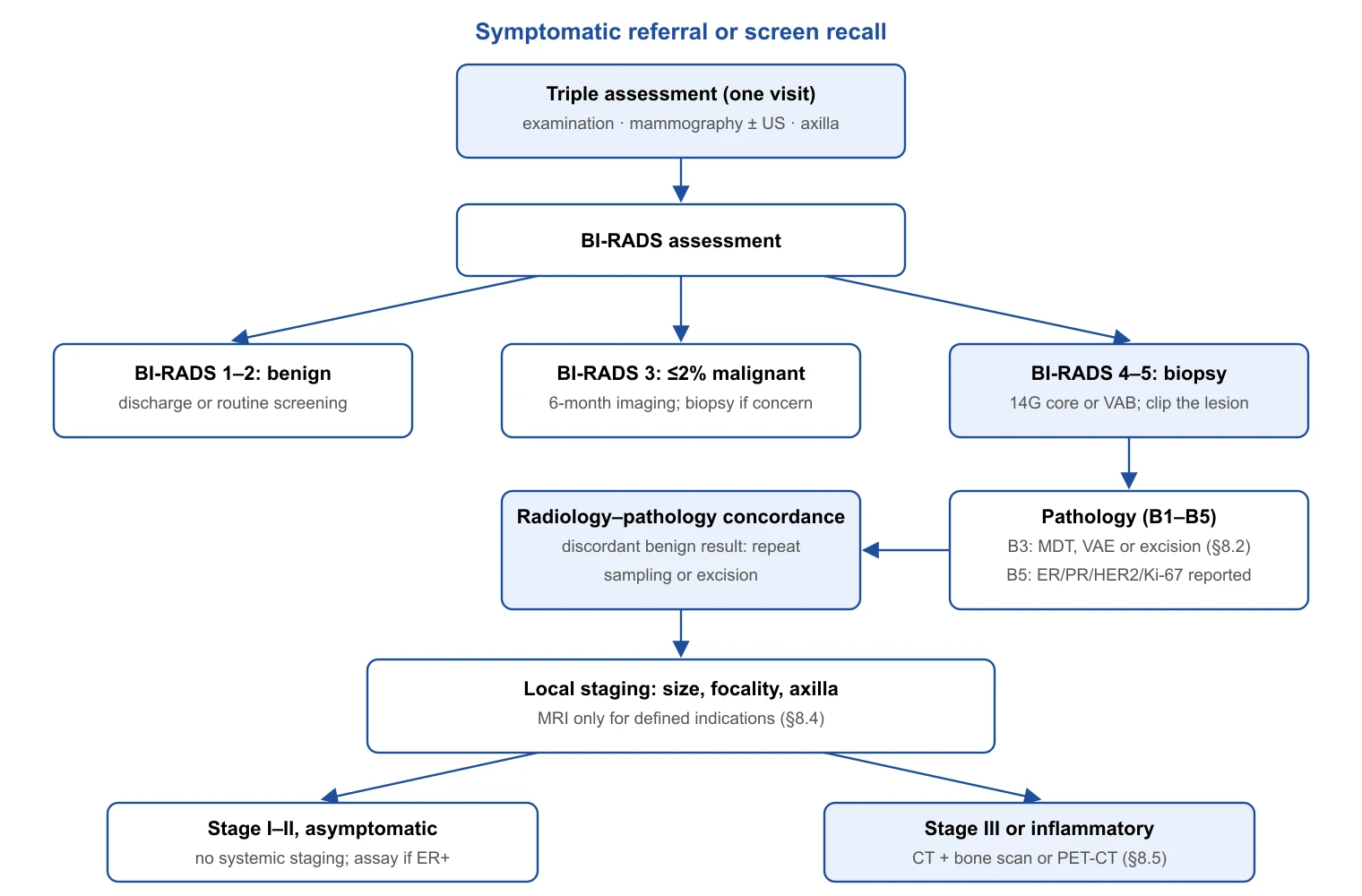
Triple assessment combines clinical examination, imaging and needle sampling. Examination alone has a pooled sensitivity of 54% and specificity of 94%, with a negative likelihood ratio of 0.47 (95% CI 0.40–0.56), so a normal examination does not exclude cancer [116]. Screening digital mammography in the Breast Cancer Surveillance Consortium (1,682,504 examinations) had a sensitivity of 86.9% (95% CI 86.3–87.6) and specificity of 88.9%, with 5.1 cancers detected per 1,000 [117]. Age and density erode this: adjusted sensitivity ranged from 62.9% in extremely dense to 87.0% in fatty breasts, and from 68.6% at age 40–44 to 83.3% at 80–89, with specificity falling from 96.9% to 89.1% across the density gradient [118]. Ultrasound recovers part of what density conceals: in ACRIN 6666 (2,809 women with dense breasts) it added 4.2 cancers per 1,000 (95% CI 1.1–7.2) to mammography, but the positive predictive value of a biopsy recommendation fell from 22.6% to 11.2%[119]. In the symptomatic clinic ultrasound is targeted to a finding, and under 40 it is the first imaging test.
Findings are reported with the ACR BI-RADS lexicon, whose fifth edition defines category 3 as carrying a ≤2% likelihood of malignancy, category 4 as >2% to <95% with subdivisions 4A (>2–10%), 4B (>10–50%) and 4C (>50–<95%), and category 5 as ≥95%[34]. In the US National Mammography Database the positive predictive value of biopsy was 7.6% for 4A, 22% for 4B and 69.3% for 4C, although subdivisions were used in only a third of category 4 reports [120].
The pathway standard matters as much as the tests. EUSOMA quality indicators require that at least 90% of women with breast cancer have preoperative mammography and ultrasound of both breasts and axillae, that at least 85% have a preoperative histological or cytological (B5/C5) diagnosis, and that at least 80% proceed from first diagnostic examination to first treatment within six weeks [121]. In England, NICE NG12 directs a suspected-cancer referral for women aged 30 and over with an unexplained breast lump and those aged 50 and over with unilateral nipple discharge, retraction or other change of concern [122]; since 1 October 2023 the two-week-wait standard has been replaced by the Faster Diagnosis Standard, under which 75% of urgently referred patients must have cancer diagnosed or excluded within 28 days [123]. The one-stop clinic makes this achievable; the only randomised comparison (670 women) found that it reduced anxiety at 24 hours but not at three weeks or three months [124], so its justification is throughput and completeness rather than a durable psychological benefit.
Tissue diagnosis
Core needle biopsy has replaced fine-needle aspiration because it is more sensitive and yields tissue for biomarkers: in 12 prospective series comparing both in the same patients (1,802 patients), sensitivity was 87% (95% CI 84–88) for core biopsy against 74% (72–77) for cytology, with similar specificity (98% vs 96%)[125]. Vacuum-assisted biopsy (VAB) reduces but does not eliminate sampling error. When core biopsy shows ductal carcinoma in situ, invasive cancer is found at excision in 25.9% (95% CI 22.5–29.5; 52 studies, 7,350 lesions), more often with a 14-gauge automated needle than with 11-gauge VAB (P = 0.006), and with high-grade, mass-forming, palpable or >20 mm lesions [126]. For atypical ductal hyperplasia (ADH) the pooled upgrade among surgically excised lesions is 29% (95% CI 26–32; meta-analysis of 6,458 lesions): 42% after ultrasound-guided and 23% after stereotactic sampling, and 14% (8–23) when imaging suggested complete removal [127]. The needle gauge, guidance and proportion of the lesion removed must therefore be known before any B3 result is interpreted.
Management of B3 lesions has moved towards vacuum-assisted excision (VAE). The Third International Consensus Conference (33 panellists, 2023) recommended open excision for ADH on core biopsy and, by a 76% majority, after VAB; for classical lobular neoplasia, radial scar, papilloma without atypia and flat epithelial atypia, observation after complete VAB removal was accepted by 90%, 82%, 100% and 100% of the panel respectively [35]. The 2024 European guideline of EUSOMA, EUSOBI, ESP and ESSO takes the same direction, leaving the choice between VAE and surgery to the multidisciplinary team according to atypia, size, sampling adequacy and patient preference [36]. The upgrade rates behind this position are summarised below.
| Lesion | Pooled upgrade at excision | Modifiers | Consensus position (2023–2024) | Source |
|---|---|---|---|---|
| Atypical ductal hyperplasia | 29% (95% CI 26–32) | US-guided 42%; stereotactic 23%; 14% if lesion removed | Open excision; observation after complete VAB accepted by a minority | Meta-analysis [127]; consensus [35] |
| Radial scar without atypia | 7% overall; 1% (0–4) after 8–11G VAB; 5% after 14G core | With atypia 29% (14G) | VAE or observation after complete VAB removal | Meta-analysis [128]; consensus [35] |
| Papilloma without atypia | 15.7% (12.8–18.5) for all non-malignant papillary lesions | Atypia and mammographic abnormality raise the rate | VAE or observation after complete VAB removal | Meta-analysis [129]; consensus [35] |
| Flat epithelial atypia | 5% (3–6); invasive 1% | 0% if >90% of calcifications removed | VAE; observation after complete removal | Meta-analysis [130]; consensus [35] |
| Classical lobular neoplasia (ALH/LCIS) | Not pooled here | Discordant imaging raises risk | Observation after complete VAB removal (90% of panel) | Consensus [35]; guideline [36] |
A marker clip should be placed in the tumour at diagnostic biopsy whenever neoadjuvant therapy is possible, because a complete clinical response leaves nothing to localise: in 373 women treated with neoadjuvant chemotherapy and breast conservation, five-year local control was 98.6% with a clip and 91.7% without (P = 0.02)[131]. Clipping of the biopsied axillary node is discussed in Part 1 of this series.
Biomarker reporting follows ASCO/CAP. Oestrogen receptor is positive at 1% or more of nuclei, with "ER low positive" (1–10%) reported separately because endocrine benefit in this group is poorly characterised [132]. HER2 immunohistochemistry 2+ is weak-to-moderate complete membrane staining in >10% of cells, with concomitant immunohistochemistry required for the uncommon in situ hybridisation groups 2–4 [133]; the 2023 update affirmed this algorithm and, because trastuzumab deruxtecan is licensed for 1+ and 2+/ISH-negative tumours, added a comment on distinguishing 0 from 1+ without creating a formal "HER2-low" category [38]. That distinction is the least reproducible score in the assay: among 18 pathologists reading 170 biopsies, concordance was 26% for 0 versus 1+ against 58% for 2+ versus 3+[134]. Ki-67 has clinical utility only at its extremes: the International Ki67 Working Group accepts ≤5% or ≥30% to estimate prognosis in T1–2 N0–1 ER-positive, HER2-negative disease and regards intermediate values as uninformative [135].
Axillary ultrasound and needle biopsy before surgery
Ultrasound-guided needle biopsy of a suspicious node is specific but only moderately sensitive: across 31 studies (6,166 women) sensitivity was 79.6% (95% CI 74.1–84.2) and specificity 98.3% (97.2–99.0), and a median of 55.2% of node-positive women were identified preoperatively [136]. For the whole pathway of ultrasound with or without biopsy, pooled sensitivity is 50% (95% CI 43–57) and one in four women with a negative assessment has a positive sentinel node [137]. The test was built for an era in which any positive node meant dissection, and that era has ended.
Two developments described in Part 1 have changed its role. In SOUND and INSEMA a negative axillary ultrasound was the entry criterion for omitting sentinel node biopsy, so a normal ultrasound now has a positive management consequence. After ACOSOG Z0011 and AMAROS, one or two macrometastatic sentinel nodes no longer mandate dissection, and a positive needle biopsy therefore risks "over-staging" low-burden disease. In the Memorial Sloan Kettering series of 141 women with cT1–2 N0 tumours by examination but a positive node biopsy, 47% had only one or two positive nodes at final pathology and would have met Z0011 criteria; only more than one abnormal node on imaging predicted three or more positive nodes (68% vs 43%, P = 0.003)[138]. In 425 Z0011-eligible women with sentinel node metastases, 71% of those with abnormal axillary imaging did not require dissection [139]. The opposing observation is that ultrasound-detected nodal disease is biologically worse: in 302 node-positive patients, those detected by ultrasound had more macrometastases and extranodal extension and worse disease-free survival (HR 2.71, 95% CI 1.49–4.92) than those detected at sentinel node biopsy [140]. Both are compatible: a positive needle biopsy enriches for high-burden disease without reliably identifying it. The 2026 European consensus keeps radiologist-performed axillary ultrasound as the standard for nodal assessment [14]; the authors' judgement is that a positive needle biopsy in a clinically node-negative patient should prompt discussion of neoadjuvant therapy or sentinel node biopsy, not automatic dissection.
Preoperative breast MRI
MRI is the most sensitive test for tumour extent, and the randomised trials show that sensitivity does not translate into better surgery. In COMICE (1,623 women scheduled for wide local excision), reoperation within six months was 19% with MRI and 19% without (OR 0.96, 95% CI 0.75–1.24)[12]. In MONET (418 women with non-palpable lesions), MRI before biopsy paradoxically raised re-excision after breast conservation from 12% to 34% (P = 0.008) without changing the mastectomy rate [141]. Meta-analysis of nine controlled studies (3,112 patients) found MRI doubled initial mastectomy (16.4% vs 8.1%, OR 2.22) and raised overall mastectomy (25.5% vs 18.2%, OR 1.54) without reducing re-excision (11.6% vs 11.4%)[142]; the 2017 update (19 studies, 85,975 patients) confirmed higher odds of mastectomy (OR 1.39, 95% CI 1.23–1.57) and of contralateral prophylactic mastectomy (OR 1.91, 1.25–2.91), with no effect on re-excision, reoperation or positive margins [13]. Nor does MRI improve oncological outcome: in an individual-patient meta-analysis of 3,169 women, eight-year local recurrence-free survival was 97% with MRI and 95% without (adjusted HR 0.88, 95% CI 0.52–1.51)[143]. The prospective MIPA cohort (5,896 women, 27 centres) shows the real-world pattern: MRI was associated with 11.3% more mastectomies and 3.2% fewer reoperations (8.5% vs 11.7%) among those conserved [144], an exchange that is, in the authors' judgement, unfavourable and paid partly through MRI-only lesions that alter the plan before they are biopsied.
MRI therefore has defined indications. Invasive lobular carcinoma is the strongest: MRI size correlates with pathology (r = 0.85) far better than mammography (r = 0.27)[145], re-excision fell from 27% to 9% (OR 3.64, 95% CI 1.30–10.20) in a two-centre cohort [146], and the meta-analyses found lower re-excision in lobular disease (10.9% vs 18.0%, OR 0.56) without a mastectomy excess in the lobular subgroup (OR 1.00)[13,142]. In occult primary cancer presenting with an axillary metastasis, MRI finds the primary in more than two thirds of patients and permits breast conservation in about a third, provided every MRI-detected lesion is biopsied [147]. After neoadjuvant therapy MRI is the best imaging test for residual disease, with an area under the curve of 0.88 across 44 studies (2,050 patients), falling to 0.83 when pathological complete response is strictly defined; with enhancement no greater than normal tissue as the threshold, sensitivity and specificity were both 0.83 [148]. MRI cannot certify a complete response, which is why the surgical-omission trials in Part 1 rely on image-guided biopsy.
The 2010 EUSOMA recommendations listed neoadjuvant response assessment, augmented or reconstructed breasts, occult primary and equivocal conventional findings among the indications [149]. The 2026 European consensus of EUSOBI, ESSO, ESP and ESTRO (13 experts, consensus on 67.4% of statements) states that not all patients should have preoperative MRI and names those likely to benefit: lobular carcinoma, HER2-overexpressing or triple-negative cancer, suspected multifocal or multicentric disease, discordant conventional imaging, high-grade DCIS on stereotactic biopsy, and all patients receiving neoadjuvant therapy (MRI before and at the end of treatment); MRI-only findings must be biopsied, and an institution that cannot arrange MRI-guided biopsy should not offer preoperative MRI [14]. Dense breasts alone are not an indication in that document. Guidelines converge on lobular histology, neoadjuvant therapy and occult primary and diverge on biological subtype, which the European consensus includes and the randomised evidence does not address.
Systemic staging
The yield of staging imaging in asymptomatic early disease is too low to justify routine use. Across 22 studies (14,824 patients) the median prevalence of distant metastases was 7.0%, lowest in stage I–II and rising with stage; specificity was 91.4% for combined conventional imaging and 98.1% for FDG-PET/CT, and the high PET/CT yields partly reflect selection of advanced disease [150]. Guidelines are unanimous that asymptomatic stage I and operable stage II disease should not be staged: ASCO's first Choosing Wisely list names PET, CT and bone scan in early breast cancer at low risk of metastasis among the five tests to avoid [151]; EUSOMA requires that at least 95% of such women are not staged and that at least 95% of women with stage III disease are [121]; ESMO 2024 and NCCN take the same position, with NCCN listing FDG-PET/CT as an option from stage III [152–154].
In locally advanced disease the question is which test. The PET-ABC randomised trial (369 patients with T3N0 or stage III ductal carcinoma) compared FDG-PET/CT with bone scan plus CT: PET/CT upstaged 23% to stage IV against 11% (absolute difference 12.3%, 95% CI 3.9–19.9; P = 0.002), and fewer PET/CT patients received curative-intent combined-modality therapy (81% vs 89.2%, P = 0.03)[155]. Histology modifies the yield: in 146 lobular carcinomas PET/CT revealed unsuspected metastases in 0 of 8 stage I, 4% of stage II and 11% of stage III patients, against 22% in stage III ductal cancers [154]. In inflammatory breast cancer, baseline PET/CT found extra-axillary nodal disease in 56% and distant lesions in 31% of 59 patients [156]. The burden of staging is the false positive: in low-prevalence groups the incidental findings worked up outnumber the metastases found, which is the quantitative basis of the Choosing Wisely recommendation [150,151].
Genomic assays and the surgeon
Multigene assays decide chemotherapy, not surgery, but increasingly they decide the sequence. The 21-gene recurrence score (Oncotype DX) has the strongest prospective evidence. In TAILORx, 1,626 of 10,253 women (15.9%) with hormone receptor-positive, HER2-negative, node-negative disease had a score of 0–10 and a five-year distant recurrence-free rate of 99.3% on endocrine therapy alone [157]. Among 6,711 women with a score of 11–25 randomised to endocrine therapy with or without chemotherapy, endocrine therapy alone was non-inferior (invasive disease-free survival HR 1.08, 95% CI 0.94–1.24; nine-year distant recurrence-free 94.5% vs 95.0%), with chemotherapy benefit confined to women aged 50 or younger with a score of 16–25 [15]. RxPONDER extended this to one to three positive nodes: among 5,018 women with a score of 25 or lower, postmenopausal patients derived no benefit (five-year invasive disease-free survival 91.9% vs 91.3%, HR 1.02, 95% CI 0.82–1.26), premenopausal patients did (89.0% vs 93.9%, HR 0.60, 0.43–0.83), with no gradient across the score [16]; whether that reflects chemotherapy or chemotherapy-induced ovarian suppression is unresolved.
MammaPrint was tested in MINDACT. At 8.7 years' median follow-up, women at high clinical and low genomic risk who received no chemotherapy had a five-year distant metastasis-free survival of 95.1% (95% CI 93.1–96.6), above the 92% non-inferiority boundary; in the randomised population the eight-year rate was 92.0% with chemotherapy versus 89.4% without (HR 0.66, 0.48–0.92), an absolute difference of 2.6 points not increased by nodal positivity and, in an underpowered exploratory analysis, confined to women aged 50 or younger (5.0 vs 0.2 points)[39]. The ultralow-risk threshold identifies tumours that may need no systemic therapy: 15% of MINDACT patients were ultralow, with eight-year distant metastasis-free interval of 97.0% and breast-cancer-specific survival of 99.6%[158], and in the pre-screening STO-3 cohort ultralow-risk women had no breast cancer deaths at 15 years on tamoxifen and a 20-year disease-specific survival of 94% without systemic therapy [159]. EndoPredict and Prosigna are prognostic in prospective–retrospective designs: EPclin low-risk patients had ten-year distant recurrence of 4% against 22–28% for high risk [160], and the PAM50 low-risk category carried a ten-year metastasis risk below 3.5%[161]. In the within-patient TransATAC comparison, the risk of recurrence score (HR 2.56, 95% CI 1.96–3.35) and EPclin (HR 2.14) added more prognostic information than the recurrence score (HR 1.69), and all signatures contributed less in node-positive disease [162,163]. Neither has chemotherapy-prediction evidence equivalent to TAILORx.
The surgical relevance is sequencing. A score on the diagnostic core directs a low or midrange result with a resectable tumour to surgery first, and a high result to neoadjuvant chemotherapy, where the response governs axillary management and radiotherapy (Part 1). The score also predicts response: in 989 National Cancer Database patients, pathological complete response occurred in 4.3% and was associated with a high score (adjusted OR 4.87, 95% CI 2.01–11.82)[164]. In the NBRST registry, "clinical luminal" tumours had a pathological complete response rate of 11% to chemotherapy, rising to 32% in the 18% reclassified as basal by molecular subtyping, while 68% of molecularly luminal tumours responded clinically to neoadjuvant endocrine therapy [165]. For the ER-positive patient with a low-risk result who needs downsizing to allow conservation, neoadjuvant endocrine therapy is the logical route, and the assay is what makes the choice defensible.
Circulating tumour DNA (ctDNA) for molecular residual disease is clinically valid but not a standard test: in 101 patients with personalised assays, detection during follow-up predicted relapse (HR 25.2, 95% CI 6.7–95.6) with a median lead time of 10.7 months [166]. Clinical utility has not followed. In c-TRAK TN (161 triple-negative patients under three-monthly surveillance), ctDNA was detected in 27.3% by 12 months, but 72% of those allocated to intervention already had metastases on staging, and none of the five who started pembrolizumab achieved sustained clearance [17]. ZEST, a phase 3 trial of niraparib for ctDNA-positive triple-negative or BRCA-mutated disease, screened 1,901 patients, found 147 (7.7%) positive, randomised 40 and was stopped; 55% of ctDNA-positive patients had radiographic recurrence at their first positive test [167]. Post-treatment ctDNA detection is often a late signal of established metastasis, and the assays are not actionable outside trials.
Diagnostic workup: what changes the operation
- Preoperative B5/C5 diagnosis in at least 85% and first treatment within six weeks in at least 80% are the EUSOMA standards [121]; a normal examination (sensitivity 54%) never excludes cancer [116].
- One in four core-biopsy diagnoses of DCIS and three in ten of ADH are upgraded at excision [126,127]; most B3 lesions other than ADH can be managed by vacuum-assisted excision [35,36].
- Clip the tumour at biopsy whenever neoadjuvant therapy is possible: five-year local control 98.6% versus 91.7% without a clip [131].
- A positive axillary needle biopsy in a clinically node-negative patient does not establish the need for dissection; about half have only one or two positive nodes [138].
- Preoperative MRI does not reduce reoperation (COMICE 19% vs 19%) or recurrence and raises mastectomy (OR 1.39); reserve it for lobular carcinoma, neoadjuvant assessment, occult primary and discordant imaging [12–14].
- No systemic staging for asymptomatic stage I–II; PET/CT or CT plus bone scan from stage III, where PET/CT upstages 23% versus 11%[151,155].
- Genomic assays on the diagnostic core decide surgery-first versus neoadjuvant therapy; ctDNA residual-disease assays remain investigational [15,17].
Summary tables
| Clinical stage | Breast and axilla | Systemic staging | Yield or performance | Source |
|---|---|---|---|---|
| Stage I, asymptomatic | Mammography + US of both breasts and axillae (≥90% of patients) | None; bloods and symptom review only | Distant metastases rare; false positives exceed true positives | [121,150,151] |
| Primary operable stage II, asymptomatic | As stage I; MRI only for defined indications | None (≥95% should not be staged) | Lobular stage II: 4% unsuspected metastases on PET/CT | [121,154] |
| Stage III (or T3N0) | As above; MRI before and after neoadjuvant therapy | CT chest/abdomen/pelvis + bone scan, or FDG-PET/CT (≥95% should be staged) | PET/CT upstages 23% vs 11% with conventional imaging | [14,121,155] |
| Inflammatory breast cancer | Skin-punch and core biopsy; MRI | FDG-PET/CT preferred | Distant lesions in 31%; extra-axillary nodes in 56% | [153,156] |
| Any stage with symptoms or abnormal bloods | As indicated | Directed imaging of the symptomatic site | Prevalence rises with clinical suspicion | [152,153] |
| Trial (assay) | n and population | Principal result | Surgical relevance |
|---|---|---|---|
| TAILORx 2015 (Oncotype DX RS 0–10)[157] | 1,626 of 10,253; HR+/HER2−, N0 | 5-yr distant recurrence-free 99.3% on endocrine therapy alone | Surgery first; no neoadjuvant chemotherapy |
| TAILORx 2018 (RS 11–25)[15] | 6,711 randomised; HR+/HER2−, N0 | Endocrine non-inferior (HR 1.08, 0.94–1.24); benefit only ≤50 yr with RS 16–25 | Surgery first for most; neoadjuvant endocrine therapy for downsizing |
| RxPONDER 2021 (RS ≤25)[16] | 5,018; HR+/HER2−, 1–3 positive nodes | Postmenopausal no benefit (HR 1.02); premenopausal benefit (HR 0.60, 0.43–0.83) | Low-burden node-positive postmenopausal disease can proceed to surgery without chemotherapy planning |
| MINDACT 2021 (MammaPrint)[39] | 6,693; 1,497 high clinical/low genomic risk randomised | 8-yr DMFS 92.0% vs 89.4% (HR 0.66); 2.6-point absolute benefit, age-dependent | Clinically high-risk but genomically low-risk tumours are not neoadjuvant chemotherapy candidates by default |
| MINDACT ultralow [158] | 1,000 (15%) ultralow | 8-yr DMFI 97.0%; BCSS 99.6% | Candidates for further de-escalation of all therapy |
| ABCSG-6/8 (EndoPredict)[160] | 1,702; ER+/HER2−, endocrine only | EPclin low 10-yr distant recurrence 4% vs 22–28% high | Prognostic; no randomised chemotherapy-prediction data |
| ABCSG-8 (Prosigna ROR)[161] | 1,478 postmenopausal, endocrine only | ROR low: 10-yr metastasis risk <3.5% | Prognostic; node-positive performance weaker [162] |
| NCDB neoadjuvant cohort (Oncotype DX)[164] | 989; HR+/HER2− treated with neoadjuvant chemotherapy | pCR 4.3% overall; high RS OR 4.87 for pCR | High RS supports neoadjuvant chemotherapy; low RS supports surgery first or neoadjuvant endocrine therapy |
| c-TRAK TN 2023 (ctDNA)[17] | 161 TNBC under surveillance | ctDNA+ 27.3% by 12 months; 72% already metastatic at detection | Not actionable; surveillance strategy under revision |
| ZEST 2024 (ctDNA, niraparib)[167] | 1,901 screened; 147 ctDNA+; 40 randomised | Stopped for low enrolment; 55% radiographically recurrent at first positive test | Not a standard test |
Breast Reconstruction
Uptake and the decision
Reconstruction after mastectomy is a routine part of the surgical conversation, but its use remains uneven. In the English National Mastectomy and Breast Reconstruction Audit (NMBRA), 21% of 16,485 women undergoing mastectomy in 2008–2009 had immediate reconstruction, with regional rates from 9% to 43%; local complications followed 10.3% of mastectomies alone and 11.0–18.2% of immediate reconstructions, and breast appearance and well-being scores at 18 months were higher after immediate reconstruction than after mastectomy alone [168]. Hospital Episode Statistics for 44,837 mastectomies showed the age gradient (32.7% under 50 years, 1.5% at 70 or over) and regional variation that survived case-mix adjustment (8.0–29.4%)[169]. In the United States, reconstruction among insured women rose from 46% in 1998 to 63% in 2007, with increasing implant and decreasing autologous use [170]; in the Nationwide Inpatient Sample, immediate reconstruction rose from 20.8% to 37.8% between 1998 and 2008 while implant use grew by 11% per year and overtook autologous methods after 2002 [171]. NICE NG101 recommends offering reconstruction to everyone after mastectomy, offering both immediate and delayed options whether or not they are available locally, and offering immediate reconstruction "including those who may need radiotherapy, unless they have comorbidities that rule out reconstructive surgery"[172]; the ASPS guideline states that patients undergoing mastectomy should be offered preoperative referral to a plastic surgeon [173].
The oncological premise is that the skin envelope, and often the nipple, can be preserved. For skin-sparing mastectomy (SSM), a meta-analysis of nine observational studies (3,739 patients) found no difference in local recurrence against conventional mastectomy (6.2% vs 4.0%; OR 1.25, 95% CI 0.81–1.94)[174]. For nipple-sparing mastectomy (NSM), a meta-analysis of 20 studies (5,594 patients) found non-significant risk differences for local recurrence, disease-free and overall survival against SSM or modified radical mastectomy [175]; its 2026 update (60 studies, 7,752 patients) gave a pooled local recurrence risk difference of −0.7% (95% CI −2.1 to 0.8) and nipple–areolar recurrence of 1.1% at 10 years [176], and a pooled analysis of 12,358 procedures reported locoregional recurrence of 2.38% at a mean 38 months with nipple necrosis in 5.9%[177]. Occult nipple involvement is present in 11.5% of specimens overall and is more likely with tumour–nipple distance under 2 cm, central or multicentric tumours, size above 5 cm, nodal metastasis, lymphovascular invasion and HER2-positive or receptor-negative disease [178]. The 2 cm criterion has been challenged: in 195 therapeutic NSM cases, nipple involvement did not differ between candidates and non-candidates at a 2 cm or 1 cm imaging cut-off [179]. Retroareolar sampling remains mandatory whichever threshold is used [177,178]. Mastectomy variants and contralateral prophylactic mastectomy were covered in Part 1 of this series.
Does immediate reconstruction delay chemotherapy? A systematic review of 14 studies (5,270 patients) found four with delays averaging 6.6–16.8 days, seven with no difference and one with a shorter interval; mean time to chemotherapy was 29–61 days after reconstruction versus 21–60 days after mastectomy alone, which the authors judged not clinically relevant in general [180]. A complication requiring reoperation does delay treatment, so the complication profile of the technique is itself an oncological variable.
Immediate, delayed and delayed-immediate reconstruction
Two prospective cohorts anchor the complication data. The UK iBRA study enrolled 2,108 women undergoing immediate implant-based reconstruction at 81 units: at 3 months, 9% (95% CI 8–10) had lost the implant, 18% (16–20) had been readmitted, 18% had returned to theatre and 25% (23–27) had been treated for infection, all above the national standards of under 5% for implant loss, reoperation and readmission and under 10% for infection [18]. The North American Mastectomy Reconstruction Outcomes Consortium (MROC) followed 2,343 women for 2 years: complications occurred in 32.9%, reoperative complications in 19.3% and wound infection in 9.8%; every autologous technique carried higher odds of any complication than expander–implant reconstruction (OR 1.87–2.05 for TRAM, DIEP and latissimus dorsi flaps; 4.71 for SIEA), whereas reconstructive failure was higher with implants (7.1%) than with flaps (0–2.8%)[19]. In the same cohort, delayed reconstruction (151 patients) had lower odds of any (OR 0.38) and major (OR 0.52) complication and lower failure (1.3% vs 6%), with no difference in any BREAST-Q domain at 2 years [41]. Delayed reconstruction is not a second-class option; its cost is the interval without a breast mound and loss of the native skin envelope.
Delayed-immediate reconstruction addresses the case in which the need for post-mastectomy radiotherapy (PMRT) is unknown until final pathology: skin-sparing mastectomy with a filled tissue expander, then definitive reconstruction within weeks if PMRT is not required, or deflation during radiotherapy and a "skin-preserving" delayed reconstruction, usually with a flap, about 3 months after its completion [40,181].
Patient-reported outcomes favour autologous tissue. In MROC at 1 year (1,632 patients), women with autologous reconstruction had higher BREAST-Q satisfaction with breasts (difference 6.3 points), sexual well-being (4.5) and psychosocial well-being (3.7), although chest physical well-being was not fully restored in either group and abdominal physical well-being fell by 13.4 points after flap harvest [182]. At 2 years (2,013 patients) the differences persisted: satisfaction with breasts 7.94 (95% CI 5.68–10.20), psychosocial 3.27 (1.25–5.29) and sexual well-being 5.53 (2.95–8.11)[20]. Against a minimal important difference of about 4 points (§9.7), the satisfaction advantage is clinically meaningful.
Implant-based reconstruction
One stage or two. A meta-analysis of 13 studies (5,216 reconstructions) found that direct-to-implant reconstruction carried higher odds of skin-flap necrosis (OR 1.43), reoperation (OR 1.25) and implant loss (OR 1.87) than two-stage expander–implant reconstruction, with similar infection and contracture, a difference the authors attributed to mastectomy flap quality [183]. In the prospective MROC comparison (99 vs 1,328 patients), no complication differed at 2 years and patient-reported outcomes were equivalent apart from better sexual well-being after direct-to-implant [184]. Direct-to-implant works where flaps are well perfused and the surgeon is prepared to convert to an expander when they are not.
Plane. Prepectoral placement avoids the animation deformity and pain of subpectoral reconstruction at the price of greater dependence on skin-flap perfusion. A 2024 meta-analysis of 40 studies found that prepectoral placement reduced animation deformity (OR 0.37, 95% CI 0.19–0.70) but increased rippling (OR 2.39, 1.53–3.72) and seroma (OR 1.55, 1.02–2.35), with similar total complications [42]; a meta-analysis of 18 direct-to-implant studies (3,135 breasts) reported lower odds of haematoma, seroma, infection and revision (OR 0.44) with prepectoral placement [185]. The only published randomised comparison, 42 women in Denmark, found no difference in any BREAST-Q domain at 12 months and was too small to exclude one [186]; the international OPBC-02/PREPEC trial (372 patients) had not reported at the time of writing [187]. The UK Pre-BRA cohort (343 women, 40 centres) gives the realistic baseline: 8.2% lost the implant, 19.5% had an infection and 16.0% needed reoperation within 3 months, comparable to subpectoral techniques in iBRA [188]; at 18 months women reported clinically meaningful falls in physical and sexual well-being, although two-thirds rated the result excellent or very good [189].
Matrices and mesh. Acellular dermal matrix (ADM) is widely used to support the lower pole, yet the only randomised trial is cautionary. In BRIOS (142 women, eight Dutch hospitals), one-stage ADM-assisted reconstruction carried higher odds per breast of surgical complications (OR 3.81, 95% CI 2.67–5.43), reoperation (OR 3.38) and removal of implant or ADM (OR 8.80), with grade 3 adverse events in 29% versus 5% of breasts [190], and no difference in any BREAST-Q domain at 1 year, the primary endpoint [191]. A systematic review of 51 studies rated the certainty of evidence for complications and implant loss with matrices as low and for infection, contracture and aesthetic outcome as very low, with no data on recurrence or quality of life [192]. Cost-utility models favour ADM against two-stage reconstruction without matrix (incremental cost about US$362) but not against an autologous dermal flap [193]. In iBRA, 65% of patients had mesh and 21% a dermal sling [18]; the choice is driven by preference rather than comparative evidence.
Surface, lymphoma and systemic symptoms. Breast implant-associated anaplastic large cell lymphoma (BIA-ALCL) is a CD30-positive, ALK-negative T-cell lymphoma of the periprosthetic capsule, almost exclusively after textured implants. In the Australian and New Zealand series of 55 cases, all patients had textured devices, 85% higher-surface-area products, and the risk was 14.1 times higher with Biocell and 10.8 times higher with polyurethane than with Siltex texturing [44]. Prospective single-surgeon follow-up of 3,546 reconstructive patients with macro-textured devices found ten cases, 1 in 355 women, at a median exposure of 11.5 years [194]; the wider Memorial Sloan Kettering cohort of 9,373 reconstructions gave 1 in 559 patients, with cumulative incidence rising from zero at 6 years to 9.4 per 1,000 at 14–16 years [195]. On 24 July 2019 the FDA requested, and Allergan undertook, a worldwide recall of Biocell textured implants and expanders, reporting 573 unique cases and 33 deaths globally, 481 of the cases attributed to Allergan devices [196]. The clinical signal is a late seroma, usually more than a year after implantation, which requires cytology and CD30 immunohistochemistry rather than drainage alone.
"Breast implant illness" is a patient-defined cluster of fatigue, arthralgia, myalgia and cognitive symptoms without a laboratory or histological marker. The honest summary is that an association with specific rheumatological diagnoses cannot be excluded, causation has not been shown, and improvement after explantation is reported but uncontrolled. In the FDA post-approval studies of 99,993 patients, silicone implants were associated with higher standardised incidence ratios for Sjögren's syndrome (8.14), scleroderma (7.00) and rheumatoid arthritis (5.96), with a caution that patient-level analysis was needed [197]. In a prospective blinded cohort study, women attributing symptoms to implants improved after explantation irrespective of the type of capsulectomy, so "en bloc" removal is not a requirement [198]; a 2025 meta-analysis (6,048 women) reported improvement in 81.9% but also substantial psychiatric and autoimmune comorbidity [199]. The FDA states that these symptoms "and what causes them are poorly understood" and that it "has not detected any association" between silicone implants and connective tissue disease [200].
Longevity. Implants are not lifetime devices [200]. In the 10-year Mentor core study, Kaplan–Meier estimates for primary reconstruction were 14.3% Baker III/IV contracture, 34.1% explantation and 52.7% any reoperation, and 59.7% reoperation after revision reconstruction [201]; the post-approval studies reported 25% reoperation at 7 years for reconstruction [197]. A woman choosing an implant in her forties should expect at least one further operation.
Autologous reconstruction
The abdomen is the usual donor site and the deep inferior epigastric perforator (DIEP) flap the usual technique. Against the free TRAM flap, a meta-analysis found that DIEP doubled fat necrosis (RR 1.94, 95% CI 1.28–2.93) and flap loss (RR 2.05, 1.16–3.61) but halved abdominal bulge or hernia (RR 0.49, 0.28–0.86); the fat-necrosis excess disappeared against muscle-sparing free TRAM [43]. A second meta-analysis found bulging after DIEP one-third as likely as after muscle-sparing TRAM on examination (RR 0.29, 0.06–1.36) but not by questionnaire (RR 1.04, 0.59–1.79)[202]. The largest comparison of techniques (1,790 patients, mean 5.5 years) found no difference in total flap loss by flap type but more partial loss, fat necrosis and hernia or bulge after pedicled TRAM [203]; in MROC, abdominal physical well-being at 2 years was 7.2 points lower after pedicled TRAM and 4.9 lower after free TRAM than after DIEP, and SIEA flaps had more donor complications (OR 2.7)[204]. The pedicled TRAM is hard to justify where microsurgical expertise exists.
Total flap loss is uncommon in experienced hands. The German national microsurgical registry (3,926 patients, 4,577 DIEP flaps, 22 centres) recorded total loss in 2.0%, partial loss in 1.1%, emergency vascular revision in 4.3% and wound revision in 8.3%; centres performing 40 or more DIEP flaps a year had shorter operations (287 vs 386 minutes) and stays (8.2 vs 9.0 days)[205]. MROC failure of 1.3% for DIEP and 2.1% for free TRAM sits in the same range [19]. Concentration in high-volume centres is measurable: the number of US hospitals performing autologous reconstruction fell by 35% between 1998 and 2010 while annual volumes in high-volume centres rose from 48 to 73, with lower charges, particularly when complications occurred [206].
When the abdomen is unavailable, the thigh, buttock and flank serve (table at the end of this section). The profunda artery perforator (PAP) flap offers a long pedicle and a scar in the gluteal crease [207], with total loss of 3% in 265 consecutive flaps [208]; the transverse upper gracilis (TUG) flap lost three of 154 flaps [209]; the lumbar artery perforator (LAP) flap suits women with little abdominal or thigh tissue, at the cost of donor seroma in 35.1%[210]. The latissimus dorsi flap needs no microsurgery, but 41% of patients report donor discomfort and most studies find some loss of shoulder motion or strength, less after muscle-sparing harvest [211].
Resource use favours implants early and flaps late. In four US state databases (15,154 women), 90-day complications were 6.5% after expander and 13.1% after autologous reconstruction, but secondary procedures within 3 years were most frequent after expanders (2,021 per 1,000 discharges) and least after flaps (949)[212]. A cost-effectiveness analysis using BREAST-Q utilities put the cost of one additional year of perfect breast-related health at US$11,941 for unilateral and US$28,017 for bilateral DIEP reconstruction against implants [213].
Fat grafting
Autologous fat transfer corrects contour defects after conservation and reconstruction, softens irradiated tissue and, in some hands, reconstructs the whole breast. The oncological concern comes from experimental data on adipose-derived stem cells; the clinical data do not support it. A meta-analysis of 59 studies (4,292 patients) found an incidence rate difference for locoregional recurrence of −0.15% per year (95% CI −0.36 to 0.07) in matched cohorts, confirmed in the pooled cohorts and case series [214]. A matched cohort with 5 years of follow-up after grafting (287 vs 300 patients) recorded eight versus 11 locoregional recurrences (HR 0.63, 95% CI 0.25–1.60)[215], and the MD Anderson matched study (719 vs 670 breasts) found 5-year locoregional recurrence of 1.6% versus 4.1%[216]. One Milan cohort is the exception: in 59 women with intraepithelial neoplasia, 5-year local events were 18% versus 3% in 118 controls (P = 0.02), concentrated in women under 50, with high-grade lesions, Ki-67 of 14% or more or previous quadrantectomy [217]. The numbers are small and the finding was not reproduced, but it is why some units still counsel caution after DCIS. No randomised trial of oncological safety has reported.
Grafting produces oil cysts, calcification and fat necrosis that must be distinguished from recurrence; in a blinded reading of 50 mammograms, grafted breasts had fewer masses requiring biopsy and lower BI-RADS scores than breasts after reduction mammaplasty [218]. In irradiated tissue, lipoaspirate injection improved or resolved LENT-SOMA grade 3–4 damage in all 20 patients of the original series [219]. Total breast reconstruction with external expansion (Brava) and serial grafting in 488 women required a mean 2.7 sessions for non-irradiated and 4.8 for irradiated mastectomies, with 12.5% drop-out, locoregional recurrence of 0.5% at 2.5 years and benign palpable masses in 12% and 37% respectively [220]; it is legitimate but demanding.
Reconstruction and radiotherapy
Radiotherapy is the variable that most changes the calculus. In MROC (622 irradiated, 1,625 unirradiated), at least one breast complication had occurred by 2 years in 38.9% of irradiated women with implants, 25.6% of irradiated women with flaps, 21.8% of unirradiated women with implants and 28.3% of unirradiated women with flaps; among irradiated patients autologous reconstruction halved the odds of complication (OR 0.47, 95% CI 0.27–0.82), and the satisfaction gap between autologous and implant reconstruction was wider after radiotherapy (63.5 vs 47.7) than without it (67.6 vs 60.5)[22]. The radiotherapy–surgery interface in general is covered in Section 10; this section addresses only the reconstructive sequence.
For two-stage implant reconstruction the question is whether to irradiate the expander or the exchanged implant. In 1,790 single-surgeon reconstructions, 6-year predicted failure was 32% after expander irradiation versus 16.4% after implant irradiation, but expander irradiation gave better aesthetic results and less grade IV contracture, with no difference in BREAST-Q scores [221]. A meta-analysis of 20 studies (2,348 patients) found pooled failure of 17.6% and Baker III/IV contracture of 37.5% regardless of timing; expander irradiation carried higher failure (20% vs 13.4%; RR 2.33, 95% CI 1.24–4.35) and lower contracture (24.5% vs 49.4%; RR 0.53, 0.26–1.09)[222], and a second meta-analysis of eight studies found a contracture reduction (RR 0.44) with a non-significant failure difference [223]. Irradiating the expander risks losing the reconstruction; irradiating the implant risks keeping a hard one. Prepectoral placement may soften the second risk (contracture after PMRT 16.1% vs 52.2% subpectoral in one series [224]), although a 2026 meta-analysis of 14 studies found no difference in implant loss between planes in irradiated patients [225]. NICE advises considering 40 Gy in 15 fractions rather than 26 Gy in 5 for chest-wall radiotherapy after implant-based reconstruction [172].
Autologous tissue tolerates radiotherapy better but not perfectly. A systematic review of 20 studies found similar pooled complications whether the flap was irradiated after transfer or placed into an irradiated field (total loss 4% vs 1%, fat necrosis 13% vs 10%), with contracture or fibrosis in 27% of irradiated flaps [226]. In MROC, 108 immediate and 67 delayed free abdominal flaps in women receiving PMRT had similar 1-year complications (25.9% vs 26.9%) and comparable BREAST-Q scores at 2 years, though immediate cases came almost entirely from one centre [227]. The conservative sequence remains delayed autologous reconstruction after radiotherapy or delayed-immediate reconstruction with an expander placeholder; immediate flap reconstruction with planned PMRT is defensible in centres with that experience, with the patient told that fibrosis may require later revision or fat grafting.
Radiotherapy and the reconstructive choice
- Expected PMRT changes the recommendation: autologous tissue halves the 2-year complication odds relative to implants in irradiated patients and preserves a wider satisfaction margin [22].
- On an implant pathway, failure is more likely when the expander is irradiated (about 20–32%) and contracture more likely when the implant is irradiated (about 50%)[221,222].
- When PMRT is uncertain until final pathology, delayed-immediate reconstruction keeps the skin envelope without committing a flap or implant to the field [40].
- Flaps placed before or after radiotherapy have similar pooled complication rates, but about one in four irradiated flaps develops fibrosis or contracture [226].
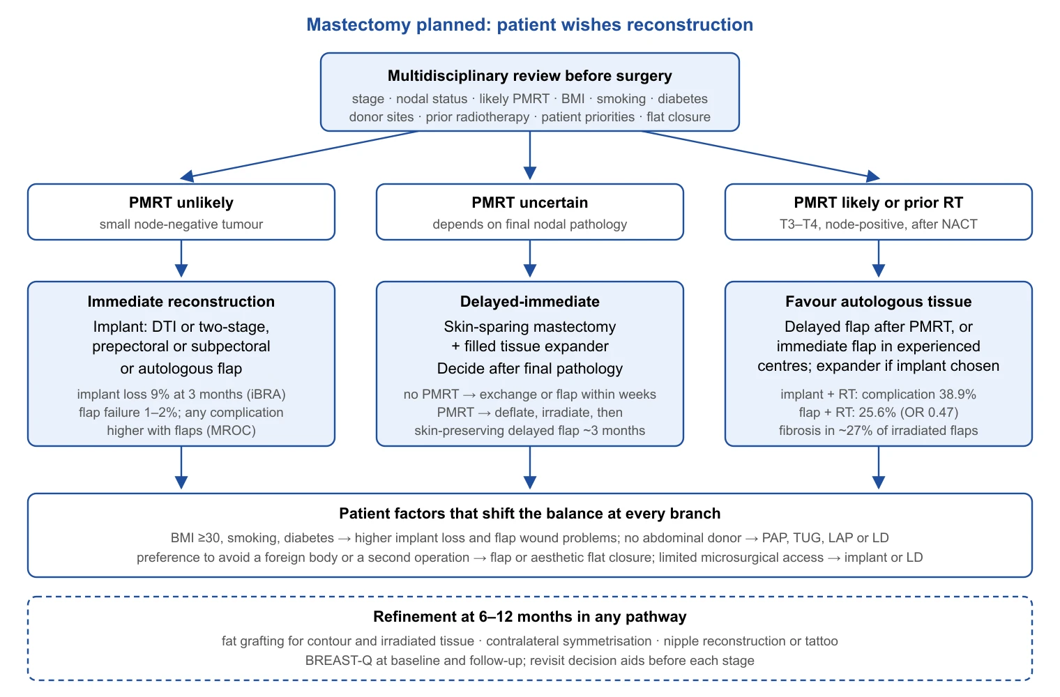
Patient-reported outcomes and shared decision-making
The BREAST-Q reconstruction module, field-tested in 1,950 women, measures satisfaction with breasts, outcome and care, and psychosocial, physical and sexual well-being on 0–100 scales with test–retest reproducibility of 0.73–0.96 [228]. The minimal important difference, estimated from 3,052 MROC patients, is 4 points for satisfaction with breasts, psychosocial and sexual well-being and 3 for physical well-being [21]; smaller differences, however statistically significant, should not drive counselling. By that standard the autologous advantage in satisfaction at 2 years (7.9 points) is meaningful, while a prepectoral–subpectoral difference has not been demonstrated [20,186].
Decision aids work. In the BRECONDA randomised trial (222 women), an online aid added to standard information reduced decisional conflict at 1 month (27.2 vs 35.5), sustained at 6 months [229]. NICE lists the topics the discussion must cover, including timing, the options each timing allows and "the uncertainty over long-term outcomes in women having radiotherapy"[172]. Mastectomy without reconstruction, with an aesthetic flat closure, is a choice rather than a default for the unfit: among 931 women who had "gone flat", it was the first choice for 73.7%, and the strongest predictor of dissatisfaction was low surgeon support for the decision (OR 3.85, 95% CI 2.59–5.72), followed by BMI of 30 or more (OR 2.74) and unilateral surgery (OR 1.99)[230]. A flat closure should be planned as deliberately as a reconstruction.
Symmetry and the nipple complete the result. Contralateral reduction, mastopexy or augmentation is often needed after unilateral reconstruction, more so with an implant that will not age with the native breast [212]. For the nipple–areola complex, a review of 75 papers found complications in 7.9% of local-flap nipple reconstructions against 46.9% of grafts, 1.6% after areolar tattoo against 10.1% after graft, and loss of projection of 45–75% that did not materially affect satisfaction [231]. Three-dimensional tattooing alone is an increasingly chosen alternative without surgical morbidity.
Complications and comparative summary
The complications below are specific to reconstruction; general complications of breast surgery and their management are covered in Section 11.
| Complication | Technique | Representative rate | Source |
|---|---|---|---|
| Implant loss at 3 months | Immediate implant, any plane | 9% (95% CI 8–10); 8.2% prepectoral | iBRA [18]; Pre-BRA [188] |
| Reconstructive failure at 2 years | Expander–implant / DTI vs flaps | 7.1% vs 0–2.8% | MROC [19] |
| Infection needing treatment, 3 months | Immediate implant | 25% (23–27) | iBRA [18] |
| Baker III/IV capsular contracture | Implant, 10 years, no RT | 14.3% primary reconstruction | Core study [201] |
| Capsular contracture after PMRT | Implant irradiated vs expander irradiated | 49.4% vs 24.5% | Meta-analysis [222] |
| Animation deformity | Subpectoral vs prepectoral | OR 0.37 favouring prepectoral | Meta-analysis [42] |
| Rippling; seroma | Prepectoral vs subpectoral | OR 2.39; OR 1.55 | Meta-analysis [42] |
| BIA-ALCL | Macro-textured implants | 1 in 355–559 patients at median 10–12 years | Cohorts [194,195] |
| Total flap loss | DIEP, national registry | 2.0%; partial 1.1% | DGPRÄC registry [205] |
| Fat necrosis | DIEP vs free TRAM | RR 1.94 (1.28–2.93) | Meta-analysis [43] |
| Abdominal bulge or hernia | DIEP vs free TRAM | RR 0.49 (0.28–0.86) | Meta-analysis [43] |
| Shoulder discomfort or weakness | Latissimus dorsi harvest | 41% any discomfort | Systematic review [211] |
| Flap fibrosis or contracture | Autologous flap irradiated | 27% | Systematic review [226] |
| Palpable benign masses | Fat grafting, irradiated breast | 37% (12% non-irradiated) | Series [220] |
| Dimension | Implant-based | Autologous | Source |
|---|---|---|---|
| Operative time and stay | Shorter; day-case to 1–2 nights | 5–6 h; 8–9 days in registry data | [205] |
| Early complications (90 days) | 6.5–6.6% | 13.1% | [212] |
| Any complication at 2 years | Reference | OR 1.9–2.1 for TRAM, DIEP, LD | [19] |
| Reconstructive failure at 2 years | 7.1% | 0–2.8% | [19] |
| Donor-site morbidity | None | Abdominal well-being −13.4 at 1 year; bulge, hernia | [43,182] |
| Satisfaction with breasts, 2 years | Reference | +7.9 points (MID 4) | [20,21] |
| Tolerance of PMRT | Complications 38.9%; failure 13–32% | Complications 25.6%; fibrosis 27% | [22,222,226] |
| Secondary procedures over 3 years | 2,021 per 1,000 discharges | 949 per 1,000 | [212] |
| Durability | Not lifetime; 52.7% reoperation by 10 years | Ages with the patient | [201] |
| Device-specific risks | BIA-ALCL (textured), rupture, systemic symptoms | Total flap loss 1–2% | [195,205] |
| Cost-effectiveness | Lower upfront cost | Cost-effective over a lifetime, especially unilateral and younger | [213] |
| Flap | Donor site | Advantages | Main drawbacks | Source |
|---|---|---|---|---|
| DIEP | Lower abdomen, rectus preserved | Workhorse; lowest abdominal morbidity; high abdominal well-being | Fat necrosis RR 1.94 vs free TRAM; microsurgery required | [43,203] |
| Muscle-sparing free TRAM | Lower abdomen, partial rectus | Similar fat necrosis to DIEP; robust perfusion | More bulge than DIEP on examination | [43,202] |
| Pedicled TRAM | Lower abdomen, whole rectus | No microsurgery | Highest hernia/bulge, fat necrosis and partial loss; abdominal well-being −7.2 | [203,204] |
| SIEA | Lower abdomen, fascia intact | No fascial incision | Donor complications OR 2.7 vs DIEP; MROC any-complication OR 4.71 | [19,204] |
| PAP | Posterior thigh | Long pedicle; hidden scar; muscle spared | Smaller volume (mean 403 g); donor wound 6.8% | [207,208] |
| TUG / TMG | Medial thigh (gracilis) | Concealed scar; bilateral harvest easy | Modest volume; thigh sensory change | [209] |
| LAP | Flank / lower back | Suits pear-shaped habitus; good tissue quality | Seroma 35.1%; short perforator, vein graft often needed | [210] |
| SGAP / IGAP | Buttock | Available when abdomen and thigh are not | Short pedicle; firm fat; positioning | [207] |
| Latissimus dorsi ± implant | Back | Reliable; no microsurgery; useful after RT | Volume usually needs an implant; shoulder discomfort 41% | [211] |
What the evidence supports
- Skin-sparing and nipple-sparing mastectomy do not increase local recurrence in selected patients; nipple–areolar recurrence is about 1% and retroareolar sampling is mandatory [174,176].
- Routine immediate implant reconstruction carries 9% implant loss and 18% reoperation at 3 months; these figures belong in the consent discussion [18].
- Autologous reconstruction has more early complications but fewer failures and a clinically meaningful satisfaction advantage at 2 years [19,20].
- Fat grafting has not increased locoregional recurrence in matched cohorts or meta-analysis [214]; macro-textured implants carry a BIA-ALCL risk of about 1 in 355–559 reconstructed patients over 10–15 years [195].
- Aesthetic flat closure is a choice to be supported; surgeon support is the strongest determinant of satisfaction with it [230].
Radiotherapy and Surgery
Radiotherapy made breast conservation oncologically acceptable, and it remains the treatment most shaped by what the surgeon did, documented and left in the breast. Part 1 of this series covered NSABP B-51/RTOG 1304 and the trials of sentinel node biopsy omission; those are referred to here but not repeated.
Why radiotherapy after breast conservation
The Early Breast Cancer Trialists' Collaborative Group (EBCTCG) meta-analysis of individual data from 10,801 women in 17 randomised trials remains the foundation. Radiotherapy after breast-conserving surgery reduced the 10-year risk of any first recurrence from 35.0% to 19.3% (absolute reduction 15.7%, 95% CI 13.7–17.7) and the 15-year risk of breast cancer death from 25.2% to 21.4% (absolute reduction 3.8%, 1.6–6.0). In node-negative disease (n = 7,287) the reductions were 31.0% to 15.6% and 20.5% to 17.2%. Roughly one breast cancer death was avoided by year 15 for every four recurrences avoided by year 10, and the proportional benefit varied little with age, grade, receptor status or tamoxifen use even though absolute benefit varied widely [232]. Two consequences follow. Radiotherapy halves recurrence in every subgroup, so the question is never whether it works but whether the absolute gain justifies its burden in a given patient; and the four-to-one relationship means that interventions which change local recurrence by a few percentage points cannot show a survival effect in trials of ordinary size, which is why the margin and omission trials below report local control. The margin consensus in Part 1 (§7.2), "no ink on tumour" for invasive disease and 2 mm for DCIS, rests on this premise: with whole-breast radiotherapy, wider excision adds nothing that radiotherapy has not already achieved [232].
Hypofractionation
Four decades of 2 Gy daily fractions gave way to 15 or 16 fractions on the strength of two trial programmes. In START-A (n = 2,236), 10-year locoregional relapse was 7.4% after 50 Gy in 25 fractions, 6.3% after 41.6 Gy and 8.8% after 39 Gy in 13 fractions; in START-B (n = 2,215), 40 Gy in 15 fractions over 3 weeks gave 4.3% versus 5.5% (HR 0.77, 95% CI 0.51–1.16), with less breast shrinkage, telangiectasia and oedema [45]. The Ontario trial (n = 1,234) reported 10-year local recurrence of 6.2% after 42.5 Gy in 16 fractions versus 6.7% after 50 Gy, with good or excellent cosmesis in 69.8% versus 71.3%[233]. ASTRO's 2018 guideline names 40 Gy in 15 or 42.5 Gy in 16 fractions as the preferred whole-breast schedule regardless of age, chemotherapy or laterality [234].
The next step was five fractions. FAST (n = 915, age ≥50, pT1–2 pN0) showed at 10 years that 28.5 Gy in five once-weekly fractions produced normal-tissue effects no different from 50 Gy in 25, whereas 30 Gy produced more [235]. FAST-Forward (n = 4,096, pT1–3 pN0–1 after conservation or mastectomy) compared 40 Gy in 15 fractions with 27 Gy and 26 Gy in five fractions over one week. Five-year ipsilateral relapse was 2.1% (95% CI 1.4–3.1) after 40 Gy, with absolute differences of −0.3% for 27 Gy and −0.7% for 26 Gy, both within the 1.6% non-inferiority margin; moderate or marked clinician-assessed normal-tissue effects at 5 years were 9.9%, 15.4% and 11.9%, so 26 Gy became the schedule [46]. At 10 years, ipsilateral recurrence was 3.6% (2.7–4.9), 2.9% and 2.1%, and moderate or marked breast or chest-wall effects 13.1%, 19.3% and 14.4%[23]. The ESTRO-ACROP consensus allows moderate hypofractionation for any patient and any volume, including chest wall with reconstruction and nodes, and five fractions for non-nodal breast or chest-wall treatment without reconstruction [236].
Whether one week suffices when the axilla must be treated is the open question. The FAST-Forward nodal substudy randomised 469 patients requiring axillary radiotherapy to the same three schedules; at a median of 7.0 years there were 32 locoregional recurrences among 466 analysed, described by the investigators as reassuring but too few for precise estimation of the 26 Gy arm alone [23]. Five-year patient-reported moderate or marked arm swelling was 13%; axillary dissection (OR 3.70, 95% CI 1.61–8.48) and BMI ≥35 kg/m² (OR 3.31) predicted it, whereas no dosimetric parameter did [237]. Fractionation has little influence on arm morbidity; the extent of axillary surgery has.
Tumour-bed boost
The EORTC 22881-10882 trial randomised 5,318 women after complete excision and 50 Gy whole-breast radiotherapy to a 16 Gy boost or none. At 20 years, ipsilateral recurrence was 16.4% without and 12.0% with the boost (HR 0.65, 99% CI 0.52–0.81), overall survival did not differ (61.1% vs 59.7%), and severe fibrosis rose from 1.8% to 5.2%[238]. The absolute benefit was largest in young women, and the trialists concluded that the boost can be avoided in most patients over 60; ASTRO recommends it for women aged 50 or younger and for high-grade disease or positive margins, and suggests omission in older women with low-grade, hormone-receptor-positive disease and clear margins [234]. For DCIS the question was unanswered until BIG 3-07/TROG 07.01 (n = 1,608, non-low-risk DCIS, margins ≥1 mm): 5-year freedom from local recurrence was 97.1% with a boost versus 92.7% without (HR 0.47, 95% CI 0.31–0.72), at the cost of more grade 2 or higher breast pain (14% vs 10%) and induration (14% vs 6%)[239].
The boost no longer needs extra visits. IMPORT HIGH (n = 2,617, pT1–3 pN0–3a) compared a sequential 16 Gy in 8 fractions with a simultaneous integrated boost to 48 Gy or 53 Gy within 15 fractions to a clip-defined tumour bed; 5-year ipsilateral recurrence was 1.9%, 2.0% and 3.2%, 48 Gy was non-inferior, 53 Gy increased moderate or marked induration (15.5% vs 11.5%) without benefit, and the median boost volume was 13 cm³ [240]. That last figure depends on the surgeon: an integrated boost to a small, clip-marked cavity is a different treatment from one to a large, unmarked oncoplastic cavity.
Partial-breast irradiation
If recurrence after conservation arises mostly at the tumour bed, irradiating the whole breast treats tissue that gains little. The trials reach different conclusions depending on technique and selection (Table). IMPORT LOW (n = 2,018, age ≥50, pT1–2 ≤3 cm, pN0–1, margins ≥2 mm) simply shortened the tangential beams to the tumour quadrant, 40 Gy in 15 fractions; 5-year local relapse was 1.1% after whole-breast, 0.2% after reduced-dose and 0.5% after partial-breast radiotherapy, both experimental arms were non-inferior, and patients reported less change in breast appearance and firmness [24]. The Florence trial (n = 520, mostly low risk) delivered 30 Gy in five once-daily IMRT fractions; 10-year ipsilateral recurrence was 3.7% versus 2.5% after whole-breast radiotherapy with boost (HR 1.56, 95% CI 0.55–4.37), overall survival 91.9% in both arms, and toxicity and cosmesis favoured the partial-breast arm [241]. Twice-daily schedules fared worse: in RAPID (n = 2,135, 38.5 Gy in 10 fractions twice daily), 8-year recurrence was 3.0% versus 2.8%, but late grade ≥2 toxicity was 32% versus 13% and adverse cosmesis 17.7% more frequent at 7 years [242]. NSABP B-39/RTOG 0413 (n = 4,216, broad eligibility including up to three positive nodes, brachytherapy or 38.5 Gy twice daily) did not meet its criterion: 10-year ipsilateral recurrence was 4.6% versus 3.9% (HR 1.22, 90% CI 0.94–1.58, outside the 0.667–1.5 equivalence window), an absolute difference under 1% that the authors considered acceptable for some women [243].
Intraoperative radiotherapy is a separate matter. TARGIT-A (n = 2,298, age ≥45, invasive ductal ≤3.5 cm) reported 5-year local recurrence of 2.11% after risk-adapted single-dose 50 kV radiotherapy versus 0.95% after external beam, within its 2.5% margin, with about 20% of the intraoperative arm receiving supplementary external beam radiotherapy on final pathology [244]. The 2020 report was contested: the German Society of Radiation Oncology expert panel pointed to the change in primary outcome, loss to follow-up and absence of analysis by risk factors, and reaffirmed that the 50 kV system cannot be recommended for routine partial-breast treatment [245]. ELIOT (n = 1,305, 21 Gy electrons in a single dose) is less ambiguous: ipsilateral recurrence occurred in 70 (11%) of 651 patients versus 16 (2%) of 654 (HR 4.62, 95% CI 2.68–7.95), 15-year rates 12.6% versus 2.4%, with no difference in overall survival [246]. ASTRO restricts intraoperative radiotherapy to "suitable" patients (age ≥50 for invasive disease, or low-risk DCIS) with counselling about the higher recurrence risk [247]; its 2024 guideline endorses external beam and brachytherapy partial-breast techniques delivered once daily or on alternate days [248]. The GEC-ESTRO low-risk group requires age ≥50, unifocal pT1–2 ≤3 cm disease, clear margins and pN0 [249].
The surgeon determines whether partial-breast irradiation is deliverable. Target definition after open-cavity surgery depends on seroma visibility and surgical clips, which the GEC-ESTRO recommendations require to be enclosed by the cavity contour [250]. Oncoplastic displacement and replacement move the cavity; a systematic review of 670 patients after volume-replacement surgery found that only two of nine studies reported boost volumes and that tumour-bed localisation after such surgery remains unresolved [251]. Clips at the cavity walls and an operative note stating what was moved and where are therefore part of the oncological operation.
| Trial | n | Schedule tested | Local relapse (test vs control) | Cosmesis / normal tissue | Source |
|---|---|---|---|---|---|
| START-A | 2,236 | 41.6 or 39 Gy/13 fr vs 50 Gy/25 | 10 y 6.3% and 8.8% vs 7.4% | Fewer effects with 39 Gy | [45] |
| START-B | 2,215 | 40 Gy/15 fr vs 50 Gy/25 | 10 y 4.3% vs 5.5% | Less shrinkage, telangiectasia, oedema | [45] |
| Ontario | 1,234 | 42.5 Gy/16 fr vs 50 Gy/25 | 10 y 6.2% vs 6.7% | Good/excellent 69.8% vs 71.3% | [233] |
| FAST | 915 | 30 or 28.5 Gy/5 weekly fr vs 50 Gy/25 | 11 events in total at 10 y | 28.5 Gy equivalent; 30 Gy worse | [235] |
| FAST-Forward | 4,096 | 26 or 27 Gy/5 fr vs 40 Gy/15 | 5 y 1.4%, 1.7% vs 2.1%; 10 y 2.1%, 2.9% vs 3.6% | Moderate/marked 11.9%, 15.4% vs 9.9% at 5 y | [23,46] |
| IMPORT LOW | 2,018 | Partial or reduced-dose 40 Gy/15 vs whole breast | 5 y 0.5%, 0.2% vs 1.1% | Less firmness and change in appearance | [24] |
| IMPORT HIGH | 2,617 | Integrated boost 48 or 53 Gy/15 vs sequential | 5 y 2.0%, 3.2% vs 1.9% | Induration 10.6%, 15.5% vs 11.5% | [240] |
| Florence | 520 | 30 Gy/5 daily fr IMRT vs 50 Gy/25 + boost | 10 y 3.7% vs 2.5% | Toxicity and cosmesis favour APBI | [241] |
| RAPID | 2,135 | 38.5 Gy/10 fr twice daily vs whole breast | 8 y 3.0% vs 2.8% | Adverse cosmesis +17.7% at 7 y | [242] |
| NSABP B-39 | 4,216 | Brachytherapy or 38.5 Gy/10 twice daily vs 50 Gy/25 | 10 y 4.6% vs 3.9% (HR 1.22) | Grade 3 toxicity 10% vs 7% | [243] |
| TARGIT-A | 2,298 | Single 50 kV intraoperative dose vs EBRT | 5 y 2.11% vs 0.95% | Not reported long term; design contested | [244,245] |
| ELIOT | 1,305 | 21 Gy electrons intraoperative vs 50 Gy + 10 Gy | 15 y 12.6% vs 2.4% (HR 4.62) | Late effects not collected | [246] |
Omitting radiotherapy
The question is not whether radiotherapy reduces recurrence in older women with favourable disease, which it does, but whether a difference of this size matters to a woman in her eighth decade who is taking endocrine therapy. CALGB 9343 (n = 636, age ≥70, T1 N0 ER-positive, tamoxifen) reported 10-year freedom from locoregional recurrence of 98% with radiotherapy and 90% without, with no difference in mastectomy rate, distant metastasis or overall survival (67% vs 66%)[252]. PRIME II (n = 1,326, age ≥65, hormone-receptor-positive, node-negative, ≤3 cm, clear margins, endocrine therapy) reported 10-year local recurrence of 9.5% (95% CI 6.8–12.3) without radiotherapy and 0.9% (0.1–1.7) with it (HR 10.4), distant recurrence as first event 1.6% versus 3.0%, and overall survival 80.8% versus 80.7%[25]. LUMINA selected by biology instead: 500 women aged ≥55 with T1 N0 grade 1–2 luminal A disease (ER ≥1%, PR >20%, HER2-negative, central Ki-67 ≤13.25%) received endocrine therapy without radiotherapy, and 5-year local recurrence was 2.3% (90% CI 1.3–3.8), within the prespecified 5% boundary [253]. LUMINA is a single-arm cohort and 5 years is early for luminal recurrence, but it shows that biological thresholds define a group in which the PRIME II recurrence rate is roughly quartered. Three randomised trials are testing genomic selection: EXPERT (NCT02889874; 1,167 planned, PAM50 low risk, primary completion expected 2027), DEBRA (NCT04852887; 1,670 planned, Oncotype recurrence score ≤18) and the Danish NATURAL trial (NCT03646955; 926 planned, partial-breast irradiation versus none)[254–256].
Omission trials assume endocrine therapy, which has costs of its own. EUROPA (NCT04134598) randomises women aged ≥70 with stage I luminal A-like disease to single-modality endocrine therapy or single-modality radiotherapy (whole or partial breast, 5–15 fractions). In the preplanned interim analysis of 207 patients, global health status at 24 months fell by 9.79 points with endocrine therapy and 3.40 with radiotherapy (adjusted difference 6.39, 95% CI 0.14–12.65), and treatment-related adverse events occurred in 85% versus 67%[257]. The 5-year recurrence co-primary endpoint is pending, so the "either/or" question is open, but it has been reframed: for a patient who will not tolerate five years of an aromatase inhibitor, one week of radiotherapy may be the less burdensome single treatment. Two surgical points follow. Omission belongs to a patient meeting all the trial criteria, with clear margins, after a conversation that quantifies the recurrence difference; and wider excision is not a substitute, because the EBCTCG analysis showed the same proportional effect of radiotherapy irrespective of extent of surgery [232].
| Trial | Population | Design, n | Local recurrence | Survival or other outcome | Source |
|---|---|---|---|---|---|
| CALGB 9343 | ≥70 y, T1 N0 ER+, tamoxifen | RCT, 636 | 10 y 10% vs 2% (locoregional) | OS 66% vs 67%; no difference in mastectomy or metastasis | [252] |
| PRIME II | ≥65 y, HR+, N0, ≤3 cm, endocrine therapy | RCT, 1,326 | 10 y 9.5% vs 0.9% | OS 80.8% vs 80.7%; distant first event 1.6% vs 3.0% | [25] |
| LUMINA | ≥55 y, T1 N0 grade 1–2, luminal A, Ki-67 ≤13.25% | Single arm, 500 | 5 y 2.3% (90% CI 1.3–3.8) | Contralateral 1.9%; any recurrence 2.7% | [253] |
| EUROPA | ≥70 y, stage I luminal A-like | RCT, 731 (interim 207) | 5 y IBTR pending | Global health status better with RT; AE 67% vs 85% | [257] |
| EXPERT | PAM50 low risk | RCT, 1,167 planned | Pending (2027) | – | [254] |
| DEBRA | Oncotype score ≤18 | RCT, 1,670 planned | Pending (2031) | – | [255] |
| NATURAL | Low risk, PBI vs none | RCT, 926 planned | Pending (2033) | – | [256] |
Post-mastectomy radiotherapy
The EBCTCG meta-analysis of 8,135 women in 22 trials of radiotherapy to the chest wall and regional nodes after mastectomy and axillary surgery (1964–86) established the modern indications. After axillary dissection, radiotherapy had no effect in 700 node-negative women (overall recurrence RR 1.06, 95% CI 0.76–1.48; breast cancer mortality RR 1.18, 0.89–1.55) and increased overall mortality (RR 1.23, 1.02–1.49). In 1,314 women with one to three positive nodes it reduced locoregional recurrence (2p < 0.00001), overall recurrence (RR 0.68, 0.57–0.82; absolute 10-year risk 45.7% reduced to 34.2%) and breast cancer mortality (RR 0.80, 0.67–0.95); among the 1,133 who also received systemic therapy, 20-year breast cancer mortality was 49.4% without and 41.5% with radiotherapy (RR 0.78, 0.64–0.94). With four or more nodes the rate ratios were 0.79 (0.69–0.90) for recurrence and 0.87 (0.77–0.99) for breast cancer mortality. Even a single positive node carried a 17.8% 10-year locoregional recurrence rate without radiotherapy versus 2.3% with it [47]. The 2016 ASCO/ASTRO/SSO update recommended post-mastectomy radiotherapy for T1–2 disease with one to three positive nodes while acknowledging that some such patients have too low a risk to benefit, and recommended inclusion of the internal mammary and supraclavicular-apical nodes [258].
Those trials predate taxanes, trastuzumab, modern endocrine therapy and CT planning, and the intermediate-risk group was the target of SUPREMO, in which 1,607 women with pT1–2 N1, pT3 N0, or pT2 N0 grade 3 or lymphovascular-invasion-positive disease, treated with mastectomy, axillary surgery and systemic therapy, were randomised to chest-wall irradiation (40–50 Gy) or none. At a median of 9.6 years, 10-year overall survival was 81.4% versus 81.9% (HR 1.04, 95% CI 0.82–1.30); chest-wall recurrence 1.1% versus 2.5% (HR 0.45, 0.20–0.99); disease-free survival 76.2% versus 75.5%; distant metastasis-free survival 78.2% versus 79.2%[26]. The 5-year quality-of-life substudy found worse chest-wall symptoms with radiotherapy and no difference in other domains [259]. The critical reading is that SUPREMO tested chest-wall irradiation alone, without regional nodal irradiation in most patients, in a population weighted towards node-negative and single-node disease, after protocol modifications that reduced its power; it cannot be used to deny nodal radiotherapy to patients in whom the trials in §10.7 showed benefit [260]. Its defensible conclusion is narrower: chest-wall irradiation alone produces a small reduction in chest-wall recurrence and no detectable survival gain at 10 years in contemporary intermediate-risk disease.
T3 N0 disease sits in the uncertain middle. SEER data on 2,525 women associated radiotherapy with higher 8-year overall survival (76.5% vs 61.8%; adjusted HR 0.63)[261]; a propensity-weighted National Cancer Database analysis of 13,901 patients found a 7-year survival benefit in pT3 N0 disease (74% vs 65%; HR 0.78, 95% CI 0.68–0.89) confined to women who did not receive chemotherapy [262]. These are observational data with the usual selection bias; a pT3 N0 tumour with additional adverse features remains a reasonable indication (authors' judgement). After neoadjuvant therapy, B-51 (Part 1, §9.6) showed that regional nodal irradiation added nothing for cN1 patients who converted to ypN0, whereas ypN+ disease remains an indication [258]. Post-mastectomy radiotherapy with reconstruction, including timing and hypofractionation over an implant, is covered in §9.6.
Regional nodal irradiation
Two trials published together in 2015 tested whether adding the internal mammary, supraclavicular and axillary nodes to breast or chest-wall radiotherapy improves outcome. MA.20 (n = 1,832, node-positive or high-risk node-negative after conservation) reported 10-year overall survival of 82.8% with nodal irradiation versus 81.8% without (HR 0.91, 95% CI 0.72–1.13), disease-free survival of 82.0% versus 77.0% (HR 0.76, 0.61–0.94), and more grade ≥2 pneumonitis (1.2% vs 0.2%) and lymphoedema (8.4% vs 4.5%)[48]. EORTC 22922/10925 (n = 4,004, node-positive or medial/central tumours) tested internal mammary and medial supraclavicular irradiation; at 15 years overall survival was 73.1% versus 70.9% (HR 0.95, 0.84–1.06), but any breast cancer recurrence (24.5% vs 27.1%; HR 0.87) and breast cancer mortality (16.0% vs 19.8%; HR 0.81, 0.70–0.94) were reduced [27]. Both trials reduce breast cancer recurrence and death without a demonstrable overall survival effect.
The Danish cohorts add what the trials could not. DBCG-IMN allocated 3,089 node-positive women to internal mammary irradiation by laterality (right-sided treated, left-sided not, because of cardiac concern). Eight-year overall survival was 75.9% versus 72.2% (adjusted HR 0.82, 95% CI 0.72–0.94)[263]; at 15 years, 60.1% versus 55.4% (HR 0.86, 0.77–0.96), with distant recurrence 35.6% versus 38.6%[264]. DBCG IMN2 (4,541 patients treated 2007–14 with taxanes, aromatase inhibitors, trastuzumab and CT-planned radiotherapy) reproduced the effect: 15-year overall survival 65.0% versus 60.8% (HR 0.85, 0.76–0.94), no subgroup in which omission could be justified, and 15-year cardiac death of 0.2% in right-sided and 0.7% in left-sided patients [265]. Allocation by laterality is not randomisation, but laterality is unrelated to prognosis, and these data make the strongest case for internal mammary irradiation in node-positive disease.
Radiotherapy can also replace surgery in the axilla. AMAROS (n = 1,425, positive sentinel node, cT1–2 N0) reported 10-year axillary recurrence of 0.93% after dissection versus 1.82% after axillary radiotherapy (HR 1.71, 95% CI 0.67–4.39), no difference in survival, 5-year lymphoedema of 24.5% versus 11.9%, and an exploratory excess of second primary cancers after radiotherapy (12.1% vs 8.3%)[29]. Whether either treatment is needed after one or two positive sentinel nodes is the question of SENOMAC (Part 1) and POSNOC (ISRCTN54765244, 1,900 planned, adjuvant therapy alone versus axillary treatment), whose primary results had not been indexed at the time of writing [266]. Lymphoedema after axillary treatment is dealt with in §12.
| Setting | Key evidence | Effect | Current position | Source |
|---|---|---|---|---|
| pN0 after axillary dissection | EBCTCG, 700 women | No reduction in recurrence or breast cancer death; overall mortality RR 1.23 | Not indicated for T1–2 N0 | [47] |
| pT3 N0 | SEER 2,525; NCDB 13,901 | OS HR 0.63 (SEER); HR 0.78 limited to no chemotherapy (NCDB) | Individualised; observational data only | [261,262] |
| 1–3 nodes, historical trials | EBCTCG, 1,314 women | Recurrence 45.7% → 34.2%; breast cancer mortality RR 0.80 | Recommended by ASCO/ASTRO/SSO, with exceptions for very low risk | [47,258] |
| Intermediate risk, contemporary therapy | SUPREMO, 1,607 | Chest-wall RT alone: OS 81.4% vs 81.9%; chest-wall recurrence 1.1% vs 2.5% | No survival gain from chest-wall RT alone; nodal RT not tested | [26,260] |
| ≥4 nodes | EBCTCG, 1,772 women | Recurrence RR 0.79; breast cancer mortality RR 0.87 | Chest wall + regional nodes | [47] |
| Nodal RT added to breast/chest wall | MA.20 1,832; EORTC 22922 4,004 | DFS 82.0% vs 77.0% (MA.20); breast cancer mortality 16.0% vs 19.8% at 15 y (EORTC); OS not significant | Offered in node-positive disease | [27,48] |
| Internal mammary nodes | DBCG-IMN 3,089; IMN2 4,541 | OS 75.9% vs 72.2% at 8 y; 65.0% vs 60.8% at 15 y (IMN2) | Included in node-positive disease | [263,265] |
| Positive sentinel node, cT1–2 | AMAROS 1,425 | Axillary recurrence 1.82% vs 0.93%; lymphoedema 11.9% vs 24.5% | Axillary RT preferred over dissection | [29] |
| cN1 → ypN0 after neoadjuvant therapy | NSABP B-51 (Part 1) | No benefit from regional nodal RT | Omission acceptable in B-51-eligible patients | [258] |
Sequencing and timing
Chemotherapy and radiotherapy. The Cochrane review of three trials (1,166 women) found no difference in local recurrence-free, relapse-free, metastasis-free or overall survival between sequences; concurrent chemoradiation increased anaemia (OR 1.54, 95% CI 1.10–2.15), telangiectasia (OR 3.85) and pigmentation [267]. Radiotherapy therefore follows chemotherapy when both are given.
Interval from surgery. When no chemotherapy intervenes, delay matters. A systematic review of waiting times found an 11% increase in local recurrence per month of delay after post-operative breast radiotherapy (RR 1.11 per month, 95% CI 1.04–1.19), without a clear effect on metastasis or survival [268]. In 18,050 SEER-Medicare women over 65 treated without chemotherapy, an interval over six weeks was associated with local recurrence (HR 1.19, 1.01–1.39), the effect was continuous (HR 1.005 per day), and the absolute 5-year excess was 0.96%[269]. Seromas needing repeated aspiration, wound breakdown and staged oncoplastic revisions that push radiotherapy beyond eight weeks carry a measurable oncological cost.
Endocrine therapy. Concurrent tamoxifen does not blunt radiotherapy: in the SWOG 8897 analysis, 10-year local recurrence was 7% with concurrent and 5% with sequential tamoxifen (HR 0.73, 95% CI 0.26–2.04) and disease-free survival 83% in both [270]. In a Japanese nationwide database of 20,280 women, hospitalisation for radiation pneumonitis did not differ between concurrent and sequential hormonal therapy (0.27% vs 0.58%)[271]. CDK4/6 inhibitors are newer: the adjuvant trials started them after radiotherapy, the ESMO-ESTRO consensus recommends caution for most combined scenarios [272], and a propensity-matched cohort of 364 node-positive patients found no difference in discontinuation (10.3% vs 9.2%), pneumonitis or grade ≥3 dermatitis between concurrent and sequential use, with a borderline excess of grade ≥3 drug eruption in the concurrent group [273].
The heart. In a case-control study of 2,168 Swedish and Danish women, major coronary events rose linearly by 7.4% per gray of mean heart dose (95% CI 2.9–14.5) with no threshold, beginning within 5 years and persisting beyond 20 [274]. Mean heart dose in left-sided regimens published 2003–13 averaged 5.4 Gy, falling to 4.2 Gy without internal mammary irradiation, 1.3 Gy with breathing control and 0.5 Gy with protons, and rising to around 8 Gy when the internal mammary chain was included [275]. Deep-inspiration breath-hold reduced heart, left anterior descending and lung dose without loss of target coverage in a meta-analysis of 12 studies (1,019 patients)[276]. At 2010–15 doses, the EBCTCG estimated absolute long-term risks of about 0.3% for lung cancer and 0.3% for cardiac death in non-smokers, but around 4% and 1% in continuing smokers, for whom harm can approach benefit [277]. Proton therapy is being tested in RadComp (NCT02603341), a pragmatic randomised trial with major cardiovascular events as the primary endpoint [278], now closed to recruitment with primary completion expected in 2036 [279]; in 1,160 participants mean heart dose was 0.89 Gy with protons versus 2.99 Gy with photons [280].
Second cancers. A meta-analysis of 762,468 patients found that radiotherapy increased second non-breast cancers five or more years later (RR 1.12, 95% CI 1.06–1.19), lung cancer (RR 1.39, rising to 1.66 at ≥15 years), oesophageal cancer (RR 1.53) and sarcoma (RR 2.53, 1.74–3.70)[281]. In SEER (563,155 women), radiotherapy raised soft-tissue sarcoma incidence from 22 to 31 per 100,000 person-years and angiosarcoma in particular (HR 7.6, 95% CI 4.9–11.9), with the hazard peaking at 10 years [282]. In the Netherlands Cancer Registry, 209 radiotherapy-associated angiosarcomas arose among 184,823 irradiated patients, about 1 per 1,000, at a median of 8 years (range 3–20), with 5- and 10-year overall survival of 40.5% and 25.2%[283]. A new violaceous lesion in an irradiated breast years after treatment is a biopsy, not a bruise.
Surgical implications of the radiotherapy evidence
- Margins are a radiotherapy decision: "no ink on tumour" (invasive) and 2 mm (DCIS) assume whole-breast radiotherapy follows. Wider excision does not replace it; omission requires the full PRIME II or LUMINA profile plus endocrine therapy.
- Mark the cavity. Integrated boost (IMPORT HIGH) and partial-breast irradiation (IMPORT LOW, Florence) depend on a clip-defined target; after oncoplastic displacement or replacement the tumour bed may be unlocatable without clips.
- Axillary surgery, not fractionation, drives arm morbidity: dissection tripled the odds of swelling in the FAST-Forward nodal substudy and no dose metric mattered; in AMAROS axillary radiotherapy halved lymphoedema with equivalent control.
- After mastectomy, nodal burden sets the indication. SUPREMO showed no survival gain from chest-wall irradiation alone; it did not test nodal radiotherapy, which MA.20, EORTC 22922 and the Danish internal mammary cohorts support in node-positive disease. Document nodal counts and extranodal extension.
- Avoid delay. Radiotherapy should begin within six to eight weeks when no chemotherapy intervenes; each month of delay adds about 11% to the relative risk of local recurrence.
- Mean heart dose is a surgical variable too: left-sided internal mammary coverage roughly doubles it, which argues for breath-hold planning rather than omission of nodal treatment that improves survival.
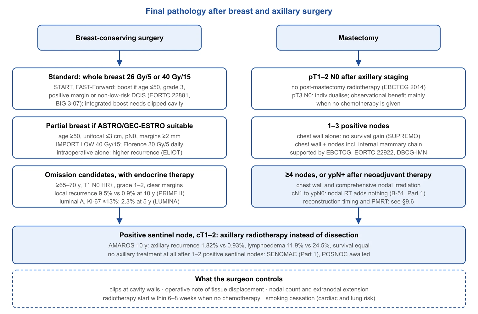
Complications of Breast and Axillary Surgery
Grading and overall rates
Complications are graded by the Clavien–Dindo classification, which ranks severity by the treatment required, from grade I (no intervention beyond simple measures) to grade IIIb (reoperation under general anaesthesia) and grade V (death)[284]. The breast-specific problem is that most events are grade I–II: seroma aspiration, a wound infection treated with oral antibiotics or a small area of flap necrosis managed with dressings. They rarely threaten life but delay adjuvant therapy and dominate the patient's experience. In 26,988 NSQIP patients, 30-day morbidity was 4.0% after mastectomy and 1.6% after breast-conserving surgery, with superficial and deep surgical-site infection the commonest events [285]. An earlier NSQIP benchmark gave wound infection in 4.34% after mastectomy and 1.97% after lumpectomy with axillary surgery, and 30-day mortality of 0.24% and 0%[286]. Among 16,063 NSQIP reconstructions, major surgical complications occurred in 8.4%, wound complications in 3.5% and medical complications in 1.6%, with immediate timing, autologous technique, obesity and smoking as independent risk factors [287]. Broader registry definitions capture more (surgical-site infection within 90 days in 15.7% of 82,102 Swedish patients)[288]. In the axilla, the randomised comparisons are unambiguous: in ACOSOG Z0011, any adverse surgical effect followed 70% of sentinel biopsy plus dissection and 25% of sentinel biopsy alone [289]; in ALMANAC, lymphoedema at 12 months was 5% versus 13% (RR 0.37, 95% CI 0.23–0.60) and sensory loss 11% versus 31% (RR 0.37, 0.27–0.50)[290]. Reconstruction-specific complications (implant loss, capsular contracture, flap failure) are covered in Section 9 and are not repeated here.
Seroma, drains and haematoma
Seroma is the commonest complication of mastectomy and axillary dissection; in the pooled control arms of twelve flap-fixation studies, 43.6% of women developed one [291]. Drains after axillary dissection reduce seroma (OR 0.46, 95% CI 0.23–0.91; seven RCTs, 960 participants) at the cost of 1.47 days longer stay (0.67–2.28), with no effect on infection, in a Cochrane review of low-quality trials [292]. In a three-arm RCT of removal thresholds (below 30, 20 or 10 mL/day), seroma rates were 31.1%, 38.3% and 52.1%, and 20 mL offered the best balance of duration and function [293]. Flap fixation (quilting sutures or glue closing the dead space) is the strongest intervention: in the meta-analysis of 1,887 patients, seroma fell from 43.6% to 22.4% (OR 0.27, 0.15–0.46) without more infection [291], and the double-blind SAM trial found fewer seroma aspirations with sutured fixation than conventional closure (7.3% vs 17.5%; OR 0.37, 0.16–0.89) with no penalty in shoulder function, cosmesis or pain [294]. Fibrin sealants do not reduce seroma incidence in either the 2006 (RR 1.14, 0.88–1.46) or the 2020 (23 RCTs, 1,640 patients) meta-analysis, the latter showing only a shorter drainage duration (−0.84 days, −1.50 to −0.19)[295,296]. Tranexamic acid is the newest candidate: a double-blind RCT of 160 modified radical mastectomies (intravenous 15 mg/kg then oral for five days) reduced drain output (590 vs 725 mL), clinical seroma (16.3% vs 31.3%, P = 0.025) and wound infection (6.3% vs 23.8%)[297], whereas a meta-analysis of topical tranexamic acid (five RCTs and two cohorts, 1,258 breasts) found a 75% reduction in haematoma (RR 0.25, 0.08–0.72) but no change in seroma [298]; an antifibrinolytic acts on bleeding more reliably than on lymph (authors' judgement: low-to-moderate certainty).
Haematoma is less common but more often returns the patient to theatre. In 477,108 Japanese breast cancer operations, postoperative bleeding occurred in 1.5% and reoperation for bleeding in 0.5%; total mastectomy (OR 2.32, 2.10–2.56), flap reconstruction (OR 1.93) and preoperative heparin (OR 1.64) raised the risk [299]. In the US home-recovery cohort described below, haematoma occurred in 0.6–1.3% of mastectomies [300].
Surgical-site infection
Preoperative antibiotic prophylaxis probably reduces surgical-site infection after breast cancer surgery without reconstruction (RR 0.67, 95% CI 0.53–0.85; eleven RCTs, 2,867 participants; moderate certainty), from 105 to 71 per 1,000 [301]. In NSQIP mastectomy patients, high BMI, smoking and diabetes were independent predictors of wound infection (ORs 1.8, 1.6 and 1.8)[285], whereas a meta-analysis of eight case–control studies confirmed BMI, diabetes, hypertension, ASA 3–4, prior biopsy, seroma, haematoma and drains but not smoking [302]. The two modifiable factors that recur everywhere are obesity and glycaemic control.
Skin-flap and nipple necrosis
Mastectomy skin-flap necrosis is under-reported in retrospective series. In a prospective study of 606 mastectomies with reconstruction, some degree of necrosis occurred in 14% (8% mild, 1% moderate, 5% severe); nipple-sparing mastectomy, longer time from incision to specimen removal and previous reduction were independent predictors, and smoking was significant on univariate analysis [303]. After nipple-sparing mastectomy specifically, a pooled analysis of 12,358 procedures gave an overall complication rate of 22.3% and partial or total nipple necrosis in 5.9%[304]. Incision choice matters: across 9,975 procedures, partial nipple–areola necrosis was 4.62% (95% CI 3.14–6.37) and total necrosis 2.49% (1.87–3.21), but the periareolar incision carried an 18.1% necrosis rate against 6.8% for the inframammary fold [305]. Indocyanine green angiography maps flap perfusion, but the Cochrane review found only nine non-randomised studies (1,589 women) and could not show a reduction in necrosis (RR 0.79, 0.40–1.56; very low certainty)[306]. A 2026 diagnostic meta-analysis explains why: pooled sensitivity 85.1% and specificity 61.4%, with a negative predictive value of 94.7–97.1% but a positive predictive value of 21.3%, so the technique reassures more reliably than it predicts [307].
Pain and enhanced recovery
Persistent pain is the complication patients report most often. In a Danish nationwide questionnaire of 3,253 women two to three years after surgery, 47% reported pain and 13% severe pain; young age (OR 3.62), radiotherapy (OR 1.50) and axillary dissection (OR 1.77) were associated [308]. A meta-analysis of 30 observational studies (19,813 patients) confirmed younger age (OR 1.36 per decade), radiotherapy (OR 1.35), axillary dissection (OR 2.41, 95% CI 1.73–3.35) and greater acute pain as high-quality predictors against a baseline risk of about 30%[309]. Preservation of the intercostobrachial nerve, where oncologically safe, probably reduces chronic post-surgical pain (RR 0.27, 0.21–0.34; absolute reduction 18%) and sensory deficit (RR 0.25) at the cost of 11 minutes of operating time (25 RCTs, 2,560 patients; moderate certainty)[310]. Regional blocks reduce opioid use: the Pecs II block lowered 24-hour opioid consumption by 13.6 mg oral morphine equivalents (95% CI −21.2 to −6.1) against systemic analgesia and matched paravertebral block [311]; erector spinae plane block reduced opioid use in six small RCTs of low certainty [312]. The 2025 Cochrane network meta-analysis (39 RCTs, 2,348 participants) found the blocks broadly equivalent, Pecs being statistically but not clinically better than paravertebral block for pain at rest at 24 hours (MD −0.32, −0.61 to −0.03; high certainty, below the one-point minimal important difference)[313]. These elements sit inside the ERAS Society 2017 recommendations for breast reconstruction (18 items: opioid-sparing analgesia, minimal fasting, antiemetic strategy, normothermia, early mobilisation)[314]. Same-day discharge follows: among 11,789 US mastectomy encounters, 40% recovered at home, with lower adjusted odds of any complication (aOR 0.81, 0.72–0.91) and fewer readmissions (2.5% vs 3.5%), acknowledging selection [300].
Shoulder dysfunction, axillary web syndrome, thromboembolism and mortality
Axillary web syndrome (cording) affected a median of 31.8% of patients across nine cohorts; whether it raises lymphoedema risk is unresolved (OR 1.19, 95% CI 0.60–2.37; very low certainty)[315]. Exercise helps and does not harm. In the Cochrane review, early rather than delayed exercise improved shoulder flexion (WMD 10.6°, 4.5–16.6) at the cost of more drainage, with no increase in lymphoedema [316]; in the UK PROSPER trial (392 women at risk of shoulder problems), a physiotherapist-led programme improved upper-limb function at 12 months (DASH 16.3 vs 23.7; adjusted difference 7.81, 3.17–12.44), reduced pain, caused no excess lymphoedema and was cost-effective [317]. Venous thromboembolism is rare: 0.16% per procedure in 4,416 operations on a pathway of mechanical prophylaxis and early mobilisation, with median onset at 14 days [318]. Risk rises with reconstruction, where the Caprini score stratifies it (11.3% at scores above 8, more than half of events after day 14)[319]. Thirty-day mortality is below 0.3% and effectively nil after breast conservation [286].
| Complication | Operation | Rate | Key modifiable factor | Source |
|---|---|---|---|---|
| Any 30-day morbidity | BCS vs mastectomy | 1.6% vs 4.0% | BMI, smoking | [285] |
| Major surgical complication | Mastectomy + immediate reconstruction | 8.4% | Obesity, smoking | [287] |
| Seroma | Mastectomy ± ALND | 43.6% vs 22.4% with flap fixation | Flap fixation (OR 0.27) | [291] |
| Haematoma reoperation | All | 0.5% | Anaemia, heparin | [299] |
| Surgical-site infection | Without reconstruction | 105 → 71 per 1,000 | Antibiotic prophylaxis (RR 0.67) | [301] |
| Nipple necrosis | NSM | 5.9% (periareolar 18.1%) | Incision choice | [304,305] |
| Persistent pain | Any, 2–3 years | 47% any, 13% severe | ALND (OR 2.41); ICBN preservation | [308–310] |
| Lymphoedema at 12 months | SLNB vs ALND | 5% vs 13% | Axillary de-escalation | [290] |
| Venous thromboembolism | All | 0.16% | Caprini score; mechanical prophylaxis | [318,319] |
| 30-day mortality | Mastectomy / BCS | 0.24% / 0% | — | [286] |
Lymphoedema: Prevention and Treatment
Definition, measurement and incidence
Breast cancer-related lymphoedema is persistent swelling of the arm from disruption of axillary lymphatic drainage. The International Society of Lymphology stages it from 0 (subclinical) through I (pitting, reversible with elevation), II (non-pitting, with fibrosis) to III (elephantiasis)[49]. Measurement determines the number reported. Tape circumference, perometry (optoelectronic volume) and bioimpedance spectroscopy (BIS, reported as an L-Dex score of extracellular fluid asymmetry) detect different stages: the Boston perometer screening programme defines lymphoedema as a relative volume change of 10% or more from a preoperative baseline [28], and PREVENT triggered intervention at a rise of 6.5 L-Dex units above baseline, lowered from 10 during the trial [30]. The 2013 meta-analysis of 72 studies gave a pooled incidence of 16.6% (95% CI 13.6–20.2), 21.4% when restricted to prospective cohorts, 5.6% after sentinel node biopsy and 19.9% after axillary dissection, rising for about two years after surgery [320]. Regional nodal irradiation adds a further increment: in 1,815 prospectively screened patients, five-year cumulative incidence was 8.0% after sentinel biopsy alone, 10.7% after sentinel biopsy plus nodal irradiation, 24.9% after dissection alone and 30.1% after dissection plus irradiation, with the type of axillary surgery the dominant factor (ALND alone vs SLNB plus irradiation, HR 2.66)[28]. In AMAROS, axillary radiotherapy in place of dissection for a positive sentinel node gave lymphoedema in 11.9% versus 24.5% at five years, with no difference in 10-year axillary recurrence (1.82% vs 0.93%) or survival [29]. Taxanes cause transient fluid retention but, in a 1,121-patient prospective cohort, did not independently increase lymphoedema (HR 1.14, P = 0.62)[321]. The best-supported risk factors are extent of axillary surgery, number of nodes removed, mastectomy and overweight or obesity [320], with cellulitis the strongest post-treatment trigger [322]. Of women who develop lymphoedema, 80% do so within two years and 89% within three [323].
| Treatment | Incidence | Measurement | Source |
|---|---|---|---|
| Any, pooled | 16.6% (prospective cohorts 21.4%) | Mixed | 72 studies [320] |
| SLNB | 5.6% pooled; 8.0% at 5 years | Mixed; perometer RVC ≥10% | [28,320] |
| SLNB + regional nodal irradiation | 10.7% at 5 years | Perometer | [28] |
| ALND | 19.9% pooled; 24.9% at 5 years | Mixed; perometer | [28,320] |
| ALND + regional nodal irradiation | 30.1% at 5 years | Perometer | [28] |
| Axillary radiotherapy vs ALND (positive SLN) | 11.9% vs 24.5% at 5 years | Clinical | AMAROS [29] |
| ALND with arm-lymphatic preservation (ARM) | 3.3% vs 15.3% at 37 months | Clinical | RCT [324] |
| ALND with immediate lymphatic reconstruction | 9.5% vs 32% (preliminary) | BIS and volume | RCT interim [325] |
Prevention
The most effective preventive measure is to avoid axillary dissection where oncologically justified; the trials that permit this (Z0011, AMAROS, SENOMAC, SOUND, INSEMA) are reviewed in Part 1 of this series. For women who still need dissection, four surgical and behavioural strategies have evidence.
Axillary reverse mapping (ARM). Injecting dye into the arm identifies the arm's lymphatics so they can be spared. In a meta-analysis of 29 studies (4,954 patients), preserving ARM lymphatics during dissection reduced lymphoedema (OR 0.27, 95% CI 0.20–0.36), and ALND with ARM outperformed standard ALND (OR 0.21, 0.14–0.31)[50]. An RCT of 1,191 patients found lymphoedema in 3.3% with arm-lymphatic preservation versus 15.3% without at 37 months, with identical regional recurrence (1.4% vs 1.2%)[324]. The oncological concern is crossover: shared arm–breast nodes were found in 0–10% of patients, with metastases in 0–20% of them, and excised ARM nodes were metastatic in 0–19%[326]; in the RCT, 6.8% of patients had metastatic arm nodes [324]. ARM nodes that are suspicious, crossover or adjacent to an involved sentinel node should be excised.
Immediate lymphatic reconstruction (LYMPHA / ILR). Anastomosing the divided arm lymphatics to a branch of the axillary vein at dissection was reported by Boccardo in 74 patients, three of whom developed lymphoedema at over four years [51]. The Cochrane review found two small RCTs (95 participants) from one centre giving RR 0.20 (0.06–0.63), graded low certainty [327]. The first independent RCT (144 randomised) reported a preliminary cumulative incidence of 9.5% with ILR versus 32% without (P = 0.014), pending completion at 174 patients with 24-month follow-up [325]. A 2026 meta-analysis of comparative studies (2,261 patients) gave 12.5% versus 24.2% (OR 0.27, 0.17–0.42), with predominantly observational data and possible publication bias [328]. Honest certainty: low-to-moderate, with a consistent direction of effect and the final RCT result awaited (authors' judgement).
Prospective surveillance with early compression. The PREVENT trial randomised 963 women to surveillance by BIS or tape measure, with a four-week compression sleeve intervention on crossing the threshold. BIS triggered intervention less often (20.1% vs 27.5%) and later (median 9.7 vs 3.9 months), and, among women who triggered intervention, progression to chronic lymphoedema requiring complete decongestive therapy was 7.9% with BIS versus 19.2% with tape (RR 0.41, 95% CI 0.13–0.81; absolute reduction 11.3%)[30]. The trial establishes the model, early detection followed by short compression, more securely than the superiority of one instrument, since both arms received the same intervention.
Exercise and prophylactic compression. The PAL trials settled the exercise question: in women with established lymphoedema, slowly progressive weight-lifting did not increase swelling (5% or more in 11% vs 12%) and halved exacerbations (14% vs 29%)[329]; in women at risk, incident lymphoedema was 11% versus 17%, and 7% versus 22% with five or more nodes removed [330]. In a high-risk Indian RCT, a prophylactic sleeve worn from surgery reduced and delayed arm swelling at one year (BIS-defined 42% vs 52%; HR 0.61, 0.43–0.85)[331]. The traditional precautions have no supporting data: across 3,041 prospective measurements, ipsilateral blood draws, injections, blood-pressure readings and air travel were not associated with arm volume change, whereas BMI of 25 or more, dissection, nodal irradiation and cellulitis were [322].
| Intervention | Effect | Certainty (as graded by source or authors' judgement) |
|---|---|---|
| Avoidance of ALND (SLNB; axillary RT for positive SLN) | 5% vs 13% at 1 year; 11.9% vs 24.5% at 5 years | High [29,290] |
| Axillary reverse mapping | OR 0.27 (0.20–0.36); RCT 3.3% vs 15.3% | Moderate; crossover-node caveat [50,324,326] |
| Immediate lymphatic reconstruction | RR 0.20; 9.5% vs 32% (interim); OR 0.27 | Low to moderate [325,327,328] |
| BIS surveillance + early compression | Progression among triggered 7.9% vs 19.2% (RR 0.41) | Moderate [30] |
| Progressive weight-lifting | 11% vs 17% incident; no harm when established | High [329,330] |
| Prophylactic compression sleeve (high risk) | HR 0.61 (0.43–0.85) at 1 year | Moderate [331] |
| Avoiding blood draws, BP, air travel | No association | No benefit shown [322] |
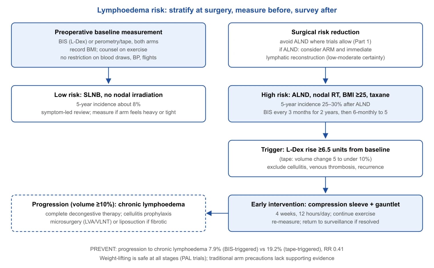
Treatment
Complete decongestive therapy (manual lymphatic drainage, multilayer bandaging, exercise and skin care, then maintenance compression) is the conventional first step, but its incremental value over compression alone is smaller than its reputation: in a Canadian RCT of 103 women, excess arm volume fell by 29.0% with decongestive therapy versus 22.6% with garments alone (difference 6.4%, 95% CI −6.8 to 20.5; P = 0.34), although absolute volume loss was greater (250 vs 143 mL, P = 0.03)[332]. Intermittent pneumatic compression adds a modest short-term benefit; a 2025 meta-analysis of 14 RCTs (1,397 patients) found reduced incidence when used preventively (RR 0.36, 0.22–0.58) and limited efficacy in chronic, fibrotic disease [333].
Surgery is reserved for lymphoedema that persists despite conservative treatment. Physiological procedures (lymphaticovenous anastomosis, LVA, and vascularised lymph node transfer, VLNT) aim to restore drainage; in a 2025 meta-analysis, pooled clinical improvement of the upper limb was 36.5% (95% CI 29.4–43.5), 29.4% after LVA and 41.7% after VLNT, from predominantly observational series [334], and the only RCT of VLNT (36 participants) reported a 39% volume reduction graded very low certainty [327]. Reductive surgery (circumferential liposuction with lifelong compression) addresses the non-pitting, fat-dominated stage II–III arm: in 105 women with a mean excess of 1,573 mL and lymphoedema of ten years' duration, reduction was complete (117% of excess volume) at five years [335]. Cellulitis both results from and worsens lymphoedema [322]; the evidence for antibiotic prophylaxis comes from the leg, where penicillin reduced recurrence (22% vs 37%; HR 0.55, 95% CI 0.35–0.86) with the effect fading when stopped [336], and is applied to the arm by extrapolation. The quality-of-life burden is measurable: of seventeen instruments in 39 studies, the lymphoedema-specific ULL-27 has the strongest psychometric properties, and exercise and decongestive therapy were associated with better scores [337].
Lymphoedema: what the evidence supports
- Axillary surgery is the dominant determinant: 5-year incidence about 8% after sentinel biopsy, 25% after dissection, 30% after dissection plus nodal irradiation [28].
- Baseline measurement, a BIS trigger and four weeks of compression reduced progression to chronic lymphoedema from 19.2% to 7.9% among women who needed intervention [30].
- Progressive resistance exercise is safe at every stage [329,330]; blood draws, cuffs and air travel on the treated side are not associated with swelling, whereas cellulitis and BMI are [322].
- Arm-lymphatic preservation and immediate lymphatic reconstruction reduce lymphoedema in RCT and pooled data; the safety of preserved crossover nodes and the completed reconstruction RCT are outstanding [324,325,328].
Emerging Surgical Technology
Wire-free localisation: the comparative evidence
Part 1 (Section 7.3) summarised the localisation options and noted that an oncological dividend was unproven; the comparative evidence since has firmed up modestly. The Cochrane review of eleven RCTs found radioguided occult lesion localisation comparable with wire for positive margins (RR 0.74, 95% CI 0.42–1.29) and re-operation (RR 0.51, 0.21–1.23), neither significant [338]. For magnetic seeds, a 2026 meta-analysis of 16 studies (8,794 patients) found no significant difference in positive or close margins (OR 0.86, 0.66–1.11) but lower re-excision (OR 0.77, 0.60–0.98) and smaller specimens, from largely non-randomised data [339]. The first RCT of magnetic seed against ROLL (not wire) showed non-inferior negative margins (93.1% vs 97.7%, P = 0.14), shorter stay and more post-localisation haematoma (17% vs 7%)[340]. The international MELODY cohort (EUBREAST-4/iBRA-NET, NCT05559411), designed to compare all techniques on margins, re-excision and preference, had not reported at the time of writing [341]. Wire-free techniques improve logistics and patient experience with, at most, a small reduction in re-excision.
Robotic nipple-sparing mastectomy
Robotic nipple-sparing mastectomy (RNSM) through a short lateral or axillary incision is practised in Europe and East Asia and, since late 2025, under a cleared labelling in the United States. The single-centre Milan RCT (80 women with cancer or a BRCA mutation) found the robotic operation 78 minutes longer, no difference in complications (P = 0.11), higher BREAST-Q and body-image scores, and no local events at a median of 28.6 months [32]. An international pooled analysis reported less nipple necrosis after RNSM (2.2% vs 7.8%, P = 0.002) and similar short-term oncological outcomes [342]; the Korean single-port series of 428 patients recorded seroma in 10.5% and reoperation in 2.8%[343]. A GRADE-assessed meta-analysis of prospective studies found longer operating time (mean difference 64 minutes, 95% CI 8.5–119.5) and no significant difference in major complications (RR 0.63, 0.27–1.47) or nipple necrosis (RR 0.56, 0.28–1.11), with low certainty throughout [344]. The US multicentre RCT under an investigational device exemption (74 patients, 131 mastectomies, 14 sites) found no conversions, serious adverse events in 3 robotic versus 8 open patients, nipple preservation at 42 days of 100% versus 98.5%, identical positive-margin counts (six each) and non-significant BREAST-Q trends favouring the robot; the operation took 141.6 versus 83.9 minutes, and accrual stopped when the device was cleared [345]. An expert consensus of 53 statements frames the procedure as evolving rather than standard [346].
Intraoperative margin assessment
A meta-analysis of 35 studies ranks the established techniques: frozen section (sensitivity 86%, specificity 96%, AUROC 0.96) and imprint cytology (91%, 95%, 0.98) outperform intraoperative ultrasound (59%, 81%) and specimen radiography (53%, 84%), with optical spectroscopy between (85%, 87%)[348]. In a 596-patient RCT, the radiofrequency spectroscopy probe (MarginProbe) reduced re-excision from 25.8% to 19.8% by lowering the false-negative rate (24.8% vs 66.1%) at the cost of more false positives (53.6% vs 16.6%)[349]. Rapid evaporative ionisation mass spectrometry (the "iKnife") classifies electrosurgical vapour by lipid profile in 1.8 seconds, with 90.9% sensitivity and 98.8% specificity on 260 validation specimens, but remains proof-of-concept [350]; optical coherence tomography has similar laboratory promise and no randomised outcome data. Fluorescence guidance is the first new modality with phase 3 data: in INSITE, cavity imaging after intravenous pegulicianine removed residual tumour left by standard lumpectomy in 27 of 357 patients (7.6%) and avoided a second operation in 9 of 62 with positive margins, with per-margin specificity of 85.2% but sensitivity of 49.3%, below the prespecified threshold, and two grade 3 drug-related events [351]; the FDA approved the agent in April 2024 [352]. None of these devices has been shown in a randomised trial to change local recurrence (authors' judgement: low-to-moderate certainty for a modest re-excision benefit).
Fluorescence sentinel node mapping, ablation and planning tools
Indocyanine green (ICG) fluorescence is an alternative to radioisotope where nuclear medicine is unavailable: in a meta-analysis of ten studies, ICG was equivalent to radioisotope for sentinel node identification (OR 2.58, 95% CI 0.35–19.08) and superior to blue dye (OR 9.07, 6.73–12.23) and to the dual technique (OR 4.22, 2.17–8.20)[353], and a 2026 RCT of 70 patients found identification of 100% with ICG alone and 97.1% with dual tracer [354]. Cryoablation without excision is investigational: in the single-arm ICE3 trial, 194 women (mean age 74.9 years) with luminal A-type cancers of mean size 7–8 mm had a five-year ipsilateral recurrence rate of 4.3% (3.7% among the 124 on endocrine therapy alone), with no serious device-related events [355]. Without a surgical comparator, in a population whose risk is already low, these data support trials rather than practice. Three-dimensional and augmented-reality planning for breast conservation is at the feasibility stage, with proof-of-concept protocols enrolling a handful of patients to test registration of supine imaging to the operative field [356]. Artificial intelligence in surgical planning is at a similar point: radiogenomic and margin-prediction models exist, but reviews cite limited interpretability, unstable performance across populations and the absence of prospective trials [357].
| Technology | Best comparative evidence | Key number | Evidence level |
|---|---|---|---|
| Magnetic seed vs wire | Meta-analysis, 8,794 patients | Re-excision OR 0.77 (0.60–0.98) | Mostly non-randomised [339] |
| Robotic NSM vs open | RCTs (80 and 74 patients) | Complications similar; +64 to +78 min; follow-up ≤36 months | Low certainty [32,344,345] |
| MarginProbe | RCT, 596 patients | Re-excision 19.8% vs 25.8% | One RCT [349] |
| Pegulicianine fluorescence | Phase 3 | Residual tumour removed in 7.6%; sensitivity 49.3% | FDA-approved 2024 [351,352] |
| iKnife mass spectrometry | Ex vivo validation | Sensitivity 90.9%, specificity 98.8% | Proof of concept [350] |
| ICG sentinel node mapping | Meta-analysis; small RCT | Equivalent to isotope; 100% vs 97.1% | Moderate [353,354] |
| Cryoablation (ICE3) | Single-arm, 194 patients | 5-year IBTR 4.3% | Investigational [355] |
| AR/3D and AI planning | Protocols, narrative reviews | — | Feasibility [356,357] |
Clinical Guidelines
The positions below are those cited in the body of this review; wording is paraphrased and the strength is given as the source states it. The St Gallen consensus is not cited in Sections 6 to 13 and is not tabulated; where a body has no position among the sources cited, the cell says so rather than inferring one.
| Decision point | North American bodies | European bodies | United Kingdom | Position |
|---|---|---|---|---|
| Screening age and interval | USPSTF 2024: biennial 40–74 (grade B); insufficient evidence at 75 and over [4] | ECIBC: biennial 50–69 (strong); conditional at 45–49 and 70–74; none at 40–44 [5] | NHS: triennial 50–70 [2] | Disagree at 40–49 and on interval |
| Supplemental imaging in dense breasts | USPSTF: I statement; FDA density notification mandatory from 2024 [4,100] | EUSOBI: MRI every 2–4 years at 50–70 with extremely dense breasts [101] | No position cited; BRAID is the UK trial [7] | Disagree |
| Germline testing and carrier surgery | NCCN: criteria-based testing, annual MRI from 25; ASBrS: test all; ASCO/ASTRO/SSO: conservation acceptable in BRCA1/2, radiotherapy avoided in TP53 [11,76,80] | No position cited | No position cited | Testing criteria disagree within North America |
| Preoperative breast MRI | No position cited | European consensus 2026: not for all; lobular, HER2-positive or triple-negative, suspected multifocality, discordant imaging, high-grade DCIS, neoadjuvant patients; MRI-only findings biopsied [14] | No position cited | Subtype indications untested in trials |
| B3 lesions | International consensus 2023: excision for ADH; observation after complete VAB removal for other B3 lesions [35] | European guideline 2024: VAE or surgery by multidisciplinary decision [36] | NICE NG12 governs referral only [122] | Agree |
| Systemic staging | ASCO Choosing Wisely: none in early disease; NCCN: PET/CT optional from stage III [151,153] | ESMO 2024: none in asymptomatic stage I–II; EUSOMA: 95% of stage III staged [121,152] | No position cited | Agree |
| Reconstruction offer and PMRT | ASPS: preoperative plastic surgery referral [173] | ESTRO-ACROP: moderate hypofractionation acceptable over reconstruction [236] | NICE NG101: offer to all, including those who may need radiotherapy; consider 40 Gy in 15 rather than 26 Gy in 5 over an implant [172] | Agree on offer; no guideline on implant plane [187] |
| Whole-breast fractionation and boost | ASTRO 2018: 40 Gy in 15 or 42.5 Gy in 16 for all; boost at 50 or younger, high grade or positive margin [234] | ESTRO-ACROP 2022: five fractions only for non-nodal treatment without reconstruction [236] | NICE NG101 as above [172] | Five-fraction scope differs |
| Partial-breast irradiation and IORT | ASTRO 2017/2024: PBI once daily or alternate days; IORT only for "suitable" patients with counselling [247,248] | GEC-ESTRO: 50 or over, unifocal pT1–2 up to 3 cm, clear margins, pN0; German panel: 50 kV IORT not recommended [245,249] | No position cited | Agree on PBI; disagree on IORT |
| Radiotherapy omission after conservation | Trial criteria only [25,253] | No guideline cited | No guideline cited | Trial-based |
| PMRT with 1–3 positive nodes | ASCO/ASTRO/SSO 2016: recommend, with exceptions for very low risk; include internal mammary and supraclavicular nodes [258] | Danish practice includes internal mammary irradiation [265]; SUPREMO reading contested [260] | No position cited | Guideline predates SUPREMO |
| Lymphoedema surveillance | No guideline cited; PREVENT model [30] | ISL 2020 staging [49] | No position cited | Trial-based |
Where they disagree, the disagreement is about screening and about scope. USPSTF starts biennial screening at 40 on a small, imprecise effect at 40–49 [4,89], whereas ECIBC does not recommend organised screening below 45 and the UK invites women every three years from 50 [2,5]. EUSOBI recommends supplemental MRI for extremely dense breasts on the strength of DENSE [6,101]; USPSTF, reading the same trial, finds the evidence insufficient because no mortality effect has been shown [4]. NCCN selects for germline testing by criteria that miss about half of carriers; the ASBrS tests everyone [10,76,80]. The 2026 European consensus names HER2-positive and triple-negative disease as MRI indications [14], which the randomised trials never stratified [12,13]. ESTRO-ACROP confines five fractions to non-nodal treatment without reconstruction and NICE prefers 15 fractions over an implant [172,236], a caution the FAST-Forward nodal substudy, with 32 events in 466 patients, has not resolved [23]. ASTRO permits intraoperative radiotherapy for suitable patients while the German panel rejects the 50 kV technique [245,247]. The ASCO/ASTRO/SSO recommendation of post-mastectomy radiotherapy for one to three nodes [258] predates SUPREMO, which showed no survival gain from chest-wall irradiation alone but did not test nodal irradiation [26,260]; the guideline stands for nodal treatment and is weakened for the chest wall alone.
Evidence Gaps
- Whether tomosynthesis reduces advanced or interval cancers is unproven; TOSYMA's 24-month interval cancer difference was not significant (OR 0.76, 0.55–1.06) and TMIST is not expected before 2028 [96,99].
- Risk-based screening was non-inferior for stage IIB or higher cancers in WISDOM but did not reduce biopsies and its margin is contested; MyPeBS is expected in 2027 [110–112].
- No trial of supplemental MRI, contrast-enhanced mammography or AI-supported reading has measured overdiagnosis; BRAID does not estimate it and MASAI detected 51% more in situ cancers [7,107].
- Preoperative MRI by biological subtype has no randomised evidence; the European consensus indication rests on expert opinion and MIPA is observational [14,144].
- ctDNA detection of residual disease is valid but not actionable; most c-TRAK TN positives were already metastatic and ZEST closed after randomising 40 patients [17,167].
- Prepectoral against subpectoral placement has one 42-patient trial; OPBC-02/PREPEC has not reported [186,187]. Acellular dermal matrix has one cautionary randomised trial and low-certainty pooled evidence [190,192].
- Immediate autologous reconstruction with planned radiotherapy rests on one largely single-centre comparison [227]; the safety of fat grafting after DCIS rests on one unreplicated cohort [217].
- Five-fraction radiotherapy when the axilla or an implant must be treated: the FAST-Forward nodal substudy recorded 32 events, too few to estimate the 26 Gy arm alone [23,237].
- Genomic selection for radiotherapy omission is untested; EXPERT, DEBRA and NATURAL report in 2027, 2031 and 2033, and EUROPA's recurrence end point is pending [254–257].
- Nodal radiotherapy with contemporary systemic therapy in one to three nodes has not been randomised; SUPREMO tested the chest wall alone and POSNOC has not reported [26,260,266]. Proton therapy's cardiac benefit awaits RadComp in 2036 [279].
- Immediate lymphatic reconstruction rests on two small single-centre trials and an interim result, with the first independent trial completing at 174 patients [325,327]; the safety of preserved crossover nodes in axillary reverse mapping is unresolved [326].
- Robotic nipple-sparing mastectomy has a longest randomised follow-up of 28.6 months [32,345], cryoablation has no surgical comparator [355] and the MELODY localisation cohort has not reported [341].
Scenario-Based Decision Framework
The eight scenarios below condense Sections 6 to 13 into common consultations. Each assumes triple assessment, multidisciplinary review and the Part 1 discussion of the operation; the approach is a framework for shared decision-making, not a prescription for an individual without examination.
Scenario 1. Supplemental screening in dense breasts. A 47-year-old with extremely dense breasts, a normal mammogram and no family history asks for an additional test. Evidence. Mammographic sensitivity falls to 62.9% in extremely dense breasts [118]. At 50–75, supplemental MRI halved interval cancers at 79.8 false positives per 1,000 in the first round [6]; abbreviated MRI and contrast-enhanced mammography detected about four times as many cancers as automated ultrasound [7]. USPSTF finds the evidence insufficient; EUSOBI recommends MRI from 50 [4,101]. Approach. Explain that she is below the age window of the trial and guideline, that supplemental imaging finds more cancers without proof of a mortality effect, and that the price is recall and biopsy; if she chooses it, abbreviated MRI or contrast-enhanced mammography rather than automated ultrasound. Caveat. No trial has estimated the overdiagnosis added by supplemental imaging [7].
Scenario 2. Atypical ductal hyperplasia on vacuum-assisted biopsy. A 58-year-old has ADH on 9-gauge stereotactic VAB of a 7 mm cluster of calcifications, with none remaining afterwards. Evidence. Pooled upgrade at excision is 29%, 23% after stereotactic sampling and 14% when imaging suggests complete removal [127]. The 2023 consensus recommends excision for ADH, by a 76% majority even after VAB; the 2024 European guideline leaves VAE against surgery to the multidisciplinary team [35,36]. Atypia carries a cumulative cancer risk of about 30% at 25 years [55]. Approach. Multidisciplinary review with gauge, guidance and proportion removed stated; excision remains the default, and observation is an option for a small, concordant, fully sampled lesion with the residual risk explained. Caveat. The 14% figure applies only when imaging shows complete removal; a 14-gauge ultrasound-guided diagnosis carries a 42% upgrade [127].
Scenario 3. Recurrence score 20 and the sequence of treatment. A 52-year-old postmenopausal woman with a 2.2 cm grade 2 ER-positive, HER2-negative cancer, normal axillary ultrasound and a recurrence score of 20 asks whether chemotherapy should come first. Evidence. For scores of 11–25, endocrine therapy alone was non-inferior (HR 1.08), with chemotherapy benefit confined to women aged 50 or younger with scores of 16–25 [15]; if one to three nodes prove positive, postmenopausal women with a score of 25 or lower still gain nothing (HR 1.02)[16]. Pathological complete response to neoadjuvant chemotherapy in this setting was 4.3%[164]; 68% of molecularly luminal tumours responded clinically to neoadjuvant endocrine therapy [165]. Approach. Surgery first, with the axillary pathway from Part 1; neoadjuvant endocrine therapy rather than chemotherapy if downsizing is needed; no systemic staging [151]. Caveat. The assay governs chemotherapy, not local treatment; nodal status at surgery still decides radiotherapy.
Scenario 4. BRCA1 carrier choosing nipple-sparing mastectomy. A 38-year-old BRCA1 carrier with a 1.8 cm triple-negative cancer more than 2 cm from the nipple wants bilateral nipple-sparing mastectomy with immediate reconstruction. Evidence. Her contralateral risk is 40% at 20 years [9], and the ASCO/ASTRO/SSO guideline supports discussion of bilateral mastectomy in carriers [11]. Nipple-sparing mastectomy carries 1.1% nipple–areolar recurrence at 10 years [176]; occult nipple involvement is more likely in receptor-negative tumours [178]; nipple necrosis follows 18.1% of periareolar and 6.8% of inframammary incisions [305]. Immediate implant reconstruction carries 9% implant loss and 18% reoperation at 3 months [18]. Approach. Settle the systemic sequence first (Part 1); bilateral nipple-sparing mastectomy through inframammary incisions with retroareolar sampling, and reconstruction chosen with the likelihood of radiotherapy in mind (Section 9.6). Caveat. If nodes prove positive, radiotherapy over an implant raises contracture to about 50%[222]; delayed-immediate reconstruction preserves the option [40].
Scenario 5. Mastectomy with likely radiotherapy: implant or DIEP, and when. A 60-year-old with a 3.5 cm cancer and two biopsy-proven nodes, BMI 31, is planned for mastectomy and will almost certainly need radiotherapy. Evidence. In irradiated patients, implants had a complication in 38.9% by 2 years against 25.6% for flaps (OR 0.47)[22]; irradiating an expander raised failure (20% vs 13.4%) and irradiating the implant raised contracture to 49.4%[222]. Flaps placed before or after radiotherapy had similar pooled complications, with fibrosis in 27%[226]; DIEP total loss is 2.0%[205]; obesity raises reconstruction complications [287]. NICE advises offering immediate reconstruction even when radiotherapy is likely [172]. Approach. Favour autologous tissue: delayed DIEP about 3 months after radiotherapy, or delayed-immediate reconstruction with an expander placeholder; immediate DIEP only in a centre with that experience and with fibrosis and revision explained. Caveat. If neoadjuvant therapy converts the axilla to ypN0, the radiotherapy indication changes (Part 1, B-51) and so does the reconstructive calculus.
Scenario 6. Omitting radiotherapy at 72. A 72-year-old with a 14 mm grade 2 ER-positive, PR-positive, HER2-negative cancer, Ki-67 8%, clear margins and a negative sentinel node asks whether she can avoid radiotherapy. Evidence. In PRIME II, omission raised 10-year local recurrence from 0.9% to 9.5% with almost identical survival [25]; in LUMINA, luminal A disease with Ki-67 of 13.25% or less had 5-year local recurrence of 2.3% on endocrine therapy alone [253]. Omission assumes five years of endocrine therapy, which in EUROPA worsened global health status more than radiotherapy (−9.79 vs −3.40 points)[257]; the alternative is one week of radiotherapy [46]. Wider excision does not substitute [232]. Approach. Quantify the difference as about one in ten against one in a hundred at 10 years; omission is acceptable if she will take endocrine therapy, and five fractions without a boost is the alternative if she will not [234]. Caveat. LUMINA is a single-arm cohort at 5 years and luminal recurrence continues to year 20 [64].
Scenario 7. Rising bioimpedance after dissection and nodal radiotherapy. A 55-year-old who had axillary dissection and regional nodal irradiation, with a preoperative L-Dex baseline, has a rise of 7 units at 6 months and no visible swelling. Evidence. Her 5-year risk is about 30%[28] and 80% of lymphoedema appears within 2 years [323]. In PREVENT, a rise of 6.5 units triggered a four-week compression sleeve, and among women who triggered it progression to chronic lymphoedema was 7.9% against 19.2% with tape measurement [30]. Cellulitis and BMI of 25 or more are associated with arm volume change; blood draws, cuffs and air travel are not [322]; weight-lifting reduced incident lymphoedema from 22% to 7% with five or more nodes removed [330]. Approach. Exclude cellulitis, venous thrombosis and recurrence; sleeve and gauntlet for four weeks, 12 hours a day; re-measure; three-monthly surveillance to 2 years, with supervised resistance exercise and weight management. Caveat. Both PREVENT arms received the same intervention, so early detection is better established than the superiority of bioimpedance; a volume change of 10% or more means established lymphoedema and complete decongestive therapy [28].
Scenario 8. Robotic nipple-sparing mastectomy. A 45-year-old with a 1.5 cm ER-positive cancer suitable for conservation has decided on mastectomy and asks for the robotic operation. Evidence. The Milan trial (80 women) found the robotic operation 78 minutes longer, no difference in complications and no local events at a median of 28.6 months [32]; the US trial (74 patients) found serious adverse events in 3 versus 8 patients, six positive margins in each arm and an operation of 141.6 versus 83.9 minutes [345]. The GRADE-assessed meta-analysis rates every outcome low certainty with follow-up of 6 to 36 months [344]. The FDA cleared the indication on 15 December 2025 on substantial equivalence, not cancer outcomes [31]. Approach. Revisit the choice of mastectomy over conservation (Part 1); if mastectomy stands, open nipple-sparing mastectomy is the operation with long-term data, and the robotic operation is offered only within a registry or trial with the follow-up gap disclosed. Caveat. Local recurrence after nipple-sparing mastectomy accrues over five to ten years; the robotic evidence covers three [344].
Conclusions
Screening and biology determine what reaches the operating theatre. Invitation to mammography reduces breast cancer mortality by about a fifth at the cost of about three overdiagnosed cancers per death prevented, and the range of published overdiagnosis estimates is mostly arithmetic [2,33]. Supplemental MRI halves interval cancers in extremely dense breasts, AI-supported reading is a safe substitute for one of two readers, and risk-based screening is feasible but not yet shown to be better [6,8,110]. The surgeon who treats screen-detected DCIS and small luminal tumours is treating the reservoir that screening uncovers [1,58].
Germline status should be known before the operation it may change, because criteria-based testing misses about half of carriers and the result alters both the operation and the radiotherapy plan [10,11]. In the workup, a test earns its place only if it changes a decision: preoperative MRI does not improve surgical outcomes outside defined indications [13,14]; a positive axillary needle biopsy no longer mandates dissection [138]; asymptomatic stage I–II disease is not staged [151]; and a recurrence score on the diagnostic core decides whether surgery comes first [15,16].
Reconstruction should be offered to everyone and described with its real numbers: 9% implant loss and 18% reoperation at 3 months for immediate implants [18], more early complications but fewer failures and a meaningful satisfaction advantage for autologous tissue [19,20]. Radiotherapy shifts the recommendation towards a flap or a delayed-immediate sequence [22,40]; macro-textured implants carry a lymphoma risk of about 1 in 355–559 patients [195]; fat grafting has not increased recurrence [214]; and flat closure is a choice to be supported [230].
Radiotherapy after conservation is now one week for most patients, one quadrant for suitable patients, and omitted only after a conversation that quantifies about one in ten against one in a hundred recurrences at 10 years in women meeting the trial criteria and taking endocrine therapy [23–25]. After mastectomy, nodal burden sets the indication: chest-wall irradiation alone adds no survival, whereas nodal and internal mammary irradiation reduce breast cancer mortality [26,27,265]. The surgeon controls the clips that define the target, the nodal count that sets the indication, and the interval before treatment starts [240,269].
Most complications are grade I–II, but they delay adjuvant treatment and dominate the patient's experience; flap fixation, antibiotic prophylaxis, inframammary incisions and intercostobrachial nerve preservation each reduce them [291,301,305,310]. Lymphoedema is determined mainly by the extent of axillary surgery, about 8% after sentinel biopsy and 25–30% after dissection [28]; avoiding dissection is the most effective prevention, and where it is needed, arm-lymphatic preservation, immediate lymphatic reconstruction, surveillance-triggered compression and resistance exercise each reduce the risk [30,324,325,330]. Emerging technology should be adopted at the pace of its evidence: wire-free localisation and margin devices offer at most small reductions in re-excision [339,349], and cryoablation and robotic mastectomy have short-term safety data only [345,355]. A regulatory clearance based on 42-day outcomes is not an oncological endorsement, and the patient should be told so [31].
Appendix A. Verification Notes and Limitations of This Review
- Welch HG, Prorok PC, O'Malley AJ, Kramer BS. Breast-cancer tumor size, overdiagnosis, and mammography screening effectiveness. N Engl J Med. 2016;375(15):1438-1447. doi:10.1056/NEJMoa1600249. PMID: 27732805.
- Independent UK Panel on Breast Cancer Screening. The benefits and harms of breast cancer screening: an independent review. Lancet. 2012;380(9855):1778-1786. doi:10.1016/S0140-6736(12)61611-0. PMID: 23117178.
- Marmot MG, Altman DG, Cameron DA, Dewar JA, Thompson SG, Wilcox M. The benefits and harms of breast cancer screening: an independent review. Br J Cancer. 2013;108(11):2205-2240. doi:10.1038/bjc.2013.177. PMID: 23744281.
- US Preventive Services Task Force, Nicholson WK, Silverstein M, Wong JB, Barry MJ, Chelmow D, et al. Screening for breast cancer: US Preventive Services Task Force recommendation statement. JAMA. 2024;331(22):1918-1930. doi:10.1001/jama.2024.5534. PMID: 38687503.
- Schünemann HJ, Lerda D, Quinn C, Follmann M, Alonso-Coello P, Rossi PG, et al. Breast cancer screening and diagnosis: a synopsis of the European Breast Guidelines. Ann Intern Med. 2020;172(1):46-56. doi:10.7326/M19-2125. PMID: 31766052.
- Bakker MF, de Lange SV, Pijnappel RM, Mann RM, Peeters PHM, Monninkhof EM, et al. Supplemental MRI screening for women with extremely dense breast tissue. N Engl J Med. 2019;381(22):2091-2102. doi:10.1056/NEJMoa1903986. PMID: 31774954.
- Gilbert FJ, Payne NR, Allajbeu I, Yit L, Vinnicombe S, Lyburn I, et al. Comparison of supplemental breast cancer imaging techniques-interim results from the BRAID randomised controlled trial. Lancet. 2025;405(10493):1935-1944. doi:10.1016/S0140-6736(25)00582-3. PMID: 40412427.
- Gommers J, Hernström V, Josefsson V, Sartor H, Schmidt D, Hjelmgren A, et al. Interval cancer, sensitivity, and specificity comparing AI-supported mammography screening with standard double reading without AI in the MASAI study: a randomised, controlled, non-inferiority, single-blinded, population-based, screening-accuracy trial. Lancet. 2026;407(10527):505-514. doi:10.1016/S0140-6736(25)02464-X. PMID: 41620232.
- Kuchenbaecker KB, Hopper JL, Barnes DR, Phillips KA, Mooij TM, Roos-Blom MJ, et al. Risks of breast, ovarian, and contralateral breast cancer for BRCA1 and BRCA2 mutation carriers. JAMA. 2017;317(23):2402-2416. doi:10.1001/jama.2017.7112. PMID: 28632866.
- Beitsch PD, Whitworth PW, Hughes K, Patel R, Rosen B, Compagnoni G, et al. Underdiagnosis of hereditary breast cancer: are genetic testing guidelines a tool or an obstacle? J Clin Oncol. 2019;37(6):453-460. doi:10.1200/JCO.18.01631. PMID: 30526229.
- Tung NM, Boughey JC, Pierce LJ, Robson ME, Bedrosian I, Dietz JR, et al. Management of hereditary breast cancer: American Society of Clinical Oncology, American Society for Radiation Oncology, and Society of Surgical Oncology guideline. J Clin Oncol. 2020;38(18):2080-2106. doi:10.1200/JCO.20.00299. PMID: 32243226.
- Turnbull L, Brown S, Harvey I, Olivier C, Drew P, Napp V, et al. Comparative effectiveness of MRI in breast cancer (COMICE) trial: a randomised controlled trial. Lancet. 2010;375(9714):563-571. doi:10.1016/S0140-6736(09)62070-5. PMID: 20159292.
- Houssami N, Turner RM, Morrow M. Meta-analysis of pre-operative magnetic resonance imaging (MRI) and surgical treatment for breast cancer. Breast Cancer Res Treat. 2017;165(2):273-283. doi:10.1007/s10549-017-4324-3. PMID: 28589366.
- Camps-Herrero J, Gullo RL, Athanasiou A, Fuchsjaeger M, Gilbert FJ, Kirova Y, et al. European consensus on pre-operative imaging in breast cancer developed jointly by EUSOBI, ESSO, ESP and ESTRO. Eur Radiol. 2026;36(10):7806-7816. doi:10.1007/s00330-026-12558-9. PMID: 42301480.
- Sparano JA, Gray RJ, Makower DF, Pritchard KI, Albain KS, Hayes DF, et al. Adjuvant Chemotherapy Guided by a 21-Gene Expression Assay in Breast Cancer. N Engl J Med. 2018;379(2):111-121. doi:10.1056/NEJMoa1804710. PMID: 29860917.
- Kalinsky K, Barlow WE, Gralow JR, Meric-Bernstam F, Albain KS, Hayes DF, et al. 21-Gene Assay to Inform Chemotherapy Benefit in Node-Positive Breast Cancer. N Engl J Med. 2021;385(25):2336-2347. doi:10.1056/NEJMoa2108873. PMID: 34914339.
- Turner NC, Swift C, Jenkins B, Kilburn L, Coakley M, Beaney M, et al. Results of the c-TRAK TN trial: a clinical trial utilising ctDNA mutation tracking to detect molecular residual disease and trigger intervention in patients with moderate- and high-risk early-stage triple-negative breast cancer. Ann Oncol. 2023;34(2):200-211. doi:10.1016/j.annonc.2022.11.005. PMID: 36423745.
- Potter S, Conroy EJ, Cutress RI, Williamson PR, Whisker L, Thrush S, et al. Short-term safety outcomes of mastectomy and immediate implant-based breast reconstruction with and without mesh (iBRA): a multicentre, prospective cohort study. Lancet Oncol. 2019;20(2):254-66. doi:10.1016/S1470-2045(18)30781-2. PMID: 30639093.
- Bennett KG, Qi J, Kim HM, Hamill JB, Pusic AL, Wilkins EG. Comparison of 2-year complication rates among common techniques for postmastectomy breast reconstruction. JAMA Surg. 2018;153(10):901-8. doi:10.1001/jamasurg.2018.1687. PMID: 29926077.
- Santosa KB, Qi J, Kim HM, Hamill JB, Wilkins EG, Pusic AL. Long-term patient-reported outcomes in postmastectomy breast reconstruction. JAMA Surg. 2018;153(10):891-9. doi:10.1001/jamasurg.2018.1677. PMID: 29926096.
- Voineskos SH, Klassen AF, Cano SJ, Pusic AL, Gibbons CJ. Giving meaning to differences in BREAST-Q scores: minimal important difference for breast reconstruction patients. Plast Reconstr Surg. 2020;145(1):11e-20e. doi:10.1097/PRS.0000000000006317. PMID: 31577663.
- Jagsi R, Momoh AO, Qi J, Hamill JB, Billig J, Kim HM, et al. Impact of radiotherapy on complications and patient-reported outcomes after breast reconstruction. J Natl Cancer Inst. 2018;110(2):djx148. doi:10.1093/jnci/djx148. PMID: 28954300.
- Brunt AM, Cafferty FH, Kirby AM, Patel J, Sydenham MA, Haviland JS, et al. Hypofractionated breast radiotherapy for 1 week versus 3 weeks (FAST-Forward): 10-year efficacy and late normal tissue effects from a multicentre, open-label, non-inferiority, phase 3, randomised controlled trial and 5-year efficacy results from a randomised axillary substudy. Lancet Oncol. 2026;27(6):686-98. doi:10.1016/S1470-2045(26)00076-8. PMID: 42134381.
- Coles CE, Griffin CL, Kirby AM, Titley J, Agrawal RK, Alhasso A, et al. Partial-breast radiotherapy after breast conservation surgery for patients with early breast cancer (UK IMPORT LOW trial): 5-year results from a multicentre, randomised, controlled, phase 3, non-inferiority trial. Lancet. 2017;390(10099):1048-60. doi:10.1016/S0140-6736(17)31145-5. PMID: 28779963.
- Kunkler IH, Williams LJ, Jack WJL, Cameron DA, Dixon JM. Breast-conserving surgery with or without irradiation in early breast cancer. N Engl J Med. 2023;388(7):585-94. doi:10.1056/NEJMoa2207586. PMID: 36791159.
- Kunkler IH, Russell NS, Anderson N, Sainsbury R, Dixon JM, Cameron D, et al. Ten-year survival after postmastectomy chest-wall irradiation in breast cancer. N Engl J Med. 2025;393(18):1771-83. doi:10.1056/NEJMoa2412225. PMID: 41191939.
- Poortmans PM, Weltens C, Fortpied C, Kirkove C, Peignaux-Casasnovas K, Budach V, et al. Internal mammary and medial supraclavicular lymph node chain irradiation in stage I-III breast cancer (EORTC 22922/10925): 15-year results of a randomised, phase 3 trial. Lancet Oncol. 2020;21(12):1602-10. doi:10.1016/S1470-2045(20)30472-1. PMID: 33152277.
- Naoum GE, Roberts S, Brunelle CL, Shui AM, Salama L, Daniell K, et al. Quantifying the impact of axillary surgery and nodal irradiation on breast cancer-related lymphedema and local tumor control: long-term results from a prospective screening trial. J Clin Oncol. 2020;38(29):3430-8. doi:10.1200/JCO.20.00459. PMID: 32730184.
- Bartels SAL, Donker M, Poncet C, Sauvé N, Straver ME, van de Velde CJH, et al. Radiotherapy or surgery of the axilla after a positive sentinel node in breast cancer: 10-year results of the randomized controlled EORTC 10981-22023 AMAROS trial. J Clin Oncol. 2023;41(12):2159-65. doi:10.1200/JCO.22.01565. PMID: 36383926.
- Ridner SH, Dietrich MS, Boyages J, Koelmeyer L, Elder E, Hughes TM, et al. A comparison of bioimpedance spectroscopy or tape measure triggered compression intervention in chronic breast cancer lymphedema prevention. Lymphat Res Biol. 2022;20(6):618-28. doi:10.1089/lrb.2021.0084. PMID: 35099283.
- US Food and Drug Administration. 510(k) premarket notification K252675: da Vinci SP Surgical System (SP1098), Intuitive Surgical, Inc.; labeling modification adding nipple sparing mastectomy procedures to the indications for use. Decision date 15 December 2025.
- Toesca A, Sangalli C, Maisonneuve P, Massari G, Girardi A, Baker JL, et al. A randomized trial of robotic mastectomy versus open surgery in women with breast cancer or BrCA mutation. Ann Surg. 2022;276(1):11-9. doi:10.1097/SLA.0000000000004969. PMID: 34597010.
- de Gelder R, Heijnsdijk EA, van Ravesteyn NT, Fracheboud J, Draisma G, de Koning HJ. Interpreting overdiagnosis estimates in population-based mammography screening. Epidemiol Rev. 2011;33:111-121. doi:10.1093/epirev/mxr009. PMID: 21709144.
- D'Orsi CJ, Sickles EA, Mendelson EB, Morris EA, et al. ACR BI-RADS Atlas, Breast Imaging Reporting and Data System. 5th ed. Reston, VA: American College of Radiology; 2013.
- Elfgen C, Leo C, Kubik-Huch RA, Muenst S, Schmidt N, Quinn C, et al. Third International Consensus Conference on lesions of uncertain malignant potential in the breast (B3 lesions). Virchows Arch. 2023;483(1):5-20. doi:10.1007/s00428-023-03566-x. PMID: 37330436.
- Rubio IT, Wyld L, Marotti L, Athanasiou A, Regitnig P, Catanuto G, et al. European guidelines for the diagnosis, treatment and follow-up of breast lesions with uncertain malignant potential (B3 lesions) developed jointly by EUSOMA, EUSOBI, ESP (BWG) and ESSO. Eur J Surg Oncol. 2024;50(1):107292. doi:10.1016/j.ejso.2023.107292. PMID: 38061151.
- Modi S, Jacot W, Yamashita T, Sohn J, Vidal M, Tokunaga E, et al. Trastuzumab deruxtecan in previously treated HER2-low advanced breast cancer. N Engl J Med. 2022;387(1):9-20. doi:10.1056/NEJMoa2203690. PMID: 35665782.
- Wolff AC, Somerfield MR, Dowsett M, Hammond MEH, Hayes DF, McShane LM, et al. Human Epidermal Growth Factor Receptor 2 Testing in Breast Cancer: ASCO-College of American Pathologists Guideline Update. J Clin Oncol. 2023;41(22):3867-3872. doi:10.1200/JCO.22.02864. PMID: 37284804.
- Piccart M, van 't Veer LJ, Poncet C, Lopes Cardozo JMN, Delaloge S, Pierga JY, et al. 70-gene signature as an aid for treatment decisions in early breast cancer: updated results of the phase 3 randomised MINDACT trial with an exploratory analysis by age. Lancet Oncol. 2021;22(4):476-488. doi:10.1016/S1470-2045(21)00007-3. PMID: 33721561.
- Kronowitz SJ. Delayed-immediate breast reconstruction: technical and timing considerations. Plast Reconstr Surg. 2010;125(2):463-74. doi:10.1097/PRS.0b013e3181c82d58. PMID: 19910850.
- Yoon AP, Qi J, Brown DL, Kim HM, Hamill JB, Erdmann-Sager J, et al. Outcomes of immediate versus delayed breast reconstruction: results of a multicenter prospective study. Breast. 2018;37:72-9. doi:10.1016/j.breast.2017.10.009. PMID: 29102781.
- Wu Y, Yu L, Huang M, Huang Y, Li C, Liang Y, et al. Comparative complications of prepectoral versus subpectoral breast reconstruction in patients with breast cancer: a meta-analysis. Front Oncol. 2024;14:1439293. doi:10.3389/fonc.2024.1439293. PMID: 39257552.
- Man LX, Selber JC, Serletti JM. Abdominal wall following free TRAM or DIEP flap reconstruction: a meta-analysis and critical review. Plast Reconstr Surg. 2009;124(3):752-64. doi:10.1097/PRS.0b013e31818b7533. PMID: 19342994.
- Loch-Wilkinson A, Beath KJ, Knight RJW, Wessels WLF, Magnusson M, Papadopoulos T, et al. Breast implant-associated anaplastic large cell lymphoma in Australia and New Zealand: high-surface-area textured implants are associated with increased risk. Plast Reconstr Surg. 2017;140(4):645-54. doi:10.1097/PRS.0000000000003654. PMID: 28481803.
- Haviland JS, Owen JR, Dewar JA, Agrawal RK, Barrett J, Barrett-Lee PJ, et al. The UK Standardisation of Breast Radiotherapy (START) trials of radiotherapy hypofractionation for treatment of early breast cancer: 10-year follow-up results of two randomised controlled trials. Lancet Oncol. 2013;14(11):1086-94. doi:10.1016/S1470-2045(13)70386-3. PMID: 24055415.
- Murray Brunt A, Haviland JS, Wheatley DA, Sydenham MA, Alhasso A, Bloomfield DJ, et al. Hypofractionated breast radiotherapy for 1 week versus 3 weeks (FAST-Forward): 5-year efficacy and late normal tissue effects results from a multicentre, non-inferiority, randomised, phase 3 trial. Lancet. 2020;395(10237):1613-26. doi:10.1016/S0140-6736(20)30932-6. PMID: 32580883.
- EBCTCG (Early Breast Cancer Trialists' Collaborative Group), McGale P, Taylor C, Correa C, Cutter D, Duane F, et al. Effect of radiotherapy after mastectomy and axillary surgery on 10-year recurrence and 20-year breast cancer mortality: meta-analysis of individual patient data for 8135 women in 22 randomised trials. Lancet. 2014;383(9935):2127-35. doi:10.1016/S0140-6736(14)60488-8. PMID: 24656685.
- Whelan TJ, Olivotto IA, Parulekar WR, Ackerman I, Chua BH, Nabid A, et al. Regional nodal irradiation in early-stage breast cancer. N Engl J Med. 2015;373(4):307-16. doi:10.1056/NEJMoa1415340. PMID: 26200977.
- Executive Committee of the International Society of Lymphology. The diagnosis and treatment of peripheral lymphedema: 2020 consensus document of the International Society of Lymphology. Lymphology. 2020;53(1):3-19. PMID: 32521126.
- Wijaya WA, Peng J, He Y, Chen J, Cen Y. Clinical application of axillary reverse mapping in patients with breast cancer: a systematic review and meta-analysis. Breast. 2020;53:189-200. doi:10.1016/j.breast.2020.08.007. PMID: 32858404.
- Boccardo F, Casabona F, De Cian F, Friedman D, Murelli F, Puglisi M, et al. Lymphatic microsurgical preventing healing approach (LYMPHA) for primary surgical prevention of breast cancer-related lymphedema: over 4 years follow-up. Microsurgery. 2014;34(6):421-4. doi:10.1002/micr.22254. PMID: 24677148.
- Lopez-Garcia MA, Geyer FC, Lacroix-Triki M, Marchió C, Reis-Filho JS. Breast cancer precursors revisited: molecular features and progression pathways. Histopathology. 2010;57(2):171-192. doi:10.1111/j.1365-2559.2010.03568.x. PMID: 20500230.
- Lim E, Vaillant F, Wu D, Forrest NC, Pal B, Hart AH, et al. Aberrant luminal progenitors as the candidate target population for basal tumor development in BRCA1 mutation carriers. Nat Med. 2009;15(8):907-913. doi:10.1038/nm.2000. PMID: 19648928.
- Hartmann LC, Radisky DC, Frost MH, Santen RJ, Vierkant RA, Benetti LL, et al. Understanding the premalignant potential of atypical hyperplasia through its natural history: a longitudinal cohort study. Cancer Prev Res (Phila). 2014;7(2):211-217. doi:10.1158/1940-6207.CAPR-13-0222. PMID: 24480577.
- Hartmann LC, Degnim AC, Santen RJ, Dupont WD, Ghosh K. Atypical hyperplasia of the breast--risk assessment and management options. N Engl J Med. 2015;372(1):78-89. doi:10.1056/NEJMsr1407164. PMID: 25551530.
- Erbas B, Provenzano E, Armes J, Gertig D. The natural history of ductal carcinoma in situ of the breast: a review. Breast Cancer Res Treat. 2006;97(2):135-144. doi:10.1007/s10549-005-9101-z. PMID: 16319971.
- Sanders ME, Schuyler PA, Simpson JF, Page DL, Dupont WD. Continued observation of the natural history of low-grade ductal carcinoma in situ reaffirms proclivity for local recurrence even after more than 30 years of follow-up. Mod Pathol. 2015;28(5):662-669. doi:10.1038/modpathol.2014.141. PMID: 25502729.
- Welch HG, Black WC. Using autopsy series to estimate the disease "reservoir" for ductal carcinoma in situ of the breast: how much more breast cancer can we find? Ann Intern Med. 1997;127(11):1023-1028. doi:10.7326/0003-4819-127-11-199712010-00014. PMID: 9412284.
- Perou CM, Sørlie T, Eisen MB, van de Rijn M, Jeffrey SS, Rees CA, et al. Molecular portraits of human breast tumours. Nature. 2000;406(6797):747-752. doi:10.1038/35021093. PMID: 10963602.
- Sørlie T, Perou CM, Tibshirani R, Aas T, Geisler S, Johnsen H, et al. Gene expression patterns of breast carcinomas distinguish tumor subclasses with clinical implications. Proc Natl Acad Sci U S A. 2001;98(19):10869-10874. doi:10.1073/pnas.191367098. PMID: 11553815.
- Cancer Genome Atlas Network. Comprehensive molecular portraits of human breast tumours. Nature. 2012;490(7418):61-70. doi:10.1038/nature11412. PMID: 23000897.
- Dent R, Trudeau M, Pritchard KI, Hanna WM, Kahn HK, Sawka CA, et al. Triple-negative breast cancer: clinical features and patterns of recurrence. Clin Cancer Res. 2007;13(15 Pt 1):4429-4434. doi:10.1158/1078-0432.CCR-06-3045. PMID: 17671126.
- Kennecke H, Yerushalmi R, Woods R, Cheang MC, Voduc D, Speers CH, et al. Metastatic behavior of breast cancer subtypes. J Clin Oncol. 2010;28(20):3271-3277. doi:10.1200/JCO.2009.25.9820. PMID: 20498394.
- Pan H, Gray R, Braybrooke J, Davies C, Taylor C, McGale P, et al. 20-Year risks of breast-cancer recurrence after stopping endocrine therapy at 5 years. N Engl J Med. 2017;377(19):1836-1846. doi:10.1056/NEJMoa1701830. PMID: 29117498.
- Bardia A, Hu X, Dent R, Yonemori K, Barrios CH, O'Shaughnessy JA, et al. Trastuzumab deruxtecan after endocrine therapy in metastatic breast cancer. N Engl J Med. 2024;391(22):2110-2122. doi:10.1056/NEJMoa2407086. PMID: 39282896.
- Schrijver WAME, Suijkerbuijk KPM, van Gils CH, van der Wall E, Moelans CB, van Diest PJ. Receptor conversion in distant breast cancer metastases: a systematic review and meta-analysis. J Natl Cancer Inst. 2018;110(6):568-580. doi:10.1093/jnci/djx273. PMID: 29315431.
- Retsky M, Demicheli R, Hrushesky W, Baum M, Gukas I. Surgery triggers outgrowth of latent distant disease in breast cancer: an inconvenient truth? Cancers (Basel). 2010;2(2):305-337. doi:10.3390/cancers2020305. PMID: 24281072.
- Krall JA, Reinhardt F, Mercury OA, Pattabiraman DR, Brooks MW, Dougan M, et al. The systemic response to surgery triggers the outgrowth of distant immune-controlled tumors in mouse models of dormancy. Sci Transl Med. 2018;10(436):eaan3464. doi:10.1126/scitranslmed.aan3464. PMID: 29643230.
- Forget P, Bouche G, Duhoux FP, Coulie PG, Decloedt J, Dekleermaker A, et al. Intraoperative ketorolac in high-risk breast cancer patients. A prospective, randomized, placebo-controlled clinical trial. PLoS One. 2019;14(12):e0225748. doi:10.1371/journal.pone.0225748. PMID: 31800611.
- Hu C, Hart SN, Gnanaolivu R, Huang H, Lee KY, Na J, et al. A population-based study of genes previously implicated in breast cancer. N Engl J Med. 2021;384(5):440-451. doi:10.1056/NEJMoa2005936. PMID: 33471974.
- Breast Cancer Association Consortium, Dorling L, Carvalho S, Allen J, González-Neira A, Luccarini C, et al. Breast cancer risk genes - association analysis in more than 113,000 women. N Engl J Med. 2021;384(5):428-439. doi:10.1056/NEJMoa1913948. PMID: 33471991.
- Yadav S, Boddicker NJ, Na J, Polley EC, Hu C, Hart SN, et al. Contralateral breast cancer risk among carriers of germline pathogenic variants in ATM, BRCA1, BRCA2, CHEK2, and PALB2. J Clin Oncol. 2023;41(9):1703-1713. doi:10.1200/JCO.22.01239. PMID: 36623243.
- Yang X, Leslie G, Doroszuk A, Schneider S, Allen J, Decker B, et al. Cancer risks associated with germline PALB2 pathogenic variants: an international study of 524 families. J Clin Oncol. 2020;38(7):674-685. doi:10.1200/JCO.19.01907. PMID: 31841383.
- Mai PL, Best AF, Peters JA, DeCastro RM, Khincha PP, Loud JT, et al. Risks of first and subsequent cancers among TP53 mutation carriers in the National Cancer Institute Li-Fraumeni syndrome cohort. Cancer. 2016;122(23):3673-3681. doi:10.1002/cncr.30248. PMID: 27496084.
- Tan MH, Mester JL, Ngeow J, Rybicki LA, Orloff MS, Eng C. Lifetime cancer risks in individuals with germline PTEN mutations. Clin Cancer Res. 2012;18(2):400-407. doi:10.1158/1078-0432.CCR-11-2283. PMID: 22252256.
- Daly MB, Pal T, Berry MP, Buys SS, Dickson P, Domchek SM, et al. Genetic/familial high-risk assessment: breast, ovarian, and pancreatic, version 2.2021, NCCN Clinical Practice Guidelines in Oncology. J Natl Compr Canc Netw. 2021;19(1):77-102. doi:10.6004/jnccn.2021.0001. PMID: 33406487.
- Hansford S, Kaurah P, Li-Chang H, Woo M, Senz J, Pinheiro H, et al. Hereditary diffuse gastric cancer syndrome: CDH1 mutations and beyond. JAMA Oncol. 2015;1(1):23-32. doi:10.1001/jamaoncol.2014.168. PMID: 26182300.
- Schmidt MK, Hogervorst F, van Hien R, Cornelissen S, Broeks A, Adank MA, et al. Age- and tumor subtype-specific breast cancer risk estimates for CHEK2*1100delC carriers. J Clin Oncol. 2016;34(23):2750-2760. doi:10.1200/JCO.2016.66.5844. PMID: 27269948.
- Marabelli M, Cheng SC, Parmigiani G. Penetrance of ATM gene mutations in breast cancer: a meta-analysis of different measures of risk. Genet Epidemiol. 2016;40(5):425-431. doi:10.1002/gepi.21971. PMID: 27112364.
- Manahan ER, Kuerer HM, Sebastian M, Hughes KS, Boughey JC, Euhus DM, et al. Consensus guidelines on genetic testing for hereditary breast cancer from the American Society of Breast Surgeons. Ann Surg Oncol. 2019;26(10):3025-3031. doi:10.1245/s10434-019-07549-8. PMID: 31342359.
- McCormack VA, dos Santos Silva I. Breast density and parenchymal patterns as markers of breast cancer risk: a meta-analysis. Cancer Epidemiol Biomarkers Prev. 2006;15(6):1159-1169. doi:10.1158/1055-9965.EPI-06-0034. PMID: 16775176.
- Boyd NF, Guo H, Martin LJ, Sun L, Stone J, Fishell E, et al. Mammographic density and the risk and detection of breast cancer. N Engl J Med. 2007;356(3):227-236. doi:10.1056/NEJMoa062790. PMID: 17229950.
- Sprague BL, Gangnon RE, Burt V, Trentham-Dietz A, Hampton JM, Wellman RD, et al. Prevalence of mammographically dense breasts in the United States. J Natl Cancer Inst. 2014;106(10):dju255. doi:10.1093/jnci/dju255. PMID: 25217577.
- Tabár L, Vitak B, Chen TH, Yen AM, Cohen A, Tot T, et al. Swedish two-county trial: impact of mammographic screening on breast cancer mortality during 3 decades. Radiology. 2011;260(3):658-663. doi:10.1148/radiol.11110469. PMID: 21712474.
- Nyström L, Andersson I, Bjurstam N, Frisell J, Nordenskjöld B, Rutqvist LE. Long-term effects of mammography screening: updated overview of the Swedish randomised trials. Lancet. 2002;359(9310):909-919. doi:10.1016/S0140-6736(02)08020-0. PMID: 11918907.
- Zackrisson S, Andersson I, Janzon L, Manjer J, Garne JP. Rate of over-diagnosis of breast cancer 15 years after end of Malmö mammographic screening trial: follow-up study. BMJ. 2006;332(7543):689-692. doi:10.1136/bmj.38764.572569.7C. PMID: 16517548.
- Miller AB, Wall C, Baines CJ, Sun P, To T, Narod SA. Twenty five year follow-up for breast cancer incidence and mortality of the Canadian National Breast Screening Study: randomised screening trial. BMJ. 2014;348:g366. doi:10.1136/bmj.g366. PMID: 24519768.
- Duffy SW, Vulkan D, Cuckle H, Parmar D, Sheikh S, Smith RA, et al. Effect of mammographic screening from age 40 years on breast cancer mortality (UK Age trial): final results of a randomised, controlled trial. Lancet Oncol. 2020;21(9):1165-1172. doi:10.1016/S1470-2045(20)30398-3. PMID: 32800099.
- Nelson HD, Fu R, Cantor A, Pappas M, Daeges M, Humphrey L. Effectiveness of breast cancer screening: systematic review and meta-analysis to update the 2009 U.S. Preventive Services Task Force recommendation. Ann Intern Med. 2016;164(4):244-255. doi:10.7326/M15-0969. PMID: 26756588.
- Gøtzsche PC, Jørgensen KJ. Screening for breast cancer with mammography. Cochrane Database Syst Rev. 2013;2013(6):CD001877. doi:10.1002/14651858.CD001877.pub5. PMID: 23737396.
- Bleyer A, Welch HG. Effect of three decades of screening mammography on breast-cancer incidence. N Engl J Med. 2012;367(21):1998-2005. doi:10.1056/NEJMoa1206809. PMID: 23171096.
- Puliti D, Duffy SW, Miccinesi G, de Koning H, Lynge E, Zappa M, et al. Overdiagnosis in mammographic screening for breast cancer in Europe: a literature review. J Med Screen. 2012;19 Suppl 1:42-56. doi:10.1258/jms.2012.012082. PMID: 22972810.
- Jørgensen KJ, Gøtzsche PC. Overdiagnosis in publicly organised mammography screening programmes: systematic review of incidence trends. BMJ. 2009;339:b2587. doi:10.1136/bmj.b2587. PMID: 19589821.
- García-Albéniz X, Hernán MA, Logan RW, Price M, Armstrong K, Hsu J. Continuation of annual screening mammography and breast cancer mortality in women older than 70 years. Ann Intern Med. 2020;172(6):381-389. doi:10.7326/M18-1199. PMID: 32092767.
- Heindel W, Weigel S, Gerß J, Hense HW, Sommer A, Krischke M, et al. Digital breast tomosynthesis plus synthesised mammography versus digital screening mammography for the detection of invasive breast cancer (TOSYMA): a multicentre, open-label, randomised, controlled, superiority trial. Lancet Oncol. 2022;23(5):601-611. doi:10.1016/S1470-2045(22)00194-2. PMID: 35427470.
- Weigel S, Hense HW, Weyer-Elberich V, Czwoydzinski J, Decker T, Kajüter H, et al. Invasive interval breast cancer rates after digital breast tomosynthesis plus synthesised mammography versus digital mammography screening (TOSYMA): 24-month follow-up of the multicentre, open-label, randomised, controlled, superiority trial. Lancet Oncol. 2026; published online; S1470-2045(26)00341-4. doi:10.1016/S1470-2045(26)00341-4. PMID: 42854706.
- Hofvind S, Holen ÅS, Aase HS, Houssami N, Sebuødegård S, Moger TA, et al. Two-view digital breast tomosynthesis versus digital mammography in a population-based breast cancer screening programme (To-Be): a randomised, controlled trial. Lancet Oncol. 2019;20(6):795-805. doi:10.1016/S1470-2045(19)30161-5. PMID: 31078459.
- Hovda T, Holen ÅS, Lång K, Albertsen JL, Bjørndal H, Brandal SHB, et al. Interval and consecutive round breast cancer after digital breast tomosynthesis and synthetic 2D mammography versus standard 2D digital mammography in BreastScreen Norway. Radiology. 2020;294(2):256-264. doi:10.1148/radiol.2019191337. PMID: 31821118.
- ECOG-ACRIN Cancer Research Group. Digital tomosynthesis mammography and digital mammography in screening patients for breast cancer (TMIST, EA1151). ClinicalTrials.gov identifier NCT03233191. Registered 2017; status active, not recruiting; estimated enrolment 108,508; primary completion March 2028.
- Tanne JH. US women must be told breast density after mammogram, says FDA. BMJ. 2024;386:q2045. doi:10.1136/bmj.q2045. PMID: 39293857.
- Mann RM, Athanasiou A, Baltzer PAT, Camps-Herrero J, Clauser P, Fallenberg EM, et al. Breast cancer screening in women with extremely dense breasts recommendations of the European Society of Breast Imaging (EUSOBI). Eur Radiol. 2022;32(6):4036-4045. doi:10.1007/s00330-022-08617-6. PMID: 35258677.
- Veenhuizen SGA, de Lange SV, Bakker MF, Pijnappel RM, Mann RM, Monninkhof EM, et al. Supplemental breast MRI for women with extremely dense breasts: results of the second screening round of the DENSE trial. Radiology. 2021;299(2):278-286. doi:10.1148/radiol.2021203633. PMID: 33724062.
- Geuzinge HA, Bakker MF, Heijnsdijk EAM, van Ravesteyn NT, Veldhuis WB, Pijnappel RM, et al. Cost-effectiveness of magnetic resonance imaging screening for women with extremely dense breast tissue. J Natl Cancer Inst. 2021;113(11):1476-1483. doi:10.1093/jnci/djab119. PMID: 34585249.
- Comstock CE, Gatsonis C, Newstead GM, Snyder BS, Gareen IF, Bergin JT, et al. Comparison of abbreviated breast MRI vs digital breast tomosynthesis for breast cancer detection among women with dense breasts undergoing screening. JAMA. 2020;323(8):746-756. doi:10.1001/jama.2020.0572. PMID: 32096852.
- Saluja KV, Saxena S, Khokhar H, Sharma AK, Meena D, Sharma A, et al. Contrast-enhanced mammography for breast cancer screening: a systematic review and meta-analysis. J Breast Imaging. 2026;8(4):354-365. doi:10.1093/jbi/wbag025. PMID: 42479629.
- Ohuchi N, Suzuki A, Sobue T, Kawai M, Yamamoto S, Zheng YF, et al. Sensitivity and specificity of mammography and adjunctive ultrasonography to screen for breast cancer in the Japan Strategic Anti-cancer Randomized Trial (J-START): a randomised controlled trial. Lancet. 2016;387(10016):341-348. doi:10.1016/S0140-6736(15)00774-6. PMID: 26547101.
- Hernström V, Josefsson V, Sartor H, Schmidt D, Larsson AM, Hofvind S, et al. Screening performance and characteristics of breast cancer detected in the Mammography Screening with Artificial Intelligence trial (MASAI): a randomised, controlled, parallel-group, non-inferiority, single-blinded, screening accuracy study. Lancet Digit Health. 2025;7(3):e175-e183. doi:10.1016/S2589-7500(24)00267-X. PMID: 39904652.
- Lång K, Josefsson V, Larsson AM, Larsson S, Högberg C, Sartor H, et al. Artificial intelligence-supported screen reading versus standard double reading in the Mammography Screening with Artificial Intelligence trial (MASAI): a clinical safety analysis of a randomised, controlled, non-inferiority, single-blinded, screening accuracy study. Lancet Oncol. 2023;24(8):936-944. doi:10.1016/S1470-2045(23)00298-X. PMID: 37541274.
- Eisemann N, Bunk S, Mukama T, Baltus H, Elsner SA, Gomille T, et al. Nationwide real-world implementation of AI for cancer detection in population-based mammography screening. Nat Med. 2025;31(3):917-924. doi:10.1038/s41591-024-03408-6. PMID: 39775040.
- Esserman LJ, Fiscalini AS, Naeim A, Van't Veer LJ, Kaster A, Scheuner MT, et al. Risk-based vs annual breast cancer screening: the WISDOM randomized clinical trial. JAMA. 2026;335(9):763-774. doi:10.1001/jama.2025.24784. PMID: 41385349.
- Monticciolo DL, Hendrick RE. Is risk-based screening justified by the WISDOM trial? J Breast Imaging. 2026;8(5):471-476. doi:10.1093/jbi/wbag048. PMID: 42501308.
- Unicancer. My Personalized Breast Screening (MyPeBS). ClinicalTrials.gov identifier NCT03672331. Registered 2018; started July 2019; status active, not recruiting; actual enrolment 53,142; primary completion August 2027.
- Warner E, Plewes DB, Hill KA, Causer PA, Zubovits JT, Jong RA, et al. Surveillance of BRCA1 and BRCA2 mutation carriers with magnetic resonance imaging, ultrasound, mammography, and clinical breast examination. JAMA. 2004;292(11):1317-1325. doi:10.1001/jama.292.11.1317. PMID: 15367553.
- Saadatmand S, Geuzinge HA, Rutgers EJT, Mann RM, de Roy van Zuidewijn DBW, Zonderland HM, et al. MRI versus mammography for breast cancer screening in women with familial risk (FaMRIsc): a multicentre, randomised, controlled trial. Lancet Oncol. 2019;20(8):1136-1147. doi:10.1016/S1470-2045(19)30275-X. PMID: 31221620.
- Ernster VL, Ballard-Barbash R, Barlow WE, Zheng Y, Weaver DL, Cutter G, et al. Detection of ductal carcinoma in situ in women undergoing screening mammography. J Natl Cancer Inst. 2002;94(20):1546-1554. doi:10.1093/jnci/94.20.1546. PMID: 12381707.
- Barton MB, Harris R, Fletcher SW. The rational clinical examination. Does this patient have breast cancer? The screening clinical breast examination: should it be done? How? JAMA. 1999;282(13):1270-1280. doi:10.1001/jama.282.13.1270. PMID: 10517431.
- Lehman CD, Arao RF, Sprague BL, Lee JM, Buist DS, Kerlikowske K, et al. National Performance Benchmarks for Modern Screening Digital Mammography: Update from the Breast Cancer Surveillance Consortium. Radiology. 2017;283(1):49-58. doi:10.1148/radiol.2016161174. PMID: 27918707.
- Carney PA, Miglioretti DL, Yankaskas BC, Kerlikowske K, Rosenberg R, Rutter CM, et al. Individual and combined effects of age, breast density, and hormone replacement therapy use on the accuracy of screening mammography. Ann Intern Med. 2003;138(3):168-175. doi:10.7326/0003-4819-138-3-200302040-00008. PMID: 12558355.
- Berg WA, Blume JD, Cormack JB, Mendelson EB, Lehrer D, Böhm-Vélez M, et al. Combined screening with ultrasound and mammography vs mammography alone in women at elevated risk of breast cancer. JAMA. 2008;299(18):2151-2163. doi:10.1001/jama.299.18.2151. PMID: 18477782.
- Elezaby M, Li G, Bhargavan-Chatfield M, Burnside ES, DeMartini WB. ACR BI-RADS Assessment Category 4 Subdivisions in Diagnostic Mammography: Utilization and Outcomes in the National Mammography Database. Radiology. 2018;287(2):416-422. doi:10.1148/radiol.2017170770. PMID: 29315061.
- Biganzoli L, Marotti L, Hart CD, Cataliotti L, Cutuli B, Kühn T, et al. Quality indicators in breast cancer care: An update from the EUSOMA working group. Eur J Cancer. 2017;86:59-81. doi:10.1016/j.ejca.2017.08.017. PMID: 28963914.
- National Institute for Health and Care Excellence. Suspected cancer: recognition and referral. NICE guideline NG12. London: NICE; 2015 (last updated 15 April 2026). Available from: https://www.nice.org.uk/guidance/ng12
- NHS England. Changes to cancer waiting times standards from 1 October 2023. Publication reference PRN00654_ii. London: NHS England; 17 August 2023.
- Dey P, Bundred N, Gibbs A, Hopwood P, Baildam A, Boggis C, et al. Costs and benefits of a one stop clinic compared with a dedicated breast clinic: randomised controlled trial. BMJ. 2002;324(7336):507. doi:10.1136/bmj.324.7336.507. PMID: 11872547.
- Wang M, He X, Chang Y, Sun G, Thabane L. A sensitivity and specificity comparison of fine needle aspiration cytology and core needle biopsy in evaluation of suspicious breast lesions: A systematic review and meta-analysis. Breast. 2017;31:157-166. doi:10.1016/j.breast.2016.11.009. PMID: 27866091.
- Brennan ME, Turner RM, Ciatto S, Marinovich ML, French JR, Macaskill P, et al. Ductal carcinoma in situ at core-needle biopsy: meta-analysis of underestimation and predictors of invasive breast cancer. Radiology. 2011;260(1):119-128. doi:10.1148/radiol.11102368. PMID: 21493791.
- Schiaffino S, Calabrese M, Melani EF, Trimboli RM, Cozzi A, Carbonaro LA, et al. Upgrade Rate of Percutaneously Diagnosed Pure Atypical Ductal Hyperplasia: Systematic Review and Meta-Analysis of 6458 Lesions. Radiology. 2020;294(1):76-86. doi:10.1148/radiol.2019190748. PMID: 31660803.
- Farshid G, Buckley E. Meta-analysis of upgrade rates in 3163 radial scars excised after needle core biopsy diagnosis. Breast Cancer Res Treat. 2019;174(1):165-177. doi:10.1007/s10549-018-5040-3. PMID: 30460464.
- Wen X, Cheng W. Nonmalignant breast papillary lesions at core-needle biopsy: a meta-analysis of underestimation and influencing factors. Ann Surg Oncol. 2013;20(1):94-101. doi:10.1245/s10434-012-2590-1. PMID: 22878621.
- Wahab RA, Lee SJ, Mulligan ME, Zhang B, Mahoney MC. Upgrade Rate of Pure Flat Epithelial Atypia Diagnosed at Core Needle Biopsy: A Systematic Review and Meta-Analysis. Radiol Imaging Cancer. 2021;3(1):e200116. doi:10.1148/rycan.2021200116. PMID: 33778758.
- Oh JL, Nguyen G, Whitman GJ, Hunt KK, Yu TK, Woodward WA, et al. Placement of radiopaque clips for tumor localization in patients undergoing neoadjuvant chemotherapy and breast conservation therapy. Cancer. 2007;110(11):2420-2427. doi:10.1002/cncr.23068. PMID: 17941034.
- Allison KH, Hammond MEH, Dowsett M, McKernin SE, Carey LA, Fitzgibbons PL, et al. Estrogen and Progesterone Receptor Testing in Breast Cancer: ASCO/CAP Guideline Update. J Clin Oncol. 2020;38(12):1346-1366. doi:10.1200/JCO.19.02309. PMID: 31928404.
- Wolff AC, Hammond MEH, Allison KH, Harvey BE, Mangu PB, Bartlett JMS, et al. Human Epidermal Growth Factor Receptor 2 Testing in Breast Cancer: American Society of Clinical Oncology/College of American Pathologists Clinical Practice Guideline Focused Update. J Clin Oncol. 2018;36(20):2105-2122. doi:10.1200/JCO.2018.77.8738. PMID: 29846122.
- Fernandez AI, Liu M, Bellizzi A, Brock J, Fadare O, Hanley K, et al. Examination of Low ERBB2 Protein Expression in Breast Cancer Tissue. JAMA Oncol. 2022;8(4):1-4. doi:10.1001/jamaoncol.2021.7239. PMID: 35113160.
- Nielsen TO, Leung SCY, Rimm DL, Dodson A, Acs B, Badve S, et al. Assessment of Ki67 in Breast Cancer: Updated Recommendations From the International Ki67 in Breast Cancer Working Group. J Natl Cancer Inst. 2021;113(7):808-819. doi:10.1093/jnci/djaa201. PMID: 33369635.
- Houssami N, Ciatto S, Turner RM, Cody HS, Macaskill P. Preoperative ultrasound-guided needle biopsy of axillary nodes in invasive breast cancer: meta-analysis of its accuracy and utility in staging the axilla. Ann Surg. 2011;254(2):243-251. doi:10.1097/SLA.0b013e31821f1564. PMID: 21597359.
- Diepstraten SC, Sever AR, Buckens CF, Veldhuis WB, van Dalen T, van den Bosch MA, et al. Value of preoperative ultrasound-guided axillary lymph node biopsy for preventing completion axillary lymph node dissection in breast cancer: a systematic review and meta-analysis. Ann Surg Oncol. 2014;21(1):51-59. doi:10.1245/s10434-013-3229-6. PMID: 24008555.
- Pilewskie M, Mautner SK, Stempel M, Eaton A, Morrow M. Does a Positive Axillary Lymph Node Needle Biopsy Result Predict the Need for an Axillary Lymph Node Dissection in Clinically Node-Negative Breast Cancer Patients in the ACOSOG Z0011 Era? Ann Surg Oncol. 2016;23(4):1123-1128. doi:10.1245/s10434-015-4944-y. PMID: 26553439.
- Pilewskie M, Jochelson M, Gooch JC, Patil S, Stempel M, Morrow M. Is Preoperative Axillary Imaging Beneficial in Identifying Clinically Node-Negative Patients Requiring Axillary Lymph Node Dissection? J Am Coll Surg. 2016;222(2):138-145. doi:10.1016/j.jamcollsurg.2015.11.013. PMID: 26711795.
- Verheuvel NC, van den Hoven I, Ooms HW, Voogd AC, Roumen RM. The role of ultrasound-guided lymph node biopsy in axillary staging of invasive breast cancer in the post-ACOSOG Z0011 trial era. Ann Surg Oncol. 2015;22(2):409-415. doi:10.1245/s10434-014-4071-1. PMID: 25205303.
- Peters NH, van Esser S, van den Bosch MA, Storm RK, Plaisier PW, van Dalen T, et al. Preoperative MRI and surgical management in patients with nonpalpable breast cancer: the MONET - randomised controlled trial. Eur J Cancer. 2011;47(6):879-886. doi:10.1016/j.ejca.2010.11.035. PMID: 21195605.
- Houssami N, Turner R, Morrow M. Preoperative magnetic resonance imaging in breast cancer: meta-analysis of surgical outcomes. Ann Surg. 2013;257(2):249-255. doi:10.1097/SLA.0b013e31827a8d17. PMID: 23187751.
- Houssami N, Turner R, Macaskill P, Turnbull LW, McCready DR, Tuttle TM, et al. An individual person data meta-analysis of preoperative magnetic resonance imaging and breast cancer recurrence. J Clin Oncol. 2014;32(5):392-401. doi:10.1200/JCO.2013.52.7515. PMID: 24395846.
- Sardanelli F, Trimboli RM, Houssami N, Gilbert FJ, Helbich TH, Álvarez Benito M, et al. Magnetic resonance imaging before breast cancer surgery: results of an observational multicenter international prospective analysis (MIPA). Eur Radiol. 2022;32(3):1611-1623. doi:10.1007/s00330-021-08240-x. PMID: 34643778.
- Mann RM, Veltman J, Barentsz JO, Wobbes T, Blickman JG, Boetes C. The value of MRI compared to mammography in the assessment of tumour extent in invasive lobular carcinoma of the breast. Eur J Surg Oncol. 2008;34(2):135-142. doi:10.1016/j.ejso.2007.04.020. PMID: 17574805.
- Mann RM, Loo CE, Wobbes T, Bult P, Barentsz JO, Gilhuijs KG, et al. The impact of preoperative breast MRI on the re-excision rate in invasive lobular carcinoma of the breast. Breast Cancer Res Treat. 2010;119(2):415-422. doi:10.1007/s10549-009-0616-6. PMID: 19885731.
- de Bresser J, de Vos B, van der Ent F, Hulsewé K. Breast MRI in clinically and mammographically occult breast cancer presenting with an axillary metastasis: a systematic review. Eur J Surg Oncol. 2010;36(2):114-119. doi:10.1016/j.ejso.2009.09.007. PMID: 19822403.
- Marinovich ML, Houssami N, Macaskill P, Sardanelli F, Irwig L, Mamounas EP, et al. Meta-analysis of magnetic resonance imaging in detecting residual breast cancer after neoadjuvant therapy. J Natl Cancer Inst. 2013;105(5):321-333. doi:10.1093/jnci/djs528. PMID: 23297042.
- Sardanelli F, Boetes C, Borisch B, Decker T, Federico M, Gilbert FJ, et al. Magnetic resonance imaging of the breast: recommendations from the EUSOMA working group. Eur J Cancer. 2010;46(8):1296-1316. doi:10.1016/j.ejca.2010.02.015. PMID: 20304629.
- Brennan ME, Houssami N. Evaluation of the evidence on staging imaging for detection of asymptomatic distant metastases in newly diagnosed breast cancer. Breast. 2012;21(2):112-123. doi:10.1016/j.breast.2011.10.005. PMID: 22094116.
- Schnipper LE, Smith TJ, Raghavan D, Blayney DW, Ganz PA, Mulvey TM, et al. American Society of Clinical Oncology identifies five key opportunities to improve care and reduce costs: the top five list for oncology. J Clin Oncol. 2012;30(14):1715-1724. doi:10.1200/JCO.2012.42.8375. PMID: 22493340.
- Loibl S, André F, Bachelot T, Barrios CH, Bergh J, Burstein HJ, et al. Early breast cancer: ESMO Clinical Practice Guideline for diagnosis, treatment and follow-up. Ann Oncol. 2024;35(2):159-182. doi:10.1016/j.annonc.2023.11.016. PMID: 38101773.
- Gradishar WJ, Moran MS, Abraham J, Abramson V, Aft R, Agnese D, et al. Breast Cancer, Version 3.2024, NCCN Clinical Practice Guidelines in Oncology. J Natl Compr Canc Netw. 2024;22(5):331-357. doi:10.6004/jnccn.2024.0035. PMID: 39019058.
- Hogan MP, Goldman DA, Dashevsky B, Riedl CC, Gönen M, Osborne JR, et al. Comparison of 18F-FDG PET/CT for Systemic Staging of Newly Diagnosed Invasive Lobular Carcinoma Versus Invasive Ductal Carcinoma. J Nucl Med. 2015;56(11):1674-1680. doi:10.2967/jnumed.115.161455. PMID: 26294295.
- Dayes IS, Metser U, Hodgson N, Parpia S, Eisen AF, George R, et al. Impact of 18F-Labeled Fluorodeoxyglucose Positron Emission Tomography-Computed Tomography Versus Conventional Staging in Patients With Locally Advanced Breast Cancer. J Clin Oncol. 2023;41(23):3909-3916. doi:10.1200/JCO.23.00249. PMID: 37235845.
- Alberini JL, Lerebours F, Wartski M, Fourme E, Le Stanc E, Gontier E, et al. 18F-fluorodeoxyglucose positron emission tomography/computed tomography (FDG-PET/CT) imaging in the staging and prognosis of inflammatory breast cancer. Cancer. 2009;115(21):5038-5047. doi:10.1002/cncr.24534. PMID: 19645022.
- Sparano JA, Gray RJ, Makower DF, Pritchard KI, Albain KS, Hayes DF, et al. Prospective Validation of a 21-Gene Expression Assay in Breast Cancer. N Engl J Med. 2015;373(21):2005-2014. doi:10.1056/NEJMoa1510764. PMID: 26412349.
- Lopes Cardozo JMN, Drukker CA, Rutgers EJT, Schmidt MK, Glas AM, Witteveen A, et al. Outcome of Patients With an Ultralow-Risk 70-Gene Signature in the MINDACT Trial. J Clin Oncol. 2022;40(12):1335-1345. doi:10.1200/JCO.21.02019. PMID: 35061525.
- Esserman LJ, Yau C, Thompson CK, van 't Veer LJ, Borowsky AD, Hoadley KA, et al. Use of Molecular Tools to Identify Patients With Indolent Breast Cancers With Ultralow Risk Over 2 Decades. JAMA Oncol. 2017;3(11):1503-1510. doi:10.1001/jamaoncol.2017.1261. PMID: 28662222.
- Filipits M, Rudas M, Jakesz R, Dubsky P, Fitzal F, Singer CF, et al. A new molecular predictor of distant recurrence in ER-positive, HER2-negative breast cancer adds independent information to conventional clinical risk factors. Clin Cancer Res. 2011;17(18):6012-6020. doi:10.1158/1078-0432.CCR-11-0926. PMID: 21807638.
- Gnant M, Filipits M, Greil R, Stoeger H, Rudas M, Bago-Horvath Z, et al. Predicting distant recurrence in receptor-positive breast cancer patients with limited clinicopathological risk: using the PAM50 Risk of Recurrence score in 1478 postmenopausal patients of the ABCSG-8 trial treated with adjuvant endocrine therapy alone. Ann Oncol. 2014;25(2):339-345. doi:10.1093/annonc/mdt494. PMID: 24347518.
- Sestak I, Buus R, Cuzick J, Dubsky P, Kronenwett R, Denkert C, et al. Comparison of the Performance of 6 Prognostic Signatures for Estrogen Receptor-Positive Breast Cancer: A Secondary Analysis of a Randomized Clinical Trial. JAMA Oncol. 2018;4(4):545-553. doi:10.1001/jamaoncol.2017.5524. PMID: 29450494.
- Dowsett M, Sestak I, Lopez-Knowles E, Sidhu K, Dunbier AK, Cowens JW, et al. Comparison of PAM50 risk of recurrence score with oncotype DX and IHC4 for predicting risk of distant recurrence after endocrine therapy. J Clin Oncol. 2013;31(22):2783-2790. doi:10.1200/JCO.2012.46.1558. PMID: 23816962.
- Pease AM, Riba LA, Gruner RA, Tung NM, James TA. Oncotype DX Recurrence Score as a Predictor of Response to Neoadjuvant Chemotherapy. Ann Surg Oncol. 2019;26(2):366-371. doi:10.1245/s10434-018-07107-8. PMID: 30542840.
- Whitworth P, Beitsch P, Mislowsky A, Pellicane JV, Nash C, Murray M, et al. Chemosensitivity and Endocrine Sensitivity in Clinical Luminal Breast Cancer Patients in the Prospective Neoadjuvant Breast Registry Symphony Trial (NBRST) Predicted by Molecular Subtyping. Ann Surg Oncol. 2017;24(3):669-675. doi:10.1245/s10434-016-5600-x. PMID: 27770345.
- Garcia-Murillas I, Chopra N, Comino-Méndez I, Beaney M, Tovey H, Cutts RJ, et al. Assessment of Molecular Relapse Detection in Early-Stage Breast Cancer. JAMA Oncol. 2019;5(10):1473-1478. doi:10.1001/jamaoncol.2019.1838. PMID: 31369045.
- Turner NC, Pimentel I, Cescon DW, et al. Circulating tumor DNA surveillance in ZEST, a randomized, phase 3, double-blind study of niraparib or placebo in patients with triple-negative breast cancer or HER2-negative BRCA-mutated breast cancer with molecular residual disease after definitive therapy. San Antonio Breast Cancer Symposium, 10-13 December 2024; abstract GS3-01.
- Jeevan R, Cromwell DA, Browne JP, Caddy CM, Pereira J, Sheppard C, et al. Findings of a national comparative audit of mastectomy and breast reconstruction surgery in England. J Plast Reconstr Aesthet Surg. 2014;67(10):1333-44. doi:10.1016/j.bjps.2014.04.022. PMID: 24908545.
- Jeevan R, Cromwell DA, Browne JP, Trivella M, Pereira J, Caddy CM, et al. Regional variation in use of immediate breast reconstruction after mastectomy for breast cancer in England. Eur J Surg Oncol. 2010;36(8):750-5. doi:10.1016/j.ejso.2010.06.008. PMID: 20609551.
- Jagsi R, Jiang J, Momoh AO, Alderman A, Giordano SH, Buchholz TA, et al. Trends and variation in use of breast reconstruction in patients with breast cancer undergoing mastectomy in the United States. J Clin Oncol. 2014;32(9):919-26. doi:10.1200/JCO.2013.52.2284. PMID: 24550418.
- Albornoz CR, Bach PB, Mehrara BJ, Disa JJ, Pusic AL, McCarthy CM, et al. A paradigm shift in U.S. breast reconstruction: increasing implant rates. Plast Reconstr Surg. 2013;131(1):15-23. doi:10.1097/PRS.0b013e3182729cde. PMID: 23271515.
- National Institute for Health and Care Excellence. Early and locally advanced breast cancer: diagnosis and management. NICE guideline NG101, section 1.5 Breast reconstruction and section 1.13 Radiotherapy. London: NICE; 2018 (updated 2023, amended 2024). Available from: https://www.nice.org.uk/guidance/ng101.
- Alderman A, Gutowski K, Ahuja A, Gray D; Postmastectomy Expander Implant Breast Reconstruction Guideline Work Group. ASPS clinical practice guideline summary on breast reconstruction with expanders and implants. Plast Reconstr Surg. 2014;134(4):648e-55e. doi:10.1097/PRS.0000000000000541. PMID: 25357060.
- Lanitis S, Tekkis PP, Sgourakis G, Dimopoulos N, Al Mufti R, Hadjiminas DJ. Comparison of skin-sparing mastectomy versus non-skin-sparing mastectomy for breast cancer: a meta-analysis of observational studies. Ann Surg. 2010;251(4):632-9. doi:10.1097/SLA.0b013e3181d35bf8. PMID: 20224371.
- De La Cruz L, Moody AM, Tappy EE, Blankenship SA, Hecht EM. Overall survival, disease-free survival, local recurrence, and nipple-areolar recurrence in the setting of nipple-sparing mastectomy: a meta-analysis and systematic review. Ann Surg Oncol. 2015;22(10):3241-9. doi:10.1245/s10434-015-4739-1. PMID: 26242363.
- Rohrich RN, Soltani H, Snee I, Fan KL, Lesnikoski BA, De La Cruz LM. A decade later: an updated systematic review and meta-analysis of overall survival, disease-free survival, local recurrence, and nipple-areolar recurrence following nipple-sparing mastectomy. Ann Surg Oncol. 2026;33(12):10504-21. doi:10.1245/s10434-026-19808-6. PMID: 42252358.
- Headon HL, Kasem A, Mokbel K. The oncological safety of nipple-sparing mastectomy: a systematic review of the literature with a pooled analysis of 12,358 procedures. Arch Plast Surg. 2016;43(4):328-38. doi:10.5999/aps.2016.43.4.328. PMID: 27462565.
- Mallon P, Feron JG, Couturaud B, Fitoussi A, Lemasurier P, Guihard T, et al. The role of nipple-sparing mastectomy in breast cancer: a comprehensive review of the literature. Plast Reconstr Surg. 2013;131(5):969-84. doi:10.1097/PRS.0b013e3182865a3c. PMID: 23629079.
- Dent BL, Miller JA, Eden DJ, Swistel A, Talmor M. Tumor-to-nipple distance as a predictor of nipple involvement: expanding the inclusion criteria for nipple-sparing mastectomy. Plast Reconstr Surg. 2017;140(1):1e-8e. doi:10.1097/PRS.0000000000003414. PMID: 28654583.
- Xavier Harmeling J, Kouwenberg CA, Bijlard E, Burger KN, Jager A, Mureau MA. The effect of immediate breast reconstruction on the timing of adjuvant chemotherapy: a systematic review. Breast Cancer Res Treat. 2015;153(2):241-51. doi:10.1007/s10549-015-3539-4. PMID: 26285643.
- Kronowitz SJ, Hunt KK, Kuerer HM, Babiera G, McNeese MD, Buchholz TA, et al. Delayed-immediate breast reconstruction. Plast Reconstr Surg. 2004;113(6):1617-28. doi:10.1097/01.prs.0000117192.54945.88. PMID: 15114121.
- Pusic AL, Matros E, Fine N, Buchel E, Gordillo GM, Hamill JB, et al. Patient-reported outcomes 1 year after immediate breast reconstruction: results of the Mastectomy Reconstruction Outcomes Consortium study. J Clin Oncol. 2017;35(22):2499-506. doi:10.1200/JCO.2016.69.9561. PMID: 28346808.
- Basta MN, Gerety PA, Serletti JM, Kovach SJ, Fischer JP. A systematic review and head-to-head meta-analysis of outcomes following direct-to-implant versus conventional two-stage implant reconstruction. Plast Reconstr Surg. 2015;136(6):1135-44. doi:10.1097/PRS.0000000000001749. PMID: 26595013.
- Srinivasa DR, Garvey PB, Qi J, Hamill JB, Kim HM, Pusic AL, et al. Direct-to-implant versus two-stage tissue expander/implant reconstruction: 2-year risks and patient-reported outcomes from a prospective, multicenter study. Plast Reconstr Surg. 2017;140(5):869-77. doi:10.1097/PRS.0000000000003748. PMID: 29068918.
- Montorfano L, Hung YC, Chaker S, Saad M, Kalmar CL, Ferri F, et al. Examination of outcome disparities in reports of prepectoral and subpectoral direct-to-implant reconstruction: a systematic review and meta-analysis. Ann Plast Surg. 2023;90(5):506-15. doi:10.1097/SAP.0000000000003524. PMID: 36975095.
- Dyrberg DL, Dalaei F, Sollie M, Bille C, Koudahl V, Sørensen JA, et al. Comparison of patient-reported quality of life following direct-to-implant prepectoral and subpectoral breast reconstruction using BREAST-Q: a randomized controlled trial. Arch Plast Surg. 2024;51(6):542-8. doi:10.1055/a-2407-9183. PMID: 39544510.
- Kappos EA, Schulz A, Regan MM, Moffa G, Harder Y, Ribi K, et al. Prepectoral versus subpectoral implant-based breast reconstruction after skin-sparing mastectomy or nipple-sparing mastectomy (OPBC-02/PREPEC): a pragmatic, multicentre, randomised, superiority trial. BMJ Open. 2021;11(9):e045239. doi:10.1136/bmjopen-2020-045239. PMID: 34475143.
- Harvey KL, Sinai P, Mills N, White P, Holcombe C, Potter S; Pre-BRA Feasibility Study Steering Group. Short-term safety outcomes of mastectomy and immediate prepectoral implant-based breast reconstruction: Pre-BRA prospective multicentre cohort study. Br J Surg. 2022;109(6):530-8. doi:10.1093/bjs/znac077. PMID: 35576373.
- Harvey KL, Johnson L, Sinai P, Mills N, White P, Holcombe C, et al. Patient-reported outcomes 3 and 18 months after mastectomy and immediate prepectoral implant-based breast reconstruction in the UK Pre-BRA prospective multicentre cohort study. Br J Surg. 2025;112(2):znaf032. doi:10.1093/bjs/znaf032. PMID: 39996483.
- Dikmans RE, Negenborn VL, Bouman MB, Winters HA, Twisk JW, Ruhé PQ, et al. Two-stage implant-based breast reconstruction compared with immediate one-stage implant-based breast reconstruction augmented with an acellular dermal matrix: an open-label, phase 4, multicentre, randomised, controlled trial. Lancet Oncol. 2017;18(2):251-8. doi:10.1016/S1470-2045(16)30668-4. PMID: 28012977.
- Negenborn VL, Young-Afat DA, Dikmans REG, Smit JM, Winters HAH, Don Griot JPW, et al. Quality of life and patient satisfaction after one-stage implant-based breast reconstruction with an acellular dermal matrix versus two-stage breast reconstruction (BRIOS): primary outcome of a randomised, controlled trial. Lancet Oncol. 2018;19(9):1205-14. doi:10.1016/S1470-2045(18)30378-4. PMID: 30104147.
- Hallberg H, Rafnsdottir S, Selvaggi G, Strandell A, Samuelsson O, Stadig I, et al. Benefits and risks with acellular dermal matrix (ADM) and mesh support in immediate breast reconstruction: a systematic review and meta-analysis. J Plast Surg Hand Surg. 2018;52(3):130-47. doi:10.1080/2000656X.2017.1419141. PMID: 29320921.
- Krishnan NM, Chatterjee A, Rosenkranz KM, Powell SG, Nigriny JF, Vidal DC. The cost effectiveness of acellular dermal matrix in expander-implant immediate breast reconstruction. J Plast Reconstr Aesthet Surg. 2014;67(4):468-76. doi:10.1016/j.bjps.2013.12.035. PMID: 24508194.
- Cordeiro PG, Ghione P, Ni A, Hu Q, Ganesan N, Galasso N, et al. Risk of breast implant associated anaplastic large cell lymphoma (BIA-ALCL) in a cohort of 3546 women prospectively followed long term after reconstruction with textured breast implants. J Plast Reconstr Aesthet Surg. 2020;73(5):841-6. doi:10.1016/j.bjps.2019.11.064. PMID: 32008941.
- Nelson JA, Dabic S, Mehrara BJ, Cordeiro PG, Disa JJ, Pusic AL, et al. Breast implant-associated anaplastic large cell lymphoma incidence: determining an accurate risk. Ann Surg. 2020;272(3):403-9. doi:10.1097/SLA.0000000000004179. PMID: 32694446.
- U.S. Food and Drug Administration. FDA takes action to protect patients from risk of certain textured breast implants; requests Allergan voluntarily recall Natrelle BIOCELL textured breast implants and tissue expanders from market. Press announcement, 24 July 2019. Available from: https://www.fda.gov/news-events/press-announcements/fda-takes-action-protect-patients-risk-certain-textured-breast-implants-requests-allergan.
- Coroneos CJ, Selber JC, Offodile AC, Butler CE, Clemens MW. US FDA breast implant postapproval studies: long-term outcomes in 99,993 patients. Ann Surg. 2019;269(1):30-6. doi:10.1097/SLA.0000000000002990. PMID: 30222598.
- Glicksman C, McGuire P, Kadin M, Lawrence M, Haws M, Newby J, et al. Impact of capsulectomy type on post-explantation systemic symptom improvement: findings from the ASERF Systemic Symptoms in Women-Biospecimen Analysis Study: part 1. Aesthet Surg J. 2022;42(7):809-19. doi:10.1093/asj/sjab417. PMID: 34915566.
- Ferreira S, Barros AS, Marques M. Breast implant illness: symptoms, outcomes with explantation and potential etiologies-a systematic review and meta-analysis. Aesthetic Plast Surg. 2025;49(23):6600-20. doi:10.1007/s00266-025-05142-x. PMID: 40788544.
- U.S. Food and Drug Administration. Risks and complications of breast implants. Updated 14 December 2023. Available from: https://www.fda.gov/medical-devices/breast-implants/risks-and-complications-breast-implants.
- Hammond DC, Canady JW, Love TR, Wixtrom RN, Caplin DA. Mentor Contour Profile Gel implants: clinical outcomes at 10 years. Plast Reconstr Surg. 2017;140(6):1142-50. doi:10.1097/PRS.0000000000003846. PMID: 29176413.
- Egeberg A, Rasmussen MK, Sørensen JA. Comparing the donor-site morbidity using DIEP, SIEA or MS-TRAM flaps for breast reconstructive surgery: a meta-analysis. J Plast Reconstr Aesthet Surg. 2012;65(11):1474-80. doi:10.1016/j.bjps.2012.07.001. PMID: 22841854.
- Macadam SA, Zhong T, Weichman K, Papsdorf M, Lennox PA, Hazen A, et al. Quality of life and patient-reported outcomes in breast cancer survivors: a multicenter comparison of four abdominally based autologous reconstruction methods. Plast Reconstr Surg. 2016;137(3):758-71. doi:10.1097/01.prs.0000479932.11170.8f. PMID: 26910656.
- Erdmann-Sager J, Wilkins EG, Pusic AL, Qi J, Hamill JB, Kim HM, et al. Complications and patient-reported outcomes after abdominally based breast reconstruction: results of the Mastectomy Reconstruction Outcomes Consortium study. Plast Reconstr Surg. 2018;141(2):271-81. doi:10.1097/PRS.0000000000004016. PMID: 29019862.
- Heidekrueger PI, Moellhoff N, Horch RE, Lohmeyer JA, Marx M, Heitmann C, et al. Overall complication rates of DIEP flap breast reconstructions in Germany-a multi-center analysis based on the DGPRÄC prospective national online registry for microsurgical breast reconstructions. J Clin Med. 2021;10(5):1016. doi:10.3390/jcm10051016. PMID: 33801419.
- Albornoz CR, Cordeiro PG, Mehrara BJ, Pusic AL, McCarthy CM, Disa JJ, et al. Economic implications of recent trends in U.S. immediate autologous breast reconstruction. Plast Reconstr Surg. 2014;133(3):463-70. doi:10.1097/PRS.0000000000000039. PMID: 24572839.
- Allen RJ, Haddock NT, Ahn CY, Sadeghi A. Breast reconstruction with the profunda artery perforator flap. Plast Reconstr Surg. 2012;129(1):16e-23e. doi:10.1097/PRS.0b013e3182363d9f. PMID: 22186541.
- Haddock NT, Teotia SS. Consecutive 265 profunda artery perforator flaps: refinements, satisfaction, and functional outcomes. Plast Reconstr Surg Glob Open. 2020;8(4):e2682. doi:10.1097/GOX.0000000000002682. PMID: 32440397.
- Schoeller T, Huemer GM, Wechselberger G. The transverse musculocutaneous gracilis flap for breast reconstruction: guidelines for flap and patient selection. Plast Reconstr Surg. 2008;122(1):29-38. doi:10.1097/PRS.0b013e318177436c. PMID: 18594364.
- Opsomer D, Vyncke T, Ryx M, Van Landuyt K, Blondeel P, Stillaert F. Donor site morbidity after lumbar artery perforator flap breast reconstruction. J Reconstr Microsurg. 2022;38(2):129-36. doi:10.1055/s-0041-1731671. PMID: 34425594.
- Lee KT, Mun GH. A systematic review of functional donor-site morbidity after latissimus dorsi muscle transfer. Plast Reconstr Surg. 2014;134(2):303-14. doi:10.1097/PRS.0000000000000365. PMID: 24732650.
- Fischer JP, Fox JP, Nelson JA, Kovach SJ, Serletti JM. A longitudinal assessment of outcomes and healthcare resource utilization after immediate breast reconstruction-comparing implant- and autologous-based breast reconstruction. Ann Surg. 2015;262(4):692-9. doi:10.1097/SLA.0000000000001457. PMID: 26366550.
- Matros E, Albornoz CR, Razdan SN, Mehrara BJ, Macadam SA, Ro T, et al. Cost-effectiveness analysis of implants versus autologous perforator flaps using the BREAST-Q. Plast Reconstr Surg. 2015;135(4):937-46. doi:10.1097/PRS.0000000000001134. PMID: 25517411.
- Krastev TK, Schop SJ, Hommes J, Piatkowski AA, Heuts EM, van der Hulst RRWJ. Meta-analysis of the oncological safety of autologous fat transfer after breast cancer. Br J Surg. 2018;105(9):1082-97. doi:10.1002/bjs.10887. PMID: 29873061.
- Krastev T, van Turnhout A, Vriens E, Smits L, van der Hulst R. Long-term follow-up of autologous fat transfer vs conventional breast reconstruction and association with cancer relapse in patients with breast cancer. JAMA Surg. 2019;154(1):56-63. doi:10.1001/jamasurg.2018.3744. PMID: 30304330.
- Kronowitz SJ, Mandujano CC, Liu J, Kuerer HM, Smith B, Garvey P, et al. Lipofilling of the breast does not increase the risk of recurrence of breast cancer: a matched controlled study. Plast Reconstr Surg. 2016;137(2):385-93. doi:10.1097/01.prs.0000475741.32563.50. PMID: 26818270.
- Petit JY, Rietjens M, Botteri E, Rotmensz N, Bertolini F, Curigliano G, et al. Evaluation of fat grafting safety in patients with intraepithelial neoplasia: a matched-cohort study. Ann Oncol. 2013;24(6):1479-84. doi:10.1093/annonc/mds660. PMID: 23393126.
- Rubin JP, Coon D, Zuley M, Toy J, Asano Y, Kurita M, et al. Mammographic changes after fat transfer to the breast compared with changes after breast reduction: a blinded study. Plast Reconstr Surg. 2012;129(5):1029-38. doi:10.1097/PRS.0b013e31824a2a8e. PMID: 22261561.
- Rigotti G, Marchi A, Galiè M, Baroni G, Benati D, Krampera M, et al. Clinical treatment of radiotherapy tissue damage by lipoaspirate transplant: a healing process mediated by adipose-derived adult stem cells. Plast Reconstr Surg. 2007;119(5):1409-22. doi:10.1097/01.prs.0000256047.47909.71. PMID: 17415234.
- Khouri RK, Rigotti G, Khouri RK Jr, Cardoso E, Marchi A, Rotemberg SC, et al. Tissue-engineered breast reconstruction with Brava-assisted fat grafting: a 7-year, 488-patient, multicenter experience. Plast Reconstr Surg. 2015;135(3):643-58. doi:10.1097/PRS.0000000000001039. PMID: 25719686.
- Cordeiro PG, Albornoz CR, McCormick B, Hudis CA, Hu Q, Heerdt A, et al. What is the optimum timing of postmastectomy radiotherapy in two-stage prosthetic reconstruction: radiation to the tissue expander or permanent implant? Plast Reconstr Surg. 2015;135(6):1509-17. doi:10.1097/PRS.0000000000001278. PMID: 25742523.
- Ricci JA, Epstein S, Momoh AO, Lin SJ, Singhal D, Lee BT. A meta-analysis of implant-based breast reconstruction and timing of adjuvant radiation therapy. J Surg Res. 2017;218:108-16. doi:10.1016/j.jss.2017.05.072. PMID: 28985836.
- Lee KT, Mun GH. Optimal sequencing of postmastectomy radiotherapy and two stages of prosthetic reconstruction: a meta-analysis. Ann Surg Oncol. 2017;24(5):1262-8. doi:10.1245/s10434-017-5819-1. PMID: 28255833.
- Sinnott CJ, Persing SM, Pronovost M, Hodyl C, McConnell D, Ott Young A. Impact of postmastectomy radiation therapy in prepectoral versus subpectoral implant-based breast reconstruction. Ann Surg Oncol. 2018;25(10):2899-908. doi:10.1245/s10434-018-6602-7. PMID: 29978367.
- Størling R, Johansen R, Lilja C, Damsgaard TE. Beam me later: prepectoral vs subpectoral breast reconstruction before postmastectomy radiotherapy - a systematic review and meta-analysis. J Plast Reconstr Aesthet Surg. 2026;113:198-210. doi:10.1016/j.bjps.2025.11.019. PMID: 41308380.
- Kelley BP, Ahmed R, Kidwell KM, Kozlow JH, Chung KC, Momoh AO. A systematic review of morbidity associated with autologous breast reconstruction before and after exposure to radiotherapy: are current practices ideal? Ann Surg Oncol. 2014;21(5):1732-8. doi:10.1245/s10434-014-3494-z. PMID: 24473643.
- Billig J, Jagsi R, Qi J, Hamill JB, Kim HM, Pusic AL, et al. Should immediate autologous breast reconstruction be considered in women who require postmastectomy radiation therapy? A prospective analysis of outcomes. Plast Reconstr Surg. 2017;139(6):1279-88. doi:10.1097/PRS.0000000000003331. PMID: 28198770.
- Pusic AL, Klassen AF, Scott AM, Klok JA, Cordeiro PG, Cano SJ. Development of a new patient-reported outcome measure for breast surgery: the BREAST-Q. Plast Reconstr Surg. 2009;124(2):345-53. doi:10.1097/PRS.0b013e3181aee807. PMID: 19644246.
- Sherman KA, Shaw LE, Winch CJ, Harcourt D, Boyages J, Cameron LD, et al. Reducing decisional conflict and enhancing satisfaction with information among women considering breast reconstruction following mastectomy: results from the BRECONDA randomized controlled trial. Plast Reconstr Surg. 2016;138(4):592e-602e. doi:10.1097/PRS.0000000000002538. PMID: 27673530.
- Baker JL, Dizon DS, Wenziger CM, Streja E, Thompson CK, Lee MK, et al. "Going flat" after mastectomy: patient-reported outcomes by online survey. Ann Surg Oncol. 2021;28(5):2493-505. doi:10.1245/s10434-020-09448-9. PMID: 33393025.
- Sisti A, Grimaldi L, Tassinari J, Cuomo R, Fortezza L, Bocchiotti MA, et al. Nipple-areola complex reconstruction techniques: a literature review. Eur J Surg Oncol. 2016;42(4):441-65. doi:10.1016/j.ejso.2016.01.003. PMID: 26868167.
- Early Breast Cancer Trialists' Collaborative Group (EBCTCG), Darby S, McGale P, Correa C, Taylor C, Arriagada R, et al. Effect of radiotherapy after breast-conserving surgery on 10-year recurrence and 15-year breast cancer death: meta-analysis of individual patient data for 10,801 women in 17 randomised trials. Lancet. 2011;378(9804):1707-16. doi:10.1016/S0140-6736(11)61629-2. PMID: 22019144.
- Whelan TJ, Pignol JP, Levine MN, Julian JA, MacKenzie R, Parpia S, et al. Long-term results of hypofractionated radiation therapy for breast cancer. N Engl J Med. 2010;362(6):513-20. doi:10.1056/NEJMoa0906260. PMID: 20147717.
- Smith BD, Bellon JR, Blitzblau R, Freedman G, Haffty B, Hahn C, et al. Radiation therapy for the whole breast: executive summary of an American Society for Radiation Oncology (ASTRO) evidence-based guideline. Pract Radiat Oncol. 2018;8(3):145-52. doi:10.1016/j.prro.2018.01.012. PMID: 29545124.
- Brunt AM, Haviland JS, Sydenham M, Agrawal RK, Algurafi H, Alhasso A, et al. Ten-year results of FAST: a randomized controlled trial of 5-fraction whole-breast radiotherapy for early breast cancer. J Clin Oncol. 2020;38(28):3261-72. doi:10.1200/JCO.19.02750. PMID: 32663119.
- Meattini I, Becherini C, Boersma L, Kaidar-Person O, Marta GN, Montero A, et al. European Society for Radiotherapy and Oncology Advisory Committee in Radiation Oncology Practice consensus recommendations on patient selection and dose and fractionation for external beam radiotherapy in early breast cancer. Lancet Oncol. 2022;23(1):e21-e31. doi:10.1016/S1470-2045(21)00539-8. PMID: 34973228.
- Ravindra S, Cafferty FH, Harris E, Gulliford S, Miles E, Patel J, et al. Factors associated with arm swelling in patients undergoing surgery and radiotherapy for breast cancer within the FAST-Forward trial nodal sub-study. Radiother Oncol. 2026;220:111547. doi:10.1016/j.radonc.2026.111547. PMID: 42067068.
- Bartelink H, Maingon P, Poortmans P, Weltens C, Fourquet A, Jager J, et al. Whole-breast irradiation with or without a boost for patients treated with breast-conserving surgery for early breast cancer: 20-year follow-up of a randomised phase 3 trial. Lancet Oncol. 2015;16(1):47-56. doi:10.1016/S1470-2045(14)71156-8. PMID: 25500422.
- Chua BH, Link EK, Kunkler IH, Whelan TJ, Westenberg AH, Gruber G, et al. Radiation doses and fractionation schedules in non-low-risk ductal carcinoma in situ in the breast (BIG 3-07/TROG 07.01): a randomised, factorial, multicentre, open-label, phase 3 study. Lancet. 2022;400(10350):431-40. doi:10.1016/S0140-6736(22)01246-6. PMID: 35934006.
- Coles CE, Haviland JS, Kirby AM, Griffin CL, Sydenham MA, Titley JC, et al. Dose-escalated simultaneous integrated boost radiotherapy in early breast cancer (IMPORT HIGH): a multicentre, phase 3, non-inferiority, open-label, randomised controlled trial. Lancet. 2023;401(10394):2124-37. doi:10.1016/S0140-6736(23)00619-0. PMID: 37302395.
- Meattini I, Marrazzo L, Saieva C, Desideri I, Scotti V, Simontacchi G, et al. Accelerated partial-breast irradiation compared with whole-breast irradiation for early breast cancer: long-term results of the randomized phase III APBI-IMRT-Florence trial. J Clin Oncol. 2020;38(35):4175-83. doi:10.1200/JCO.20.00650. PMID: 32840419.
- Whelan TJ, Julian JA, Berrang TS, Kim DH, Germain I, Nichol AM, et al. External beam accelerated partial breast irradiation versus whole breast irradiation after breast conserving surgery in women with ductal carcinoma in situ and node-negative breast cancer (RAPID): a randomised controlled trial. Lancet. 2019;394(10215):2165-72. doi:10.1016/S0140-6736(19)32515-2. PMID: 31813635.
- Vicini FA, Cecchini RS, White JR, Arthur DW, Julian TB, Rabinovitch RA, et al. Long-term primary results of accelerated partial breast irradiation after breast-conserving surgery for early-stage breast cancer: a randomised, phase 3, equivalence trial. Lancet. 2019;394(10215):2155-64. doi:10.1016/S0140-6736(19)32514-0. PMID: 31813636.
- Vaidya JS, Bulsara M, Baum M, Wenz F, Massarut S, Pigorsch S, et al. Long term survival and local control outcomes from single dose targeted intraoperative radiotherapy during lumpectomy (TARGIT-IORT) for early breast cancer: TARGIT-A randomised clinical trial. BMJ. 2020;370:m2836. doi:10.1136/bmj.m2836. PMID: 32816842.
- Piroth MD, Strnad V, Krug D, Fastner G, Baumann R, Combs SE, et al. Long-term results of the TARGIT-A trial: more questions than answers. Breast Care (Basel). 2022;17(1):81-4. doi:10.1159/000515386. PMID: 35355706.
- Orecchia R, Veronesi U, Maisonneuve P, Galimberti VE, Lazzari R, Veronesi P, et al. Intraoperative irradiation for early breast cancer (ELIOT): long-term recurrence and survival outcomes from a single-centre, randomised, phase 3 equivalence trial. Lancet Oncol. 2021;22(5):597-608. doi:10.1016/S1470-2045(21)00080-2. PMID: 33845035.
- Correa C, Harris EE, Leonardi MC, Smith BD, Taghian AG, Thompson AM, et al. Accelerated partial breast irradiation: executive summary for the update of an ASTRO evidence-based consensus statement. Pract Radiat Oncol. 2017;7(2):73-9. doi:10.1016/j.prro.2016.09.007. PMID: 27866865.
- Shaitelman SF, Anderson BM, Arthur DW, Bazan JG, Bellon JR, Bradfield L, et al. Partial breast irradiation for patients with early-stage invasive breast cancer or ductal carcinoma in situ: an ASTRO clinical practice guideline. Pract Radiat Oncol. 2024;14(2):112-32. doi:10.1016/j.prro.2023.11.001. PMID: 37977261.
- Polgár C, Van Limbergen E, Pötter R, Kovács G, Polo A, Lyczek J, et al. Patient selection for accelerated partial-breast irradiation (APBI) after breast-conserving surgery: recommendations of the Groupe Européen de Curiethérapie-European Society for Therapeutic Radiology and Oncology (GEC-ESTRO) breast cancer working group based on clinical evidence (2009). Radiother Oncol. 2010;94(3):264-73. doi:10.1016/j.radonc.2010.01.014. PMID: 20181402.
- Major T, Gutiérrez C, Guix B, van Limbergen E, Strnad V, Polgár C; Breast Cancer Working Group of GEC-ESTRO. Recommendations from GEC ESTRO Breast Cancer Working Group (II): target definition and target delineation for accelerated or boost partial breast irradiation using multicatheter interstitial brachytherapy after breast conserving open cavity surgery. Radiother Oncol. 2016;118(1):199-204. doi:10.1016/j.radonc.2015.12.006. PMID: 26776444.
- Garreffa E, Meattini I, Coles CE, Agrawal A. Use of tumour bed boost radiotherapy in volume replacement oncoplastic breast surgery: a systematic review. Crit Rev Oncol Hematol. 2023;186:103996. doi:10.1016/j.critrevonc.2023.103996. PMID: 37061072.
- Hughes KS, Schnaper LA, Bellon JR, Cirrincione CT, Berry DA, McCormick B, et al. Lumpectomy plus tamoxifen with or without irradiation in women age 70 years or older with early breast cancer: long-term follow-up of CALGB 9343. J Clin Oncol. 2013;31(19):2382-7. doi:10.1200/JCO.2012.45.2615. PMID: 23690420.
- Whelan TJ, Smith S, Parpia S, Fyles AW, Bane A, Liu FF, et al. Omitting radiotherapy after breast-conserving surgery in luminal A breast cancer. N Engl J Med. 2023;389(7):612-9. doi:10.1056/NEJMoa2302344. PMID: 37585627.
- Breast Cancer Trials, Australia and New Zealand. EXamining PErsonalised Radiation Therapy for low-risk early breast cancer (EXPERT; ANZ 1601/BIG 16-02). ClinicalTrials.gov identifier NCT02889874. Accessed 27 October 2026.
- NRG Oncology. De-escalation of breast radiation trial for hormone sensitive, HER2-negative, Oncotype recurrence score ≤18 breast cancer (DEBRA; NRG BR007). ClinicalTrials.gov identifier NCT04852887. Accessed 27 October 2026.
- Danish Breast Cancer Cooperative Group. Partial breast versus no irradiation for women with early breast cancer (NATURAL). ClinicalTrials.gov identifier NCT03646955. Accessed 27 October 2026.
- Meattini I, De Santis MC, Visani L, Scorsetti M, Fozza A, Meduri B, et al. Single-modality endocrine therapy versus radiotherapy after breast-conserving surgery in women aged 70 years and older with luminal A-like early breast cancer (EUROPA): a preplanned interim analysis of a phase 3, non-inferiority, randomised trial. Lancet Oncol. 2025;26(1):37-50. doi:10.1016/S1470-2045(24)00661-2. PMID: 39675376.
- Recht A, Comen EA, Fine RE, Fleming GF, Hardenbergh PH, Ho AY, et al. Postmastectomy radiotherapy: an American Society of Clinical Oncology, American Society for Radiation Oncology, and Society of Surgical Oncology focused guideline update. J Clin Oncol. 2016;34(36):4431-42. doi:10.1200/JCO.2016.69.1188. PMID: 27646947.
- Velikova G, Russell NS, Williams LJ, Pollock R, Dixon JM, Loncaster J, et al. Postmastectomy chest wall radiotherapy for breast cancer (SUPREMO): 5-year quality-of-life results from a randomised, controlled, phase 3 trial. Lancet Oncol. 2026;27(8):959-72. doi:10.1016/S1470-2045(26)00122-1. PMID: 42508432.
- Machiels M, Kaidar-Person O, Marta GN, Meattini I, Poortmans P. Reconsidering the role of PMRT in low to intermediate risk breast cancer: applying results from previous standards of treatment in the current multimodal practice. Radiother Oncol. 2026;217:111416. doi:10.1016/j.radonc.2026.111416. PMID: 41633436.
- Johnson ME, Handorf EA, Martin JM, Hayes SB. Postmastectomy radiation therapy for T3N0: a SEER analysis. Cancer. 2014;120(22):3569-74. doi:10.1002/cncr.28865. PMID: 24985911.
- Almahariq MF, Quinn TJ, Siddiqui ZA, Thompson AB, Jawad MS, Chen PY, et al. Post-mastectomy radiotherapy is associated with improved overall survival in T3N0 patients who do not receive chemotherapy. Radiother Oncol. 2020;145:229-37. doi:10.1016/j.radonc.2020.01.022. PMID: 32065903.
- Thorsen LB, Offersen BV, Danø H, Berg M, Jensen I, Pedersen AN, et al. DBCG-IMN: a population-based cohort study on the effect of internal mammary node irradiation in early node-positive breast cancer. J Clin Oncol. 2016;34(4):314-20. doi:10.1200/JCO.2015.63.6456. PMID: 26598752.
- Thorsen LBJ, Overgaard J, Matthiessen LW, Berg M, Stenbygaard L, Pedersen AN, et al. Internal mammary node irradiation in patients with node-positive early breast cancer: fifteen-year results from the Danish Breast Cancer Group Internal Mammary Node study. J Clin Oncol. 2022;40(36):4198-206. doi:10.1200/JCO.22.00044. PMID: 35394824.
- Nielsen AWM, Thorsen LBJ, Özcan D, Matthiessen LW, Maae E, Milo MLH, et al. Internal mammary node irradiation in 4541 node-positive breast cancer patients treated with newer systemic therapies and 3D-based radiotherapy (DBCG IMN2): a prospective, nationwide, population-based cohort study. Lancet Reg Health Eur. 2025;49:101160. doi:10.1016/j.lanepe.2024.101160. PMID: 39810969.
- Goyal A, Mann GB, Fallowfield L, Duley L, Reed M, Dodwell D, et al. POSNOC-POsitive Sentinel NOde: adjuvant therapy alone versus adjuvant therapy plus Clearance or axillary radiotherapy: a randomised controlled trial of axillary treatment in women with early-stage breast cancer who have metastases in one or two sentinel nodes. BMJ Open. 2021;11(12):e054365. doi:10.1136/bmjopen-2021-054365. PMID: 34857578.
- Hickey BE, Francis DP, Lehman M. Sequencing of chemotherapy and radiotherapy for early breast cancer. Cochrane Database Syst Rev. 2013;(4):CD005212. doi:10.1002/14651858.CD005212.pub3. PMID: 23633328.
- Chen Z, King W, Pearcey R, Kerba M, Mackillop WJ. The relationship between waiting time for radiotherapy and clinical outcomes: a systematic review of the literature. Radiother Oncol. 2008;87(1):3-16. doi:10.1016/j.radonc.2007.11.016. PMID: 18160158.
- Punglia RS, Saito AM, Neville BA, Earle CC, Weeks JC. Impact of interval from breast conserving surgery to radiotherapy on local recurrence in older women with breast cancer: retrospective cohort analysis. BMJ. 2010;340:c845. doi:10.1136/bmj.c845. PMID: 20197326.
- Pierce LJ, Hutchins LF, Green SR, Lew DL, Gralow JR, Livingston RB, et al. Sequencing of tamoxifen and radiotherapy after breast-conserving surgery in early-stage breast cancer. J Clin Oncol. 2005;23(1):24-9. doi:10.1200/JCO.2005.01.198. PMID: 15545669.
- Konishi T, Fujiogi M, Michihata N, Kumazawa R, Ohbe H, Matsui H, et al. Interstitial lung disorders following postoperative radiotherapy with concurrent or sequential hormonal therapy for breast cancer: a nationwide database study in Japan. Breast Cancer. 2022;29(4):688-97. doi:10.1007/s12282-022-01346-0. PMID: 35217987.
- van Aken ESM, Gandhi AK, O'Cathail SM, Borst G, Barriuso J, Fokas E, et al. ESMO-ESTRO consensus statements on the safety of combining radiotherapy with CDK4/6, HER2, PARP, or mTOR inhibitors. Radiother Oncol. 2026;215:111330. doi:10.1016/j.radonc.2025.111330. PMID: 41389857.
- Liu J, Lin X, Yu J, Bi Z, Shen D, Han N, et al. Concurrent versus sequential postoperative radiotherapy and CDK4/6 inhibitors in breast cancer with positive regional nodes: a multi-center safety and toxicity study. Int J Radiat Oncol Biol Phys. 2026 (in press). doi:10.1016/j.ijrobp.2026.10.015. PMID: 42913864.
- Darby SC, Ewertz M, McGale P, Bennet AM, Blom-Goldman U, Brønnum D, et al. Risk of ischemic heart disease in women after radiotherapy for breast cancer. N Engl J Med. 2013;368(11):987-98. doi:10.1056/NEJMoa1209825. PMID: 23484825.
- Taylor CW, Wang Z, Macaulay E, Jagsi R, Duane F, Darby SC. Exposure of the heart in breast cancer radiation therapy: a systematic review of heart doses published during 2003 to 2013. Int J Radiat Oncol Biol Phys. 2015;93(4):845-53. doi:10.1016/j.ijrobp.2015.07.2292. PMID: 26530753.
- Lai J, Hu S, Luo Y, Zheng R, Zhu Q, Chen P, et al. Meta-analysis of deep inspiration breath hold (DIBH) versus free breathing (FB) in postoperative radiotherapy for left-side breast cancer. Breast Cancer. 2020;27(2):299-307. doi:10.1007/s12282-019-01023-9. PMID: 31707586.
- Taylor C, Correa C, Duane FK, Aznar MC, Anderson SJ, Bergh J, et al. Estimating the risks of breast cancer radiotherapy: evidence from modern radiation doses to the lungs and heart and from previous randomized trials. J Clin Oncol. 2017;35(15):1641-9. doi:10.1200/JCO.2016.72.0722. PMID: 28319436.
- Bekelman JE, Lu H, Pugh S, Baker K, Berg CD, Berrington de González A, et al. Pragmatic randomised clinical trial of proton versus photon therapy for patients with non-metastatic breast cancer: the Radiotherapy Comparative Effectiveness (RadComp) Consortium trial protocol. BMJ Open. 2019;9(10):e025556. doi:10.1136/bmjopen-2018-025556. PMID: 31619413.
- Abramson Cancer Center at Penn Medicine. Pragmatic randomized trial of proton vs. photon therapy for patients with non-metastatic breast cancer: a Radiotherapy Comparative Effectiveness (RADCOMP) Consortium trial. ClinicalTrials.gov identifier NCT02603341. Accessed 27 October 2026.
- Wright JL, Ko K, Kenworthy W, Bosch W, Straube W, Zou W, et al. Associations between cardiac radiation dose-volume metrics and health-related quality of life for patients with breast cancer on the RadComp trial. Int J Radiat Oncol Biol Phys. 2026;126(4):1158-71. doi:10.1016/j.ijrobp.2026.06.3055. PMID: 42479004.
- Grantzau T, Overgaard J. Risk of second non-breast cancer after radiotherapy for breast cancer: a systematic review and meta-analysis of 762,468 patients. Radiother Oncol. 2015;114(1):56-65. doi:10.1016/j.radonc.2014.10.004. PMID: 25454172.
- Mery CM, George S, Bertagnolli MM, Raut CP. Secondary sarcomas after radiotherapy for breast cancer: sustained risk and poor survival. Cancer. 2009;115(18):4055-63. doi:10.1002/cncr.24462. PMID: 19526590.
- Rombouts AJM, Huising J, Hugen N, Siesling S, Poortmans PM, Nagtegaal ID, et al. Assessment of radiotherapy-associated angiosarcoma after breast cancer treatment in a Dutch population-based study. JAMA Oncol. 2019;5(2):267-9. doi:10.1001/jamaoncol.2018.6643. PMID: 30676608.
- Dindo D, Demartines N, Clavien PA. Classification of surgical complications: a new proposal with evaluation in a cohort of 6336 patients and results of a survey. Ann Surg. 2004;240(2):205-13. doi:10.1097/01.sla.0000133083.54934.ae. PMID: 15273542.
- de Blacam C, Ogunleye AA, Momoh AO, Colakoglu S, Tobias AM, Sharma R, et al. High body mass index and smoking predict morbidity in breast cancer surgery: a multivariate analysis of 26,988 patients from the National Surgical Quality Improvement Program database. Ann Surg. 2012;255(3):551-5. doi:10.1097/SLA.0b013e318246c294. PMID: 22330036.
- El-Tamer MB, Ward BM, Schifftner T, Neumayer L, Khuri S, Henderson W. Morbidity and mortality following breast cancer surgery in women: national benchmarks for standards of care. Ann Surg. 2007;245(5):665-71. doi:10.1097/01.sla.0000245833.48399.9a. PMID: 17457156.
- Fischer JP, Nelson JA, Au A, Tuggle CT, Serletti JM, Wu LC. Complications and morbidity following breast reconstruction: a review of 16,063 cases from the 2005-2010 NSQIP datasets. J Plast Surg Hand Surg. 2014;48(2):104-14. doi:10.3109/2000656X.2013.819003. PMID: 23865900.
- Adwall L, Fredriksson I, Hultin H, Mani M, Norlén O. Postoperative complications after breast cancer surgery and effect on recurrence and survival: population-based cohort study. BJS Open. 2024;8(6):zrae137. doi:10.1093/bjsopen/zrae137. PMID: 39673757.
- Lucci A, McCall LM, Beitsch PD, Whitworth PW, Reintgen DS, Blumencranz PW, et al. Surgical complications associated with sentinel lymph node dissection (SLND) plus axillary lymph node dissection compared with SLND alone in the American College of Surgeons Oncology Group Trial Z0011. J Clin Oncol. 2007;25(24):3657-63. doi:10.1200/JCO.2006.07.4062. PMID: 17485711.
- Mansel RE, Fallowfield L, Kissin M, Goyal A, Newcombe RG, Dixon JM, et al. Randomized multicenter trial of sentinel node biopsy versus standard axillary treatment in operable breast cancer: the ALMANAC Trial. J Natl Cancer Inst. 2006;98(9):599-609. doi:10.1093/jnci/djj158. PMID: 16670385.
- Velotti N, Limite G, Vitiello A, Berardi G, Musella M. Flap fixation in preventing seroma formation after mastectomy: an updated meta-analysis. Updates Surg. 2021;73(4):1307-14. doi:10.1007/s13304-021-01049-9. PMID: 33886107.
- Thomson DR, Sadideen H, Furniss D. Wound drainage after axillary dissection for carcinoma of the breast. Cochrane Database Syst Rev. 2013;(10):CD006823. doi:10.1002/14651858.CD006823.pub2. PMID: 24158902.
- Wen N, Ouyang C, Hu X, Hou L, He L, Liu C, et al. What is the optimal strategy for drain removal after mastectomy and axillary surgery in breast cancer patients? A multicenter, three-arm randomized clinical trial. J Surg Res. 2022;277:148-56. doi:10.1016/j.jss.2022.02.034. PMID: 35490603.
- de Rooij L, van Kuijk SMJ, Granzier RWY, Hintzen KFH, Heymans C, Theunissen LLB, et al. Reducing seroma formation and its sequelae after mastectomy by closure of the dead space: a multi-center, double-blind randomized controlled trial (SAM-Trial). Ann Surg Oncol. 2021;28(5):2599-608. doi:10.1245/s10434-020-09225-8. PMID: 33078318.
- Carless PA, Henry DA. Systematic review and meta-analysis of the use of fibrin sealant to prevent seroma formation after breast cancer surgery. Br J Surg. 2006;93(7):810-9. doi:10.1002/bjs.5432. PMID: 16775816.
- Chang YT, Shih SL, Loh EW, Tam KW. Effects of fibrin sealant on seroma reduction for patients with breast cancer undergoing axillary dissection: meta-analysis of randomized controlled trials. Ann Surg Oncol. 2020;27(13):5286-95. doi:10.1245/s10434-020-08747-5. PMID: 32564232.
- Ahmed FM, Shankar G, Jain A, G B. Effect of tranexamic acid in reducing seroma formation and drain output after modified radical mastectomy: a double blind randomized control trial. Indian J Surg Oncol. 2025;16(1):190-7. doi:10.1007/s13193-024-02050-5. PMID: 40114901.
- Sobhanmanesh S, Ayeni FE, Cheung DSM. Topical tranexamic acid in breast surgery: a systematic review and meta-analysis of postoperative bleeding outcomes. ANZ J Surg. 2026; Epub ahead of print. doi:10.1111/ans.70968. PMID: 42760100.
- Konishi T, Fujiogi M, Shigemi D, Matsui H, Fushimi K, Tanabe M, et al. Risk factors for postoperative bleeding following breast cancer surgery: a nationwide database study of 477,108 cases in Japan. World J Surg. 2022;46(12):3062-71. doi:10.1007/s00268-022-06746-z. PMID: 36155832.
- Kim LS, Moore MS, Schneider E, Canner J, Ayyala H, Chen J, et al. National patterns of hospital admission versus home recovery following mastectomy for breast cancer. Ann Surg Oncol. 2024;31(13):9088-99. doi:10.1245/s10434-024-16107-w. PMID: 39322830.
- Gallagher M, Jones DJ, Bell-Syer SV. Prophylactic antibiotics to prevent surgical site infection after breast cancer surgery. Cochrane Database Syst Rev. 2019;9(9):CD005360. doi:10.1002/14651858.CD005360.pub5. PMID: 31557310.
- Xue DQ, Qian C, Yang L, Wang XF. Risk factors for surgical site infections after breast surgery: a systematic review and meta-analysis. Eur J Surg Oncol. 2012;38(5):375-81. doi:10.1016/j.ejso.2012.02.179. PMID: 22421530.
- Matsen CB, Mehrara B, Eaton A, Capko D, Berg A, Stempel M, et al. Skin flap necrosis after mastectomy with reconstruction: a prospective study. Ann Surg Oncol. 2016;23(1):257-64. doi:10.1245/s10434-015-4709-7. PMID: 26193963.
- Headon HL, Kasem A, Mokbel K. The oncological safety of nipple-sparing mastectomy: a systematic review of the literature with a pooled analysis of 12,358 procedures. Arch Plast Surg. 2016;43(4):328-38. doi:10.5999/aps.2016.43.4.328. PMID: 27462565.
- Daar DA, Abdou SA, Rosario L, Rifkin WJ, Santos PJ, Wirth GA, et al. Is there a preferred incision location for nipple-sparing mastectomy? A systematic review and meta-analysis. Plast Reconstr Surg. 2019;143(5):906e-19e. doi:10.1097/PRS.0000000000005502. PMID: 30789474.
- Pruimboom T, Schols RM, Van Kuijk SM, Van der Hulst RR, Qiu SS. Indocyanine green angiography for preventing postoperative mastectomy skin flap necrosis in immediate breast reconstruction. Cochrane Database Syst Rev. 2020;4(4):CD013280. doi:10.1002/14651858.CD013280.pub2. PMID: 32320056.
- de Haan J, Neeter LMFH, Pijpe A, Moormann GRHM, van Zuijlen PPM, Harlaar NJ, et al. Systematic review and meta-analysis on the diagnostic accuracy of indocyanine green angiography for skin viability assessment: evaluation in post-mastectomy flaps. J Plast Reconstr Aesthet Surg. 2026;123:201-13. doi:10.1016/j.bjps.2026.09.018. PMID: 42879877.
- Gärtner R, Jensen MB, Nielsen J, Ewertz M, Kroman N, Kehlet H. Prevalence of and factors associated with persistent pain following breast cancer surgery. JAMA. 2009;302(18):1985-92. doi:10.1001/jama.2009.1568. PMID: 19903919.
- Wang L, Guyatt GH, Kennedy SA, Romerosa B, Kwon HY, Kaushal A, et al. Predictors of persistent pain after breast cancer surgery: a systematic review and meta-analysis of observational studies. CMAJ. 2016;188(14):E352-61. doi:10.1503/cmaj.151276. PMID: 27402075.
- Li Z, Wang F, Kou X, Couban RJ, Busse JW, Wang L. Intercostobrachial nerve preservation during breast cancer surgery to prevent chronic postsurgical pain: a systematic review and meta-analysis of randomized controlled trials. Eur J Surg Oncol. 2025;51(9):110214. doi:10.1016/j.ejso.2025.110214. PMID: 40472725.
- Versyck B, van Geffen GJ, Chin KJ. Analgesic efficacy of the Pecs II block: a systematic review and meta-analysis. Anaesthesia. 2019;74(5):663-73. doi:10.1111/anae.14607. PMID: 30957884.
- Li HF, Shen QH, Zhou XY, Shen X. Analgesic effects of erector spinae plane block for patients after breast surgery: a systematic review and meta-analysis. J Int Med Res. 2021;49(3):300060521999568. doi:10.1177/0300060521999568. PMID: 33706565.
- Clephas PRD, Orbach-Zinger S, Gosteli-Peter MA, Hoshen M, Halpern S, Hilber ND, et al. Regional analgesia techniques for postoperative pain after breast cancer surgery: a network meta-analysis. Cochrane Database Syst Rev. 2025;6(6):CD014818. doi:10.1002/14651858.CD014818.pub2. PMID: 40464297.
- Temple-Oberle C, Shea-Budgell MA, Tan M, Semple JL, Schrag C, Barreto M, et al. Consensus review of optimal perioperative care in breast reconstruction: Enhanced Recovery after Surgery (ERAS) Society recommendations. Plast Reconstr Surg. 2017;139(5):1056e-71e. doi:10.1097/PRS.0000000000003242. PMID: 28445352.
- Brunelle CL, Serig A. Is axillary web syndrome a risk factor for breast cancer-related lymphedema of the upper extremity? A systematic review and meta-analysis. Breast Cancer Res Treat. 2024;208(3):471-90. doi:10.1007/s10549-024-07518-0. PMID: 39414730.
- McNeely ML, Campbell K, Ospina M, Rowe BH, Dabbs K, Klassen TP, et al. Exercise interventions for upper-limb dysfunction due to breast cancer treatment. Cochrane Database Syst Rev. 2010;(6):CD005211. doi:10.1002/14651858.CD005211.pub2. PMID: 20556760.
- Bruce J, Mazuquin B, Canaway A, Hossain A, Williamson E, Mistry P, et al. Exercise versus usual care after non-reconstructive breast cancer surgery (UK PROSPER): multicentre randomised controlled trial and economic evaluation. BMJ. 2021;375:e066542. doi:10.1136/bmj-2021-066542. PMID: 34759002.
- Andtbacka RH, Babiera G, Singletary SE, Hunt KK, Meric-Bernstam F, Feig BW, et al. Incidence and prevention of venous thromboembolism in patients undergoing breast cancer surgery and treated according to clinical pathways. Ann Surg. 2006;243(1):96-101. doi:10.1097/01.sla.0000193832.40178.0a. PMID: 16371742.
- Pannucci CJ, Bailey SH, Dreszer G, Fisher Wachtman C, Zumsteg JW, Jaber RM, et al. Validation of the Caprini risk assessment model in plastic and reconstructive surgery patients. J Am Coll Surg. 2011;212(1):105-12. doi:10.1016/j.jamcollsurg.2010.08.018. PMID: 21093314.
- DiSipio T, Rye S, Newman B, Hayes S. Incidence of unilateral arm lymphoedema after breast cancer: a systematic review and meta-analysis. Lancet Oncol. 2013;14(6):500-15. doi:10.1016/S1470-2045(13)70076-7. PMID: 23540561.
- Swaroop MN, Ferguson CM, Horick NK, Skolny MN, Miller CL, Jammallo LS, et al. Impact of adjuvant taxane-based chemotherapy on development of breast cancer-related lymphedema: results from a large prospective cohort. Breast Cancer Res Treat. 2015;151(2):393-403. doi:10.1007/s10549-015-3408-1. PMID: 25940996.
- Ferguson CM, Swaroop MN, Horick N, Skolny MN, Miller CL, Jammallo LS, et al. Impact of ipsilateral blood draws, injections, blood pressure measurements, and air travel on the risk of lymphedema for patients treated for breast cancer. J Clin Oncol. 2016;34(7):691-8. doi:10.1200/JCO.2015.61.5948. PMID: 26644530.
- Norman SA, Localio AR, Potashnik SL, Simoes Torpey HA, Kallan MJ, Weber AL, et al. Lymphedema in breast cancer survivors: incidence, degree, time course, treatment, and symptoms. J Clin Oncol. 2009;27(3):390-7. doi:10.1200/JCO.2008.17.9291. PMID: 19064976.
- Yuan Q, Wu G, Xiao SY, Hou J, Ren Y, Wang H, et al. Identification and preservation of arm lymphatic system in axillary dissection for breast cancer to reduce arm lymphedema events: a randomized clinical trial. Ann Surg Oncol. 2019;26(11):3446-54. doi:10.1245/s10434-019-07569-4. PMID: 31240591.
- Coriddi M, Dayan J, Bloomfield E, McGrath L, Diwan R, Monge J, et al. Efficacy of immediate lymphatic reconstruction to decrease incidence of breast cancer-related lymphedema: preliminary results of randomized controlled trial. Ann Surg. 2023;278(4):630-7. doi:10.1097/SLA.0000000000005952. PMID: 37314177.
- Ahmed M, Rubio IT, Kovacs T, Klimberg VS, Douek M. Systematic review of axillary reverse mapping in breast cancer. Br J Surg. 2016;103(3):170-8. doi:10.1002/bjs.10041. PMID: 26661686.
- Markkula SP, Leung N, Allen VB, Furniss D. Surgical interventions for the prevention or treatment of lymphoedema after breast cancer treatment. Cochrane Database Syst Rev. 2019;2(2):CD011433. doi:10.1002/14651858.CD011433.pub2. PMID: 30779124.
- Read T, Arora R, Velli G, Ho A, Cheng M, Watson L. Prophylactic immediate lymphatic reconstruction for the prevention of secondary lymphedema: a systematic review and meta-analysis of comparative studies. Microsurgery. 2026;46(7):e70295. doi:10.1002/micr.70295. PMID: 42803604.
- Schmitz KH, Ahmed RL, Troxel A, Cheville A, Smith R, Lewis-Grant L, et al. Weight lifting in women with breast-cancer-related lymphedema. N Engl J Med. 2009;361(7):664-73. doi:10.1056/NEJMoa0810118. PMID: 19675330.
- Schmitz KH, Ahmed RL, Troxel AB, Cheville A, Lewis-Grant L, Smith R, et al. Weight lifting for women at risk for breast cancer-related lymphedema: a randomized trial. JAMA. 2010;304(24):2699-705. doi:10.1001/jama.2010.1837. PMID: 21148134.
- Paramanandam VS, Dylke E, Clark GM, Daptardar AA, Kulkarni AM, Nair NS, et al. Prophylactic use of compression sleeves reduces the incidence of arm swelling in women at high risk of breast cancer-related lymphedema: a randomized controlled trial. J Clin Oncol. 2022;40(18):2004-12. doi:10.1200/JCO.21.02567. PMID: 35108031.
- Dayes IS, Whelan TJ, Julian JA, Parpia S, Pritchard KI, D'Souza DP, et al. Randomized trial of decongestive lymphatic therapy for the treatment of lymphedema in women with breast cancer. J Clin Oncol. 2013;31(30):3758-63. doi:10.1200/JCO.2012.45.7192. PMID: 24043733.
- Su L, Huang H, Tong Y, Dong L, Gu C, Zhuang S, et al. Intermittent pneumatic compression devices for the prevention and treatment of breast cancer-related lymphedema: a systematic review and meta-analysis. Support Care Cancer. 2025;33(12):1113. doi:10.1007/s00520-025-10159-8. PMID: 41272206.
- Hahn BA, Kleeven A, Richir MC, Witkamp AJ, Kuijpers AMJ, de Jong T, et al. Objectifying clinical outcomes after lymphaticovenous anastomosis and vascularized lymph node transfer in the treatment of extremity lymphedema: a systematic review and meta-analysis. Microsurgery. 2025;45(3):e70050. doi:10.1002/micr.70050. PMID: 40066947.
- Hoffner M, Ohlin K, Svensson B, Manjer J, Hansson E, Troëng T, et al. Liposuction gives complete reduction of arm lymphedema following breast cancer treatment: a 5-year prospective study in 105 patients without recurrence. Plast Reconstr Surg Glob Open. 2018;6(8):e1912. doi:10.1097/GOX.0000000000001912. PMID: 30324078.
- Thomas KS, Crook AM, Nunn AJ, Foster KA, Mason JM, Chalmers JR, et al. Penicillin to prevent recurrent leg cellulitis. N Engl J Med. 2013;368(18):1695-703. doi:10.1056/NEJMoa1206300. PMID: 23635049.
- Pusic AL, Cemal Y, Albornoz C, Klassen A, Cano S, Sulimanoff I, et al. Quality of life among breast cancer patients with lymphedema: a systematic review of patient-reported outcome instruments and outcomes. J Cancer Surviv. 2013;7(1):83-92. doi:10.1007/s11764-012-0247-5. PMID: 23212603.
- Chan BK, Wiseberg-Firtell JA, Jois RH, Jensen K, Audisio RA. Localization techniques for guided surgical excision of non-palpable breast lesions. Cochrane Database Syst Rev. 2015;(12):CD009206. doi:10.1002/14651858.CD009206.pub2. PMID: 26718728.
- Alkhawaja R, Ali NF, Alyusuf ZY, Mahdi AM, Eid R, Abdulla HA. Clinical outcomes of magnetic seed and wire-guided localisation for non-palpable breast lesions: a systematic review and meta-analysis. Surg Oncol. 2026;67:102486. doi:10.1016/j.suronc.2026.102486. PMID: 42320423.
- Corsi F, Pelizzola M, Bossi D, Zanella V, Quaglini S, Sottotetti F, et al. A prospective randomized trial comparing radioguided occult lesion localization (ROLL) and magnetic seed localization for the localization of nonpalpable breast lesions. Ann Surg Oncol. 2026;33(7):6403-13. doi:10.1245/s10434-026-19587-0. PMID: 41936682.
- Banys-Paluchowski M, Kühn T, Masannat Y, Rubio I, de Boniface J, Ditsch N, et al. Localization techniques for non-palpable breast lesions: current status, knowledge gaps, and rationale for the MELODY study (EUBREAST-4/iBRA-NET, NCT 05559411). Cancers (Basel). 2023;15(4):1173. doi:10.3390/cancers15041173. PMID: 36831516.
- Park HS, Lee J, Lai HW, Park JM, Ryu JM, Lee JE, et al. Surgical and oncologic outcomes of robotic and conventional nipple-sparing mastectomy with immediate reconstruction: international multicenter pooled data analysis. Ann Surg Oncol. 2022;29(11):6646-57. doi:10.1245/s10434-022-11865-x. PMID: 35583693.
- Choi JE, Lee YW, Gwark S, Paek SH, Chang YW, Choi HJ, et al. Single-port robot-assisted nipple-sparing mastectomy: main analysis of a retrospective multicenter study (SPROUT, KoREa-BSG 09). Ann Surg Oncol. 2026;33(6):5533-40. doi:10.1245/s10434-026-19262-4. PMID: 41724918.
- Mirza W, Iqbal H, Khan ME, Khan A, Khan HM. Robotic-assisted versus conventional nipple-sparing mastectomy with immediate implant-based breast reconstruction: a GRADE-assessed systematic review and meta-analysis. World J Surg Oncol. 2026;24(1):266. doi:10.1186/s12957-026-04473-6. PMID: 42337566.
- Kopkash K, Piltin M, Jakub J, Javid S, Flanagan MR, Aft R, et al. A prospective, multicenter randomized controlled trial (RCT) of the da Vinci SP surgical system versus open surgery in nipple-sparing mastectomy (NSM) procedures for patients with breast cancer. Ann Surg Oncol. 2026;33(11):9662-73. doi:10.1245/s10434-026-20201-6. PMID: 42463607.
- Ryu JM, Mok CW, Toesca A, Lai HW, Kuo WL, Cheng FT, et al. Consensus statement on robotic nipple sparing mastectomy expert panel. J Breast Cancer. 2025;28(3):180-92. doi:10.4048/jbc.2025.0030. PMID: 40432351.
- US Food and Drug Administration. Caution when using robotically-assisted surgical devices in women's health including mastectomy and other cancer-related surgeries: FDA safety communication. Silver Spring (MD): FDA; 28 February 2019, updated 20 August 2021.
- St John ER, Al-Khudairi R, Ashrafian H, Athanasiou T, Takats Z, Hadjiminas DJ, et al. Diagnostic accuracy of intraoperative techniques for margin assessment in breast cancer surgery: a meta-analysis. Ann Surg. 2017;265(2):300-10. doi:10.1097/SLA.0000000000001897. PMID: 27429028.
- Schnabel F, Boolbol SK, Gittleman M, Karni T, Tafra L, Feldman S, et al. A randomized prospective study of lumpectomy margin assessment with use of MarginProbe in patients with nonpalpable breast malignancies. Ann Surg Oncol. 2014;21(5):1589-95. doi:10.1245/s10434-014-3602-0. PMID: 24595800.
- St John ER, Balog J, McKenzie JS, Rossi M, Covington A, Muirhead L, et al. Rapid evaporative ionisation mass spectrometry of electrosurgical vapours for the identification of breast pathology: towards an intelligent knife for breast cancer surgery. Breast Cancer Res. 2017;19(1):59. doi:10.1186/s13058-017-0845-2. PMID: 28535818.
- Smith BL, Hunt KK, Carr D, Blumencranz PW, Hwang ES, Gadd MA, et al. Intraoperative fluorescence guidance for breast cancer lumpectomy surgery. NEJM Evid. 2023;2(7):EVIDoa2200333. doi:10.1056/EVIDoa2200333. PMID: 38320161.
- US Food and Drug Administration. FDA approves imaging drug to assist in detection of cancerous tissue following lumpectomy (pegulicianine, Lumisight). Silver Spring (MD): FDA; April 2024.
- Kedrzycki MS, Leiloglou M, Ashrafian H, Jiwa N, Thiruchelvam PTR, Elson DS, et al. Meta-analysis comparing fluorescence imaging with radioisotope and blue dye-guided sentinel node identification for breast cancer surgery. Ann Surg Oncol. 2021;28(7):3738-48. doi:10.1245/s10434-020-09288-7. PMID: 33156466.
- Chandra NB, Suhani S, Joshi M, Seenu V, Rathore R, Vyas S, et al. Evaluating single agent indocyanine green as an alternative to dual dye-tracer mapping in sentinel lymph node biopsy for breast cancer: a two-arm open-label randomized controlled trial. World J Surg. 2026;50(2):395-403. doi:10.1002/wjs.70231. PMID: 41521033.
- Fine RE, Gilmore RC, Tomkovich KR, Dietz JR, Berry MP, Hernandez LE, et al. Cryoablation without excision for early-stage breast cancer: ICE3 trial 5-year follow-up on ipsilateral breast tumor recurrence. Ann Surg Oncol. 2024;31(11):7273-83. doi:10.1245/s10434-024-16181-0. PMID: 39283572.
- Giro A, Passildas J, Thivat E, Molnar I, Pinard C, Marraoui W, et al. Study protocol for assessment of the feasibility of an intraoperative localization technique using augmented reality in breast-conserving surgery for non-palpable cancer. Health Sci Rep. 2026;9(9):e73096. doi:10.1002/hsr2.73096. PMID: 42661930.
- Pan G. Artificial intelligence for perioperative precision in surgical oncology. Front Surg. 2026;13:1865406. doi:10.3389/fsurg.2026.1865406. PMID: 42699500.