
Pilonidal Disease: An Evidence-Based Review of Causes, Diagnosis, Treatment, Recovery and Recurrence Prevention
From abscess to flap surgery: when observation is enough, when to drain, why off-midline closure is preferred, what minimally invasive methods and laser epilation deliver, and how recurrence depends on follow-up length. Guidelines, Cochrane and randomised trials.
- Pilonidal Disease
- Proctology
- Pilonidal Sinus
- Evidence-Based Medicine
Executive Summary
Pilonidal disease is a common, acquired inflammatory disease of the natal cleft that mainly affects adolescents and young adults. Hair fragments entering enlarged midline follicles trigger a foreign-body reaction, a hair-containing cavity and, often, lateral sinus tracts[1–3]. Management has moved away from wide midline excision towards two families of treatment: tissue-sparing minimally invasive procedures for limited disease and off-midline (cleft-flattening) reconstructions for extensive or recurrent disease[4–6].
Key evidence-based conclusions
- Asymptomatic pits do not need surgery. European, Dutch and German guidance advise against prophylactic treatment[4–6].
- An acute abscess needs drainage, not antibiotics alone. Drainage is a strong ASCRS recommendation (1B)[7]; curettage of the cavity at drainage improved healing (96% vs 78.7%) and reduced recurrence (11% vs 42%) in one RCT[8]. After an abscess settles, watchful waiting is acceptable[6,9].
- If the wound is closed, close it off the midline. Cochrane 2024 (33 RCTs, 3,667 participants): recurrence 1.5% vs 6.8% (RR 0.22, 95% CI 0.11–0.45) and infection 3.8% vs 11.7% (RR 0.32, 0.22–0.49) for off-midline versus conventional midline closure, both moderate certainty[10].
- No single flap is clearly best. Karydakis versus Limberg showed no difference in recurrence (RR 1.14, 0.61–2.14; low certainty)[10]; the cleft lift performs similarly in randomised comparison with Limberg[11].
- Minimally invasive methods trade recurrence for recovery. Return to work is typically 2–6 days[12–14], but recurrence rises with follow-up (pit-picking 15.6% at 5 years in pooled non-randomised data)[15], and a UK cohort found 10 percentage points more recurrence than after major excision[16].
- Laser epilation has the best single trial among adjuncts but mixed overall evidence. In adolescents and young adults, 1-year recurrence was 10.4% vs 33.6%[17]; the effect was heterogeneous[18], and guidelines disagree[4–6].
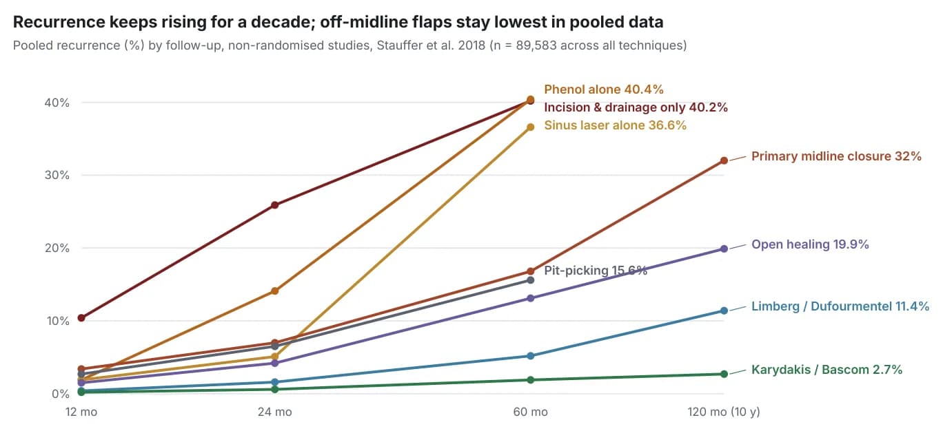
- Recurrence must be judged against follow-up length. Pooled recurrence across techniques rises from 2.0% at 1 year to 16.9% at 10 years[15]; short-term series understate it.
Abstract
Background. Pilonidal disease causes pain, discharge, lost school and work time and frequent recurrence. Treatment options range from observation to complex flap reconstruction, and recommendations differ between societies.
Objective. To synthesise current evidence on aetiology, diagnosis, treatment selection, recovery and recurrence prevention, and to identify which strategies suit which presentations.
Methods. Structured searches of PubMed/MEDLINE, the Cochrane Library, publisher databases, guideline repositories and trial registries through 11 October 2026, prioritising guidelines, systematic reviews, randomised trials and long-term cohorts published since 2016, plus landmark earlier studies. Quantitative claims were checked against the original abstracts or full texts.
Results. Five national or international guidelines and several consensus statements were reviewed[4–7,19]. Moderate-certainty evidence favours off-midline over midline closure for recurrence, infection and healing time[10]. Flap techniques do not differ consistently from one another[10,20,21]. Minimally invasive procedures (pit-picking, phenol, endoscopic and laser ablation) offer return to work within days but higher and time-dependent recurrence, supported mostly by cohort data[12,15,16,22]. One large RCT supports adjuvant laser epilation in young patients[17].
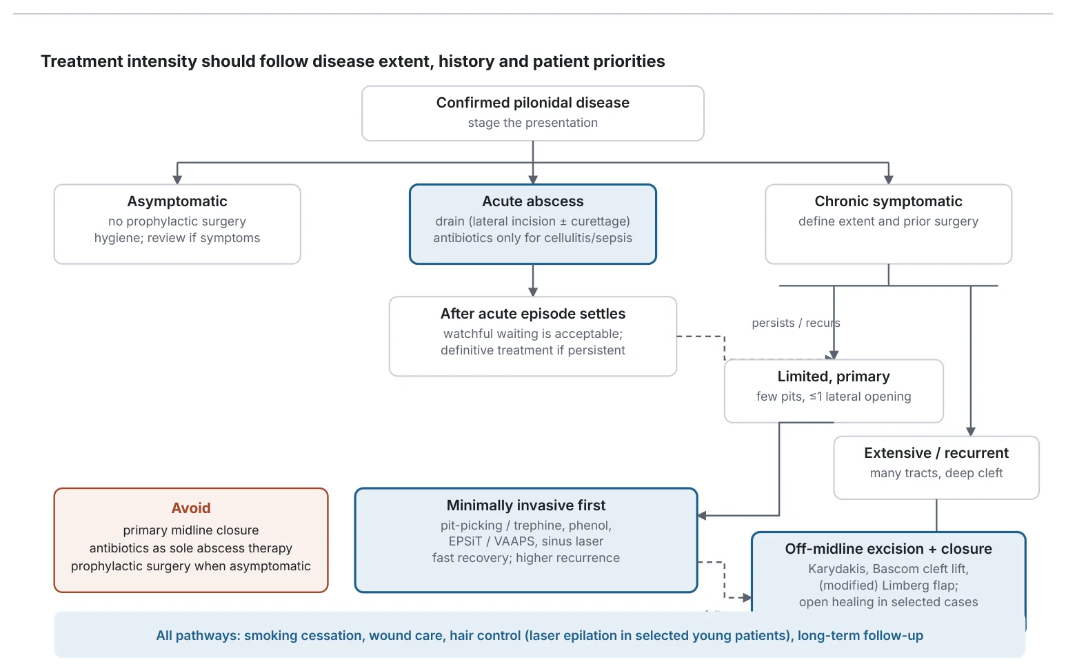
Conclusions. Treatment should be staged: no surgery for asymptomatic disease, drainage for abscess, minimally invasive treatment for limited primary disease when patients accept a higher recurrence risk, and off-midline reconstruction for extensive or recurrent disease. Primary midline closure should be avoided. Standardised definitions and long-term randomised comparisons between minimally invasive and flap techniques are the main evidence gaps.
Methods
Design. Structured narrative review with quantitative synthesis of published pooled estimates. We did not perform a de novo PRISMA 2020 systematic review, risk-of-bias assessment of every primary study, or new meta-analysis; GRADE ratings are quoted from the source reviews where available, and otherwise given as the authors' judgement and labelled as such.
Sources and dates. PubMed/MEDLINE listings, the Cochrane Library, publisher sites (Springer, Wiley, Oxford Academic, JAMA Network, LWW, Nature), Europe PMC, OpenAlex, guideline repositories (AWMF, NICE) and trial registries (ClinicalTrials.gov and mirrors). Search terms included pilonidal disease, pilonidal sinus, pilonidal abscess, recurrence, off-midline closure, cleft lift, Bascom, Karydakis, Limberg, pit-picking, trephine, EPSiT, VAAPS, SiLaC, laser hair removal, phenol and fibrin glue. Last search: 11 October 2026.
Inclusion. Clinical practice guidelines and consensus statements; systematic reviews and meta-analyses; RCTs; prospective and large retrospective cohorts, especially those with ≥2 years of follow-up; health-economic studies. Priority was given to 2016–2026 publications. Exclusion. Single case reports (except for malignancy), non-English abstracts without extractable data, and studies whose numbers could not be traced to the source.
Verification and limitations. Each quantitative claim was checked against the source abstract or full text; where only a secondary source was available this is stated in the text. Some PubMed and PMC pages were inaccessible during the search because of automated-access restrictions, so a small number of bibliographic details (issue numbers, PMIDs) rest on publisher or OpenAlex records. Embase, Scopus, Web of Science and WHO ICTRP were not searched directly. Three input drafts supplied for this report were cross-checked; errors found in them are listed in Appendix A.
Definitions and Terminology
| Term | Definition used in this review |
|---|---|
| Pilonidal disease (PD); pilonidal sinus disease | Inflammatory disease of the sacrococcygeal natal cleft characterised by midline pits, subcutaneous hair-containing cavities and sinus tracts. "Pilonidal cyst" is a lay term; there is usually no true epithelium-lined cyst. |
| Primary pit (primary opening) | Small midline opening at the base of the cleft, regarded as an enlarged follicle. |
| Secondary opening | Opening off the midline, usually cranial and lateral, where a tract reaches the skin. |
| Asymptomatic PD | Pits found incidentally without pain, swelling or discharge. |
| Acute pilonidal abscess | Painful, fluctuant collection of pus in the cavity, often with surrounding erythema. |
| Chronic pilonidal sinus | Persistent or intermittent discharge from one or more openings without an acute abscess; the most common form. |
| Complex PD | Multiple or branching tracts, extensive lateral extension, or a large cavity. No classification has been validated well enough to be recommended universally[16,23]. |
| Recurrent PD | New disease after a period of complete healing. Must be distinguished from persistent disease (never healed) and from wound failure (dehiscence, non-healing). Studies define these inconsistently. |
| Off-midline closure | Any excision whose final suture line lies away from the midline, usually flattening the cleft (Karydakis, Bascom cleft lift, Limberg/Dufourmentel and their modifications). |
| Minimally invasive (tissue-sparing) treatment | Procedures removing only pits and cavity contents through small openings: pit-picking, trephine, phenol, EPSiT/VAAPS, sinus laser ablation (SiLaT/SiLaC), fibrin glue. |
Epidemiology
The classic population incidence is 26 per 100,000 per year, from a Norwegian series of 322 patients; in that cohort men were affected 2.2 times as often as women, mean age at presentation was 21 years (men) and 19 years (women), 38% reported a family history and 44% had sedentary work[24]. In Germany, inpatient case rates rose by about one-third between 2005 and 2017 (men 43 to 56, women 14 to 18 per 100,000), with a stable male-to-female ratio of about 3:1[25]; the 2026 German guideline reports an adult male predominance of about 4:1 and a much more even sex distribution before puberty[6]. ASCRS estimates about 70,000 cases per year in the United States[7].
Prevalence is high in Turkish young-adult populations: 8.8% of 1,000 soldiers in one study[26], 6.1% in another[27], and 6.6% among about 19,000 students[28]. In a Turkish cohort of 10,324 patients, 24.8% were women, and the female share was much higher among teenagers than adults[29]. The sex ratio therefore depends on the age group and setting studied, which is why sources quote figures between about 2:1 and 4:1.
Anatomy
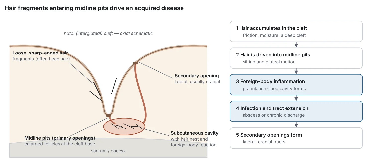
The disease occupies the intergluteal (natal) cleft over the sacrum and coccyx. Primary pits lie strictly in the midline at the base of the cleft; cavities extend into the subcutaneous fat and tracts usually run cranially and laterally to secondary openings (Figure 1). Tezel's "navicular area" concept describes the boat-shaped midline zone where the cleft is deepest and where disease and wound complications concentrate; it underpins the rationale for moving incisions out of the midline[30]. The midline wound is exposed to moisture, shear forces and hair, which explains why midline suture lines heal poorly[10,31].
Causes and Pathophysiology
Acquired-disease model. The historical congenital (neuroectodermal remnant) theory has been replaced by an acquired model. Karydakis attributed the disease to hair insertion and used the same mechanism to explain recurrence in a series of 6,545 patients[1]. Bascom proposed that disease starts in enlarged, obstructed hair follicles (pits), with hair accumulation a secondary event[2]. The two views are complementary rather than exclusive.
Hair penetration and foreign-body reaction. Hair found in sinus nests is frequently short, rootless and sharp-ended, consistent with cut fragments rather than in-growing local hair. Head (occipital) hair was the stiffest hair in 13 of 20 patients and was demonstrated in sinus nests in at least 5 of 13 where origin could be assigned[3]. Hair therefore usually penetrates the skin; the lay term "ingrown hair" is misleading. Inserted hair and keratin provoke a granulomatous foreign-body reaction and a granulation-lined cavity.
Mechanical stress and friction. Sitting and gluteal movement generate shear and suction forces in the cleft that are thought to drive hair into pits; prolonged sitting was a strong independent risk factor in a large case-control study[32], and sitting at work was associated with disease in an early military series[33].
Natal cleft depth. A deep cleft has long been regarded as a vulnerability factor and is the rationale for cleft-flattening operations. However, a 2025 case-control study (95 patients, 105 controls) found no influence of fold depth on disease development[34]. Depth may matter more for wound healing and recurrence after surgery than for disease onset; this remains unresolved.
Bacteria and secondary infection. The cavity becomes secondarily colonised, largely by skin and anaerobic flora, causing abscesses and chronic discharge. Microbiome data are limited to small series; infection is a consequence rather than the primary cause.
Chronic inflammation and sinus formation. In 1,962 patients, sinus number did not increase with disease duration, suggesting that tracts form early, possibly before symptoms[35]. Chronic inflammation then sustains discharge and recurrent abscesses until the cavity and pits are removed or the disease regresses with age.
Risk Factors
| Factor | Key data | Strength |
|---|---|---|
| Stiff / dense body hair | Adjusted OR 9.23 for stiff body hair (587 cases, 2,780 controls)[32]; high-grade gluteal hirsutism in soldiers[27] | Moderate (case-control) |
| Prolonged sitting | Adjusted OR 4.03 for sitting time[32]; drivers over-represented (58/88 vs 308/912)[26] | Moderate (observational) |
| Infrequent bathing | Adjusted OR 6.33 for number of baths[32] | Low–moderate |
| Family history | Present in 38%[24]; 12% of 578 patients had an affected relative, with earlier onset and about 50% long-term recurrence[36]; 18/88 vs 32/912 in soldiers[26] | Moderate |
| Obesity / body weight | BMI OR 1.3 per unit[32]; weight >90 kg 34/88 vs 32/912[26]; 37% overweight[24] | Low–moderate; stronger for wound complications |
| Smoking | Prior abscess 48% vs 26%, 1-year recurrence after pit-picking 36% vs 21%, wound complications after Karydakis 29% vs 10%[37]; SSI after midline closure OR 2.6[38] | Moderate for outcomes after surgery |
| Deep natal cleft | Conflicting: no effect on disease development in a 2025 case-control study[34] | Uncertain |
| Sweating, local folliculitis | Folliculitis elsewhere 22/88 vs 64/912 in soldiers[26] | Low |
| Male sex, age 15–30 | Male predominance 2.2–4:1 depending on setting[6,24,25] | Strong (descriptive) |
ORs from a single case-control study should not be multiplied together; the 219-fold risk reported for the combination of hairiness, >6 h daily sitting and ≤2 baths per week[32] illustrates interaction but is imprecise.
Clinical Presentation
Three forms are recognised: asymptomatic disease, acute abscess and chronic sinus disease, the last being the most frequent[6,7]. Median symptom duration before referral was two years in the Norwegian series[24]. Severity is judged clinically by the number of pits, the presence and position of lateral openings, cavity size, previous operations and the presence of abscess or cellulitis.
| Presentation | Symptoms | Examination | Immediate need |
|---|---|---|---|
| Asymptomatic | None | Midline pits only | Reassurance, hygiene; no surgery |
| Acute abscess | Rapid onset pain, swelling; sometimes fever | Tender fluctuant mass, erythema, usually off the midline | Drainage (± curettage); assess for sepsis |
| Chronic sinus | Intermittent discharge, staining, tenderness, odour | Pits plus one or more secondary openings, granulation | Elective definitive treatment if symptomatic |
| Recurrent / complex | Discharge or abscess after prior surgery; non-healing wound | Scars, multiple openings, deep cleft | Specialist assessment; consider imaging |
Diagnostic Approach
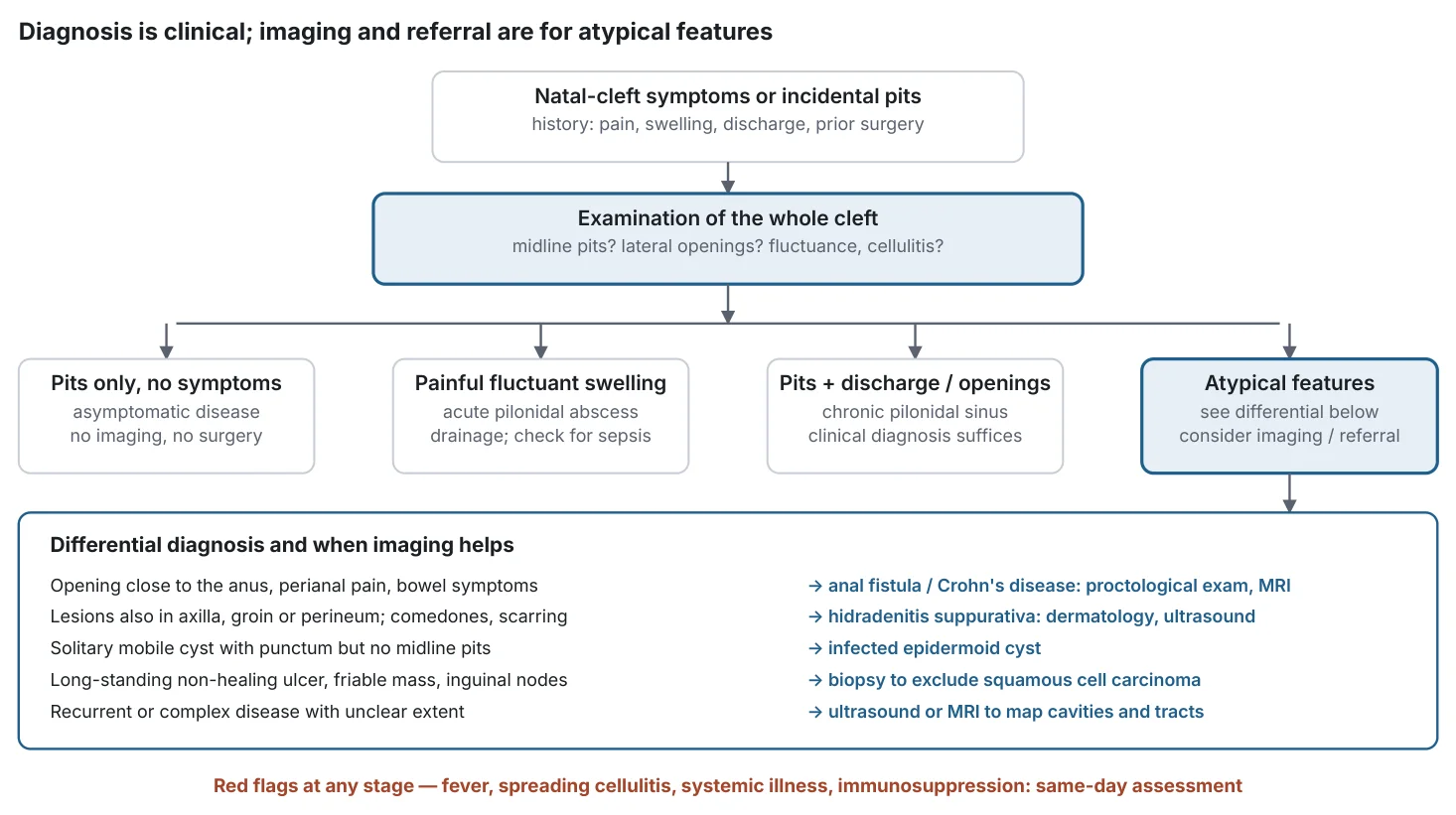
Diagnosis is clinical, based on history and a disease-specific examination (ASCRS 1C)[7]. Routine blood tests and imaging are unnecessary in typical cases. Ultrasound can localise an abscess or help separate PD from hidradenitis; MRI is reserved for complex or recurrent disease or diagnostic doubt, particularly when a fistula-in-ano is suspected[6]. MRI interobserver agreement for key features is moderate to substantial (kappa 0.47–0.71)[39]. Specialist referral is appropriate for recurrent or complex disease, openings near the anus, atypical lesions, or chronic non-healing wounds.
Differential Diagnosis
Anal fistula and Crohn's disease: openings close to the anus, perianal symptoms or bowel symptoms; proctological examination and MRI.
Hidradenitis suppurativa: multiple sites (axillae, groin, perineum), double comedones, bridged scars; the two diseases can coexist. Infected epidermoid cyst or furuncle: solitary lesion without midline pits. Malignancy: squamous cell carcinoma arising in long-standing disease is rare (calculated incidence about 0.17%), typically after a mean of about 20 years of disease, at a mean age of 54; a non-healing, friable or fungating ulcer or inguinal lymphadenopathy requires biopsy[40,41]. Others: sacrococcygeal teratoma or dermoid in children, tuberculosis and actinomycosis (rare).
Natural History Without Treatment
Asymptomatic sinuses do not heal spontaneously but many never become symptomatic; the German guideline therefore advises no prophylactic treatment[6]. Once symptomatic, disease commonly follows a relapsing course of abscesses and discharge. A classic military study suggested that disease regresses naturally and seldom presents after age 30[33]; this supports conservative management of minimal disease in older patients, although no reliable regression rate can be quoted. After drainage of a first abscess, 80.5% of 149 patients were asymptomatic at mean 58 months, of whom 17.5% later developed recurrence, while 19.5% required early definitive surgery[9]. Other series report about 60% healing after drainage alone[7] and 40.2% pooled recurrence at 5 years[15]. Untreated disease can therefore resolve, but the probability is uncertain and depends on extent.
When Observation Is Appropriate
Asymptomatic pits: observation is the recommended approach (ESCP, expert opinion; Dutch and German guidelines)[4–6]. After a drained abscess that has fully settled: watchful waiting is acceptable (German S3, grade 0)[6]; definitive treatment is offered if symptoms persist or recur[9]. Minimal, infrequent symptoms in a patient who prefers to avoid intervention: reasonable with counselling, hair and hygiene measures, and a low threshold for review. Active treatment is indicated for abscess, persistent discharge, recurrent episodes, impaired quality of life, or any suspicious, long-standing non-healing lesion.
Nonoperative Treatments
| Option | Main indication | What it does / does not do | Status |
|---|---|---|---|
| Observation, hygiene | Asymptomatic pits; settled abscess | Avoids morbidity; does not remove existing pits | Established |
| Laser epilation | Adjunct after treatment in hirsute young patients; recurrent disease | Reduces hair load; does not drain an abscess or remove a cavity | Optional (mixed guidance) |
| Shaving / depilatory cream | Short-term hair control | Cheap; retrospective association of post-operative razor use with more recurrence | Optional; not routine |
| Antibiotics | Cellulitis, systemic infection, immunosuppression | Not curative; do not replace drainage | Established (selective) |
| Incision and drainage ± curettage | Acute abscess | Relieves sepsis; recurrence common without definitive treatment | Established |
| Phenol (liquid or crystallised) | Limited primary disease, few pits | Office procedure; repeat applications sometimes needed | Established in some countries |
| Fibrin glue | Selected limited disease; adjunct | Very sparse evidence | Optional / investigational |
| Pit-picking / trephine | Limited primary disease | Removes pits and cavity contents through small openings | Established |
| EPSiT / VAAPS | Limited or selected recurrent disease | Endoscopic debridement and cautery of the cavity | Established in specialist centres |
| Sinus laser ablation (SiLaT/SiLaC) | Limited disease; selected recurrence | Radial fibre ablates tract lining | Emerging; mostly single-arm data |
| Open excision (secondary intention) | Selected extensive or infected disease | Low recurrence vs midline closure but months of wound care | Established, declining |
| Primary midline closure | — (not recommended) | Higher recurrence and infection than off-midline closure | Discouraged |
| Off-midline flaps (Karydakis, cleft lift, Limberg) | Extensive, complex or recurrent disease; deep cleft | Flattens cleft, lateral scar; lowest long-term recurrence | Established |
Hygiene, friction and moisture
Keeping the cleft clean and dry and avoiding prolonged uninterrupted sitting are low-risk, plausible measures supported by observational risk-factor data[32]. They do not eliminate an established cavity and have not been tested in randomised trials as stand-alone treatment.
Hair removal: laser, shaving and depilation
Laser epilation. The strongest single study is an unblinded single-centre RCT in 302 patients aged 11–21 years (median 17)[17]. Adding five laser sessions every 4–6 weeks to standard care reduced 1-year recurrence from 45/134 (33.6%) to 10/96 (10.4%), a risk difference of −23.2 percentage points (95% CI −33.2 to −13.1), without improving health-related quality of life. Follow-up was incomplete and unequal (63.6% vs 88.7%). A secondary analysis found significant heterogeneity of effect: benefit was concentrated in non-Hispanic white and privately insured patients (4.0% vs 33.3% recurrence), with no benefit in publicly insured patients (36.8% vs 29.7%)[18], suggesting adherence, access or skin/hair-type effects. A meta-analysis of three RCTs (about 448 patients) reported an OR of 0.32 (0.16–0.64) for recurrence[42], while an earlier RCT cited by the Dutch guideline reported more recurrence with laser (20% vs 4%)[5]. A systematic review of 14 studies (963 patients) found recurrence of 9.3% with laser, 23.4% with razor or depilatory cream, and 19.7% with no hair removal[43]; another found 0–28% recurrence after laser with 1–11 sessions[44].
Shaving. In a retrospective study, patients who shaved with a razor after surgery had higher long-term recurrence (30.1% vs 19.7%)[45]. This is a harm signal from non-randomised data, not proof. The German guideline considers routine shaving obsolete[6]; ASCRS nonetheless lists shaving or laser for non-abscess disease[7].
Depilatory creams have not been studied separately in adequately sized trials and can cause irritant dermatitis.
| Method | Best evidence | Effect on recurrence | Practical issues | Guideline position |
|---|---|---|---|---|
| Laser epilation (alexandrite, diode, Nd:YAG) | RCT n=302[17]; SR of 3 RCTs[42]; SR 14 studies[43] | Reduced at 1 year in young patients (10.4% vs 33.6%); heterogeneous; one RCT showed harm | Several sessions (5 in the RCT; 1–11 reported); cost about €600–1,500[5]; less effective on light or fine hair | ASCRS: shaving or laser (1C)[7]; ESCP: no recommendation[4]; German 2026: recommended under age 22 (B)[6]; Dutch: caution after a first operation, consider in recurrence[5]; SICCR: post-operative epilation in hirsute patients (1C)[19] |
| Razor shaving | Retrospective n=504[45] | Associated with higher recurrence after surgery (30.1% vs 19.7%) | Microtrauma; cut hair fragments | German: obsolete[6]; ESCP: post-op hair removal not necessary[4] |
| Depilatory cream | Pooled with razors in SR[43] | No evidence of benefit | Chemical irritation | Not specifically recommended |
| No hair removal | SR arm[43] | 19.7% recurrence (pooled) | — | Acceptable per ESCP[4] |
Antibiotics and pain management
Antibiotics are indicated for cellulitis, systemic signs or immunosuppression but are not curative and must not delay drainage[4]. ASCRS rates the benefit of prophylactic antibiotics as unclear (2B)[7]. A systematic review found no benefit of a single prophylactic dose (7 studies, 690 patients) and none for gentamicin-collagen sponges (4 studies, 402 patients)[46]; a meta-analysis of three sponge RCTs showed a non-significant 20% absolute SSI reduction (p=0.06)[47]. ESCP and SICCR find no indication for routine post-operative antibiotics[4,19]; the 2026 German guideline advises against routine prophylaxis for open and minimally invasive procedures (A) but permits a single dose for closed procedures[6]. Pain after minimally invasive procedures is usually managed with simple oral analgesia (EPSiT first-week VAS 1.35)[12]; open wounds and flaps need multimodal analgesia for the first days.
Phenol
Phenol (liquid or crystallised) is instilled into the cleaned cavity to destroy granulation tissue. A pooled review reported 87% (±10%) success at about 2 years, healing in 20 days, return to work in 2.3 days and 8.9% morbidity, with better results when there were 1–3 pits[13]. In an RCT of 100 adolescents, crystallised phenol and excision with primary closure had similar 24-month recurrence (8% vs 10%) with much shorter procedures (12.4 vs 42.3 minutes)[48]. A Dutch RCT comparing phenolisation with radical excision in primary disease found no significant difference in long-term recurrence and faster return to daily activities after phenol (reported 2.6% vs 5.6% recurrence; 5.2 vs 14.5 days)[49]. Pooled non-randomised recurrence, however, reached 40.4% at 5 years in a smaller number of patients[15]. ASCRS rates phenol 1B[7]; ESCP low-certainty[4]; the German guideline makes no recommendation because phenol is not licensed for this use in Germany[6].
Fibrin glue
A Cochrane review found only very low-certainty evidence: glue alone versus Bascom surgery (one trial, 39 participants) showed no clear difference in recurrence at about 4.6 years (RR 1.43, 0.27–7.61) but a 34.8-day faster return to normal activity[50]. ASCRS 2B and ESCP very-low-certainty statements allow its use in selected patients[4,7].
Acute Abscess Management
- Recognise and assess: fluctuance, cellulitis extent, fever, diabetes or immunosuppression.
- Drain: incision and drainage is recommended for any abscess (ASCRS 1B)[7]; ESCP advises a lateral (off-midline) incision[4]. Curettage of hair and granulation tissue at drainage improved healing (96% vs 78.7%) and reduced recurrence (11% vs 42%) in a 150-patient RCT[8]. ESCP also allows EPSiT in place of drainage (expert opinion)[4], and the German guideline allows single-stage definitive treatment of small abscesses[6].
- Antibiotics: add for spreading cellulitis, systemic infection or high-risk hosts; not as sole therapy[4,7].
- Definitive treatment timing: no evidence-based interval exists. Guidelines advise definitive treatment once inflammation has resolved[23]; the German guideline suggests about 10–14 days when surgery is planned[6], while one retrospective study found fewer complications when surgery was delayed to about 3 months rather than 1 month[51]. Watchful waiting after full resolution is acceptable[6,9].
- Systemic infection (sepsis, necrotising infection) requires urgent surgical debridement, intravenous antibiotics and inpatient care.
Minimally Invasive Procedures
Pit-picking and trephine techniques
Bascom's pit-picking excises the midline pits with small incisions and drains the cavity through a lateral incision; Gips' trephine technique uses skin punches. In 1,358 trephine patients (85.8% followed for a mean of 6.9 years), recurrence was 6.5% at 1 year, 13.2% at 5 years and 16.2% at 10 years; healing took 3.4 weeks[52]. A Danish cohort (158 patients, median 8 years) found 27% cumulative 10-year recurrence among healed patients and 68% overall success; smoking (HR 5.30) and three or more pits (HR 5.11) predicted failure[53]. A German series reported 74% recurrence-free at a median 30 months[54], whereas another German cohort found 62% 5-year recurrence after pit-picking versus 22% after Limberg flap[55]. Outcomes therefore vary widely between centres and case selection. Pooled non-randomised recurrence was 2.7%, 6.5% and 15.6% at 1, 2 and 5 years[15].
Endoscopic treatment (EPSiT and VAAPS)
EPSiT uses a fistuloscope to visualise the cavity, remove hair and granulation tissue and cauterise the lining[56]. A meta-analysis (9 studies, 497 patients) found a weighted failure rate of 6.3% (3.6–9.1), complications 1.1%, healing in 32.9 days, return to work in 2.9 days and first-week pain VAS 1.35[12]. In paediatric patients recurrence was 8% at ≥12 months and 15% at ≥36 months[57]. In the only RCT against a flap (VAAPS versus Bascom cleft lift, 145 patients), time off work was 1.6 versus 8.2 days[58], and 5-year follow-up (97% complete) reported similar recurrence[59]. VAAPS outperformed sinusectomy in a propensity-matched study (recurrence 7.5% vs 25%)[60]. A meta-analysis of controlled studies found fewer complications (RR 0.33) and less pain with endoscopic treatment but no significant difference in recurrence (RR 0.75, 0.30–1.90)[61]. NICE permits EPSiT with standard arrangements[62]; ASCRS rates endoscopic approaches 2B[7].
Sinus laser ablation (SiLaT/SiLaC)
A radial laser fibre (usually 1470 nm) is passed through the tract to shrink and obliterate it, typically after pit excision and curettage. This must not be confused with laser epilation. A systematic review of 10 studies (971 patients, median follow-up 12 months) reported 94.4% primary healing, 3.8% weighted recurrence and 10% minor complications[22]; a single-arm meta-analysis (13 studies, 1,214 patients) reported 84.4% healing and 7.6% recurrence[63]. Longer series show more recurrence: 14.9% in a 200-patient SiLaC cohort[64]; in recurrent disease pooled healing fell from 87.2% (≤12 months) to 74.5% (>12 months)[65]. In a matched cohort, further surgery was needed after 30.6% of SiLaC versus 19.4% of phenol procedures at a mean of 47 months[66]. A multicentre retrospective study (306 patients, median 68 months) found that adding laser to pit-picking did not change 5-year recurrence (12.7% vs 13.8%) but shortened return to work (3 vs 6 days) at higher cost[14]. One small randomised study of SiLaC with pit excision versus Limberg flap reported 4/50 versus 6/50 recurrences at 2 years[67]. An indirect meta-analysis found similar outcomes for SiLaC and EPSiT (recurrence 11% vs 9%)[68].
Comparative evidence for outpatient and local-anaesthesia procedures
The UK PITSTOP study (667 patients) found that major excisional procedures had lower recurrence than minor procedures (adjusted risk difference −10.1%, 95% CI −18.1 to −2.1) but more complications, 25.9 more days to normal activities and much longer healing; about a quarter of both groups were unhealed at 6 months[16]. In a Dutch national audit, patient-reported 1-year recurrence was 30.6% after minimally invasive techniques versus 6.7% after off-midline closure[69]. An Israeli cohort reported comparable long-term disease-free rates after minimal surgery and wide excision (81.5% vs 85%)[70]. These are non-randomised comparisons with likely selection bias.
| Method | Healing | Return to work / activity | Recurrence (follow-up) | Evidence base |
|---|---|---|---|---|
| Pit-picking / trephine | 3–4 wk[52] | ≈3–6 d[14] | 6.5% 1 y → 13.2% 5 y → 16.2% 10 y[52]; 27% 10 y[53]; 62% 5 y[55] | Large cohorts, long follow-up; wide inter-centre variation |
| EPSiT | ≈33 d[12] | ≈3 d[12] | Failure 6.3% (≈1–2 y)[12]; 15% ≥3 y in children[57] | Meta-analysis of cohorts |
| VAAPS | n.r. | 1.6 d vs 8.2 d for cleft lift[58] | Similar to cleft lift at 5 y[59] | One single-centre RCT |
| Sinus laser (SiLaT/SiLaC) | ≈20–30 d[64,68] | ≈3 d (with pit-picking)[14] | 3.8–7.6% at ≈12 mo[22,63]; 12.7% 5 y[14]; 14.9%[64] | Single-arm meta-analyses; one small randomised study |
| Phenol | ≈20 d[13] | ≈2 d[13] | 87% success ≈2 y[13]; 8% 2 y (RCT)[48]; 40.4% 5 y pooled[15] | Two RCTs vs excision; heterogeneous technique |
| Fibrin glue | n.r. | 35 d sooner than Bascom[50] | No clear difference at 4.6 y (n=39)[50] | One small trial; very low certainty |
n.r. = not reported. Values are means or medians from different populations and are not directly comparable.
Complete Review of Surgical Techniques
Excision with healing by secondary intention (open)
Wide excision left open avoids a midline suture line. Compared with primary closure, open healing reduced recurrence (RR 0.60, 0.42–0.87) at the cost of slower healing (Cochrane, 26 trials, 2,530 participants)[31]. Median healing in an RCT of open wounds was 84–93 days, with return to activities at 27–29 days[71]. Pooled non-randomised recurrence was 13.1% at 5 and 19.9% at 10 years[15]. Marsupialisation (suturing wound edges to the cavity base) reduces wound size; ESCP allows it in selected patients[4].
Primary midline closure
Midline closure heals fast when it succeeds but places the scar in the cleft. Against off-midline closure it has more recurrence, infection and dehiscence[10]; against Limberg flap, 11/100 versus 0/100 recurrences at 28 months in an RCT[72]. Long-term recurrence reached 17.4% at 5, 20.5% at 10 and 44% at 20 years in a German cohort[73]. ESCP (moderate evidence), German, Dutch and Italian guidance advise against it[4–6,19]; ASCRS 2019 still lists primary repair as an option while recommending that off-midline closure be considered[7].
Off-midline techniques
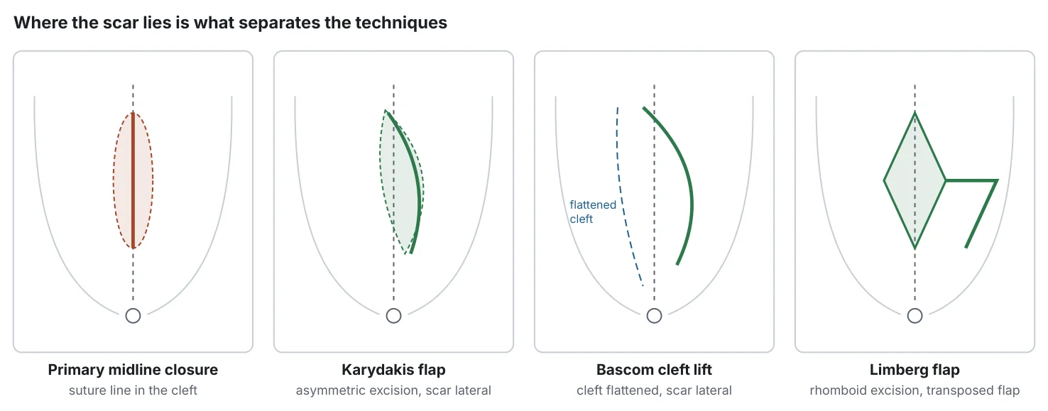
Karydakis flap: eccentric elliptical excision with mobilisation of a medial flap so that the suture line lies lateral to the midline and the cleft is shallower[1]. Bascom cleft lift: excision of the diseased skin with a lateral incision and advancement of a skin flap across the midline to flatten the cleft, with minimal removal of fat[74]. Limberg (rhomboid) flap: rhomboid excision of the disease and transposition of a fasciocutaneous flap from the buttock; the modified Limberg moves the lower apex off the midline. Dufourmentel and other rotation flaps follow similar principles.
| Feature | Karydakis | Bascom cleft lift | Limberg / modified Limberg |
|---|---|---|---|
| Principle | Eccentric excision, medial flap advancement | Lateral incision, skin flap advanced across cleft; little fat removed | Rhomboid excision, transposition flap |
| Tissue removed | Moderate | Least | Most |
| Recurrence, RCT/meta-analysis | Karydakis vs Limberg: 5.1% vs 4.5%, RR 1.14 (0.61–2.14), 9 RCTs, low certainty[10]; OR 1.07 (0.59–1.92)[20]. Cleft lift vs Limberg RCT (n=122): no difference in early recurrence[11] | Modified Limberg lower than classic Limberg (RR 3.7 for Limberg vs modified)[75] | |
| Recurrence, pooled non-RCT[15] | Karydakis + Bascom combined: 0.2% (1 y), 1.9% (5 y), 2.7% (10 y) | Limberg/Dufourmentel: 0.4% (1 y), 5.2% (5 y), 11.4% (10 y) | |
| Wound complications | More seroma than Limberg (OR 2.03)[20]; more dehiscence than modified Limberg (RR 3.36)[75] | Infection 2.6%, haematoma 1.1%, minor separation 10.9% (n=700)[76]; seroma 15% (n=74)[77] | Fewer seromas than Karydakis[20,75] |
| Return to work | No difference Karydakis vs Limberg (MD −0.23 d, low certainty)[10]; cleft lift less pain and better role function on day 10 than Limberg[11] | ||
| Recurrent / salvage disease | Used | Strong cohort record: all 31 refractory patients healed[74]; revision 5.3% in salvage cases[76] | Used, especially for large defects |
| Cosmesis | Lateral linear scar | Lateral scar; preserves contour | Larger, more visible scar (no validated comparative data) |
No high-quality RCT compares cleft lift directly with Karydakis. Network meta-analyses rank modified Limberg, off-midline closure and cleft lift highest for recurrence, with moderate or low certainty[75,78].
Recurrent, complex disease and large defects
Flap techniques are recommended for complex or recurrent disease (ASCRS 1B; Dutch, SICCR)[5,7,19]. Cleft lift is widely used as a salvage operation[74,76,79]. Large defects may need rotation or transposition flaps, or open healing with negative-pressure wound therapy (NPWT). Endoscopic or laser treatment can be offered to selected patients with limited recurrent disease[4,65].
Anaesthesia and perioperative protocols
Anaesthesia: minimally invasive procedures are usually done under local anaesthesia as day cases; flaps under spinal or general anaesthesia, often as day cases or with a short stay. Drains: no significant reduction in infection (8.3% vs 11.4%; OR 0.71, 0.48–1.03) or recurrence in a meta-analysis of 1,202 patients[80]; an 803-patient RCT found no difference[81]. Use is individualised (ASCRS)[7]. Antibiotic prophylaxis: see Section 14.3. NPWT: faster early wound shrinkage but no difference in median healing (84 vs 93 days) in open wounds[71]; no benefit on closed off-midline wounds (dehiscence 48% vs 36%)[82]. ESCP limits NPWT to selected patients after wound breakdown[4]; the ENVELOP trial is ongoing[83]. Smoking cessation before elective surgery is advisable given its association with wound complications and recurrence[37].
Primary Versus Recurrent Disease
Recurrence is time-dependent. In a German cohort followed up to 20 years, only about 60% of recurrences had occurred by year 5[84,85], and recurrence after a second operation appeared sooner. Predictors include previous recurrence, surgical-site infection and wound separation (PISI-Turkey, 1,662 patients, 12-month recurrence 6.26%)[86], family history[36], smoking and number of pits[53,54]. Primary limited disease suits minimally invasive treatment; after one or more failed excisions, off-midline reconstruction (particularly cleft lift) has the strongest record[74,76,79], while selected limited recurrences can be treated endoscopically or with laser[4,65].
Comparative Effectiveness
Comparisons follow four rules: recurrence is reported with its follow-up; primary and recurrent disease are kept apart where data allow; short-term wound failure is distinguished from long-term recurrence; and techniques are not ranked from indirect comparisons of heterogeneous, low-quality studies. Where those conditions cannot be met, the evidence is labelled insufficient.
| Conservative / nonoperative | Minimally invasive | Off-midline excision and closure | Open excision | |
|---|---|---|---|---|
| Typical candidate | Asymptomatic; settled abscess; minimal symptoms | Limited primary disease; selected recurrences | Extensive, complex or recurrent disease; deep cleft | Selected extensive or heavily infected disease |
| Anaesthesia / setting | None | Local, day case | Spinal or general; day case or short stay | Spinal or general |
| Return to work | — | ≈2–6 days[12–14] | ≈1–2 weeks[58] | ≈4 weeks to normal activity[71] |
| Time to healing | — | ≈3–5 weeks[12,52] | ≈2–3 weeks if uncomplicated[10,82] | ≈3 months[71] |
| Recurrence | Disease persists; abscess risk unknown | Higher; rises over years (≈13–16% at 5 years in pooled and large cohort data)[15,52] | Lowest in pooled data (Karydakis/Bascom 1.9% at 5 y non-RCT; 10.2% RCT subset)[15] | Lower than midline closure[31]; ≈13% at 5 y pooled[15] |
| Main drawback | Does not treat cavity | Recurrence; variable results across centres | Seroma, dehiscence; surgeon expertise | Months of dressings |
| Outcome | Off-midline | Conventional midline | Effect (95% CI) | Trials (n) | Certainty |
|---|---|---|---|---|---|
| Recurrence | 1.5% | 6.8% | RR 0.22 (0.11–0.45) | 13 (1,492) | Moderate |
| Surgical-site infection | 3.8% | 11.7% | RR 0.32 (0.22–0.49) | 13 (1,568) | Moderate |
| Wound dehiscence | 3.9% | 8.9% | RR 0.44 (0.27–0.71) | 11 (1,389) | Low |
| Time to healing | 5.23 days shorter off-midline | MD −5.23 (−7.55 to −2.92) | 3 (300) | Moderate | |
| Return to work | 3.72 days sooner off-midline | MD −3.72 (−6.11 to −1.33) | 6 (820) | Low | |
| Proportion healed | 100% | 88.5% | RR 1.13 (0.92–1.39) | 2 (207) | Very low |
| Off-midline versus tension-free midline closure | |||||
| Recurrence | 5.4% | 7.8% | RR 0.69 (0.30–1.61) | 6 (551) | Very low |
| Healed at 3 months | 94.7% | 76.4% | RR 1.24 (1.06–1.46) | 1 (115) | Low |
No quality-of-life data were available. An earlier Cochrane review reached the same conclusion (recurrence Peto OR 4.54, 2.30–8.96, for midline versus off-midline)[31].
| Technique / comparison | Recurrence | Follow-up | Evidence (quality) |
|---|---|---|---|
| Off-midline vs midline closure | 1.5% vs 6.8%[10] | Trial-dependent | 13 RCTs (GRADE moderate) |
| Karydakis vs Limberg | 5.1% vs 4.5%[10] | Trial-dependent | 9 RCTs (GRADE low) |
| Limberg vs primary midline closure | 0/100 vs 11/100[72] | Median 28 mo | 1 RCT (low)ᵃ |
| Open vs primary closure | RR 0.60 (0.42–0.87) favouring open[31] | Trial-dependent | 17 RCTs (moderate)ᵃ |
| Karydakis / Bascom (pooled) | RCT subset 1.5% 1 y, 2.4% 2 y, 10.2% 5 y; non-RCT 1.9% 5 y, 2.7% 10 y[15] | 1–10 y | Merged data (low)ᵃ |
| Primary midline closure (pooled) | RCT subset 2.1% 1 y, 21.9% 5 y; non-RCT 16.8% 5 y, 32.0% 10 y, 67.9% 20 y[15] | 1–20 y | Merged data (low)ᵃ |
| Midline closure vs open (cohort) | 17.4% vs 8.3% (5 y); 20.5% vs 11.2% (10 y); 44% vs 28% (20 y)[73] | Up to 20 y | Cohort, n=583 (low)ᵃ |
| Bascom cleft lift (cohorts) | 0/27 followed refractory cases[74]; 2/261 (≈0.8%)[79]; 3/74[77]; 6.3% vs 11.8% for excision[87] | Median 20, 20, 52 and 12 mo | Cohorts (very low–low)ᵃ |
| Pit-picking / trephine | 6.5% 1 y, 13.2% 5 y, 16.2% 10 y[52]; 27% 10 y[53]; 62% 5 y[55] | Up to 10 y | Cohorts (low)ᵃ |
| EPSiT | Failure 6.3%[12]; 8% ≥1 y, 15% ≥3 y in children[57] | Mostly ≤2 y | Cohort meta-analysis (very low)ᵃ |
| VAAPS vs cleft lift | Similar at 5 y[59] | 5 y, 97% complete | 1 RCT, n=145 (low)ᵃ |
| Sinus laser ablation | 3.8–7.6%[22,63]; 12.7% at 5 y[14] | Median 12 mo; 68 mo | Single-arm meta-analyses, retrospective cohort (very low)ᵃ |
| Phenol vs excision | 8% vs 10% (2 y)[48]; no significant difference (≈4 y)[49] | 2–4 y | 2 RCTs (low)ᵃ |
| Laser epilation adjunct vs none | 10.4% vs 33.6%[17] | 1 y | 1 RCT, attrition imbalance (low–moderate)ᵃ |
| Paediatric pooled | Off-midline 10.2%, endoscopic 11.4%, midline 17.5%, open 20.1%[88] | Variable | Meta-analysis of mainly observational studies (low)ᵃ |
ᵃ Authors' judgement using GRADE principles; not rated by the source. Definitions of recurrence differ (new disease after healing vs any failure), and many series do not separate persistence from recurrence.
| Procedure class | Return to work / normal activity | Complete wound healing | Main modifiers |
|---|---|---|---|
| Phenol | ≈2 d[13] | ≈20 d[13] | Repeat applications |
| Pit-picking / trephine (± laser) | ≈3–6 d[14] | ≈2–4 wk[14,52] | Number of pits, lateral cavity |
| EPSiT / VAAPS | 1.6–2.9 d[12,58] | ≈4–5 wk[12] | Cavity size; small wound left open |
| Sinus laser ablation | ≈3 d with pit-picking[14] | ≈3–4 wk[64,68] | Infection (≈10%) |
| Off-midline flap / cleft lift | ≈8 d (cleft lift RCT arm)[58]; 3.7 d sooner than midline closure[10] | Median 14–21 d[10,82] | Seroma, dehiscence; physical job |
| Primary midline closure | Slower than off-midline[10] | Median 20 d, rising to 62 d with infection[38] | Infection, dehiscence, smoking |
| Open excision | ≈27–29 d to normal activities[71]; sick leave ≈35 d after wide excision[89] | Median 84–93 d[71] | Wound size, dressing access |
Ranges are typical values from different study populations, not guarantees. A quarter of patients in a UK cohort were still unhealed at 6 months regardless of procedure class[16].
| Procedure | Reported complications | Typical management |
|---|---|---|
| Incision and drainage | Persistent or recurrent disease (≈40% at 5 y pooled)[15]; ≈20% need early definitive surgery[9] | Elective definitive treatment |
| EPSiT | Complications 1.1% (pooled)[12] | Local wound care |
| Sinus laser ablation | Minor complications ≈10%[22]; infection 9.5% in one series[64] | Drainage, antibiotics if cellulitis |
| Phenol | Morbidity 8.9% (abscess, cellulitis, skin irritation)[13] | Protect skin during application; wound care |
| Primary midline closure | SSI 11.7%, dehiscence 8.9%[10] | Open wound, dressings; later cleft lift if non-healing |
| Off-midline flaps | SSI 3.8%, dehiscence 3.9%[10]; seroma more frequent after Karydakis (OR 2.03 vs Limberg)[20]; cleft lift seroma 15%[77], minor separation 10.9%[76] | Aspiration of seroma, local wound care, rarely revision |
| Open excision | Prolonged healing, pain at dressing changes, chronic non-healing wound | Dressings; NPWT in selected cases; cleft lift for chronic wounds |
| Excision in children (US data) | Major complication or re-excision within 1 y: 8.7% of 1,932[90] | Favour tissue-sparing methods |
| Comparison | Direction of effect | Certainty | Source of rating |
|---|---|---|---|
| Off-midline vs conventional midline closure | Less recurrence, infection; faster healing | Moderate | Cochrane GRADE[10] |
| Karydakis vs Limberg | No clear difference | Low | Cochrane GRADE[10] |
| Open healing vs primary closure | Less recurrence, slower healing | Low–moderate | Authors[31] |
| Minimally invasive vs excisional surgery | Faster recovery, more recurrence | Low | Authors (non-randomised)[16,69] |
| VAAPS vs cleft lift | Faster return to work; similar 5-y recurrence | Low | Authors (single RCT)[58,59] |
| Phenol vs excision | Similar recurrence, faster recovery | Low | Authors (2 small RCTs)[48,49] |
| Laser epilation adjunct vs standard care | Less 1-y recurrence in young patients; inconsistent | Low–moderate | Authors[5,17,18] |
| Fibrin glue vs Bascom | Uncertain | Very low | Cochrane[50] |
| Drain vs no drain | No significant difference | Low | Authors[80,81] |
| NPWT vs standard dressing | No difference in healing | Low | Authors[71,82] |
| Antibiotic prophylaxis vs none | No consistent benefit | Low | Authors[46] |
Recovery and Return to Daily Activities
Recovery depends more on whether a wound is left open, closed in the midline or closed off the midline than on the procedure's name. Figure 5 and Table 10 give evidence-based anchors. For individual activities (sitting, driving, sport, swimming) no trial defines safe intervals; the guidance below is criteria-based common practice, and the operating surgeon's instructions take precedence.
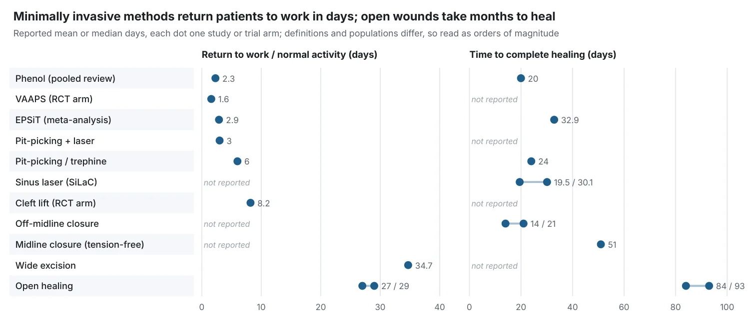
Pain: after minimally invasive procedures pain is usually mild and short-lived (EPSiT first-week VAS ≈1.4)[12]; flaps typically cause moderate pain for several days; open wounds hurt most at dressing changes. Increasing pain after initial improvement suggests infection or seroma. Walking and light activity: encouraged from the first day after all procedures. Sitting: comfortable sitting usually returns within days after minimally invasive treatment. After flap surgery, surgeons commonly limit prolonged direct sitting until the wound is secure, because shear can contribute to seroma or dehiscence; this is not trial-tested. Driving: when the patient can sit comfortably and perform an emergency stop without pain and without sedating analgesics. Work: desk work after about 2–6 days (minimally invasive) or 1–2 weeks (uncomplicated flap); physical work later; open wounds often require about a month off (Table 10). Exercise and weight training: resume gradually once the wound is healed or the surgeon confirms stability; repeated hip flexion and heavy lifting stress closed wounds. Swimming and bathing: showering is usually permitted early; immersion (baths, pools, sea) is generally deferred until the wound is fully closed to limit infection risk.
Postoperative Wound Care
Dressings and hygiene: keep the wound clean and dry, rinse in the shower, pat dry; remove hair from the wound edges (clipping or laser rather than razor; Section 22). Open wounds: regular dressing changes; hair must not enter the granulating wound. NPWT may help selected large wounds but does not shorten median healing[71]. Closed wounds and flaps: monitor for seroma, infection and separation; drains are optional[80]. Lifestyle: smoking cessation[37,53]; adequate nutrition and weight management. Signs requiring review: increasing pain, redness, swelling, purulent or foul discharge, fever, wound opening, or bleeding that does not stop with pressure. Follow-up: early review for wound problems, then patient-initiated review; because recurrence continues for 10–20 years, patients should know to return early with symptoms[73,84].
Delayed healing: assess for retained hair, residual tracts or a persistent midline wound in a deep cleft. Options include curettage, epilation of wound edges, and cleft lift, which has a strong record for chronic non-healing wounds[74,87].
Recurrence Prevention
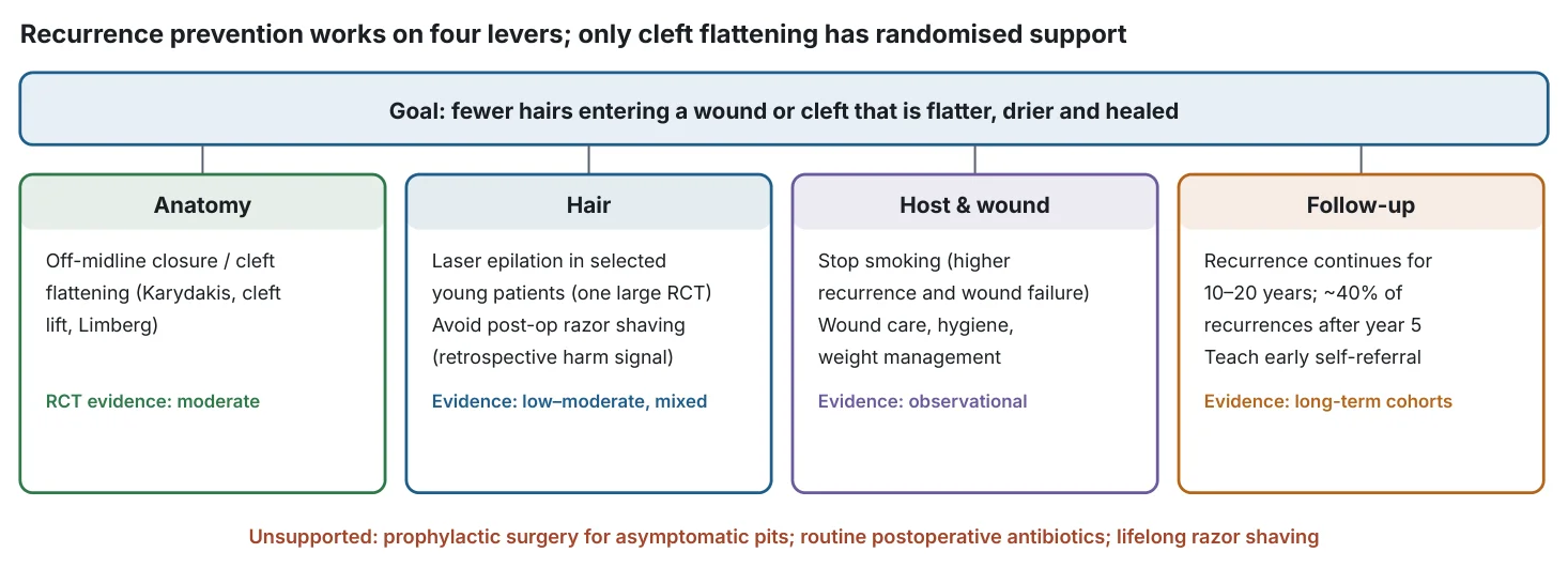
The most robust preventive measure is technical: if excision and closure are performed, the scar should lie off the midline in a flattened cleft[10]. Hair control is the second lever: laser epilation reduced 1-year recurrence in young patients[17], but effects were inconsistent across subgroups and trials[5,18], and the optimal timing (before treatment, after wound healing, or both) has not been compared directly; in the main RCT, laser was given as a series of sessions as an adjunct to standard care[17]. Razor shaving after surgery is not advised[6,45]. Smoking cessation, hygiene and avoidance of prolonged sitting are reasonable but supported only by observational data[32,37]. Unsupported practices include prophylactic excision of asymptomatic pits, routine postoperative antibiotics and indefinite razor shaving[4,46].
Complications and Red Flags
Acute abscess progression and cellulitis: untreated abscesses can spread; sepsis is uncommon but requires urgent care. Postoperative infection, dehiscence, seroma and haematoma: Table 11. Chronic non-healing wounds and persistent drainage: about 25% of patients were unhealed at 6 months in a UK cohort[16]. Recurrence: Table 9 and Figure 6. Chronic pain: poorly quantified in the literature; persistent pain should prompt assessment for residual disease. Flap-specific: flap-tip necrosis, seroma, wound separation and contour asymmetry. Malignant transformation: rare (calculated incidence 0.17%), mean latency ≈20 years, male-to-female ratio 7.75:1, mean age 54[40]; recurrence after treatment of carcinoma reached 39% in a pooled series of 59 patients[41]. Biopsy any long-standing, atypical or non-healing lesion.
Red flags requiring urgent assessment
Fever or rigors; rapidly spreading redness; severe pain out of proportion; crepitus or skin discoloration; systemic illness; diabetes or immunosuppression with infection; heavy bleeding; a long-standing ulcer that bleeds easily or is enlarging; enlarged groin lymph nodes.
Special Populations
Children and adolescents: no formal APSA guideline exists; an APSA systematic review favours minimal procedures first and advises against midline closure[91]. ESCP manages adolescents as adults[4]; the 2026 German guideline prefers minimally invasive procedures in children[6]. Excision carried an 8.7% one-year rate of major complication or re-excision in US paediatric data[90]. Early recurrences occurred irrespective of the treatment approach in a German multicentre paediatric analysis[92]. The main laser-epilation RCT was conducted in this age group[17]. Women: lower incidence but a substantial share of teenage cases[29]; the German guideline notes higher recurrence in women[6]. Obesity: associated with disease and wound complications[26,32]; flap closure and smoking cessation deserve emphasis, though no RCT compares techniques specifically in obese patients. Military personnel, drivers and sedentary workers: high prevalence[26,27]; rapid return to duty may favour minimally invasive options when disease is limited. Family history: earlier onset and high long-term recurrence; lower threshold for cleft-flattening surgery and hair control[36]. Hidradenitis or Crohn's disease: require disease-specific assessment and often multidisciplinary care.
Cost and Accessibility
Costs are driven by operating time, inpatient stay, dressing visits, time off work and repeat treatment. In Sweden, minimal excision with primary suture cost €2,231 versus €6,222 per patient for the comparison approach, with 1.0 versus 34.7 days of sick leave and no significant difference in 5-year recurrence (32% vs 23%)[89]. Adding laser ablation to pit-picking raised cost (US$1,212 vs US$888) without improving 5-year recurrence[14]. Laser epilation adds about €600–1,500 per course[5] and is often not reimbursed; the RCT benefit was absent in publicly insured patients[18], raising equity concerns. Endoscopic and laser equipment, phenol licensing (not licensed in Germany)[6] and surgeon expertise in flap techniques limit availability. No formal cost-effectiveness analysis comparing minimally invasive and flap strategies over a 10-year horizon was identified.
Clinical Guidelines
Five guideline-level documents were identified: ASCRS 2019[7] (which replaced the 2013 practice parameters[93]), the ESCP 2024 guideline[4], the German S3 guideline (2016, 2020 update and 2026 revision)[6,94,95], the Italian SICCR guidance (2015 guideline and 2021 consensus)[19,96], and the Dutch national guideline 2024[5]. There is no SAGES pilonidal guideline[23]. NICE interventional guidance covers EPSiT[62], and an international Delphi consensus endorsed by ALSGBI supports EPSiT[97]; Turkish experts published Delphi recommendations in 2025[98].
| Topic | ASCRS 2019[7] | ESCP 2024[4] | German S3 2026[6] | Dutch 2024[5] | SICCR 2021[19] |
|---|---|---|---|---|---|
| Asymptomatic | Not addressed | No treatment (EO) | No prophylactic treatment | Do not operate | — |
| Abscess | I&D (1B) | Lateral I&D; curettage can be considered (low); EPSiT possible (EO) | Drain; small abscess may be treated in one stage; watchful waiting after healing (0) | Drainage | I&D standard; excision or MI alternatives (1B) |
| Antibiotics | Individualise (2B) | Not curative; post-op not indicated (very low) | No routine prophylaxis for open/MI (A); single dose for closed | — | No effect on SSI (1B) |
| Minimally invasive | Phenol 1B; glue 2B; EPSiT/VAAPS 2B | Could be used for limited disease (very low); pit-picking, phenol, glue, endoscopic, laser listed | Pit-picking (B); endoscopic/laser no advantage over other MI methods | First option for simple disease, with counselling on recurrence | Treatment of choice for limited disease (1B) |
| Midline closure | Primary repair an option; consider off-midline (1B) | Avoid (moderate) | Should not be done (B) | Avoid | Off-midline preferred (1B) |
| Flaps | For complex/recurrent disease (1B) | Off-midline closure preferred when excising (moderate) | Karydakis and modified Limberg (A); cleft lift (0) | For complex or recurrent disease; no preferred flap | No single best off-midline technique |
| Open healing | Option (1B) | Could be considered (moderate) | — | Only with caution | Complex cases only (1B) |
| Laser epilation | Shaving or laser (1C) | No recommendation; post-op hair removal unnecessary (low) | Recommended under age 22 (B) | Caution after first operation; consider in recurrence (very low) | Post-op in hirsute patients (1C) |
| NPWT | Narrative only | After wound breakdown (very low) | Not routine | Inconclusive | After wide open excision |
EO = expert opinion; MI = minimally invasive. Grading systems differ (ASCRS GRADE 1–2/A–C; ESCP evidence level; AWMF A/B/0; SICCR ACCP-style), so cross-guideline grade comparisons are approximate. The ASCRS hair-removal recommendation is printed as 1C although its text describes a weak recommendation.
Why guidelines conflict. (1) Laser epilation: evidence consists of few, heterogeneous trials, mostly in young patients, and panels weigh cost and access differently. (2) Minimally invasive first-line: supported mostly by cohorts with short follow-up; panels differ in how they trade recurrence against morbidity. (3) Midline closure: ASCRS predates the 2024 Cochrane review and European guidance. (4) Phenol: the German position reflects licensing, not efficacy. (5) Abscess timing and NPWT: no adequate trials.
Emerging Treatments
| Technology | Current evidence | Readiness | Realistic 5-year / 10-year outlook |
|---|---|---|---|
| Endoscopic treatment (EPSiT, paediatric PEPSiT) | Cohort meta-analysis[12]; one RCT (VAAPS)[58]; Delphi support[97] | In clinical use | Comparative RCTs against flaps likely within 5 years |
| 1470 nm sinus laser (SiLaC) | Single-arm meta-analyses[22,63]; RCTs ongoing: LA POPA (pit-picking ± laser, 5-year follow-up, completion ≈2032)[99]; laser vs crystallised phenol[100] | In clinical use; evidence immature | Definitive answer on added value expected around 2030–2032 |
| 1940 nm and other new laser wavelengths | Small observational studies registered | Experimental | Speculative |
| Negative-pressure wound therapy | Neutral RCTs[71,82]; ENVELOP RCT (n=200, 2024–2027)[83] | Selective use | Clarified role for large open wounds within 5 years |
| Platelet-rich plasma | ESCP low-certainty statement[4]; phenol ± PRP RCT registered, no results[101] | Investigational | Uncertain |
| Advanced dressings and biomaterials (e.g. autologous fat, matrices) | No RCT evidence identified | Experimental | Speculative |
| Risk prediction and AI-assisted selection | No validated AI/ML model; regression-based predictors only (prior recurrence, SSI, wound separation)[86] | Research stage | Validated risk scores plausible in 5–10 years; AI tools speculative |
| Standardised classification and outcome sets | Wysocki classification inter-rater kappa 0.52[16]; no validated system[23] | Development | Core outcome set would make trials comparable |
| New hair-reduction approaches | No pilonidal-specific trials beyond conventional laser | Speculative | Speculative |
"Speculative" denotes technologies without registered pilonidal trials as of the search date; they should not be regarded as standard care.
Evidence Gaps
- No adequately powered RCT compares minimally invasive treatment (pit-picking, EPSiT, laser) with off-midline flaps using ≥5-year follow-up and patient-reported outcomes.
- No high-quality RCT compares Bascom cleft lift with Karydakis.
- Recurrence, persistence and wound failure are defined inconsistently; there is no validated disease classification or core outcome set.
- Most minimally invasive series have ≤12 months of follow-up despite recurrence continuing for 10–20 years[15,84].
- Laser epilation: optimal timing, number of sessions, device, skin and hair type, cost-effectiveness and generalisability beyond adolescents are unresolved[17,18].
- No evidence-based interval exists between abscess drainage and definitive surgery.
- Quality of life, chronic pain, cosmesis and long-term costs are rarely measured; no Cochrane comparison reported quality of life[10].
- Likely publication and selection bias favour single-centre enthusiast series, especially for new technologies.
Scenario-Based Treatment Decision Framework
| Scenario | Reasonable options | Evidence and limitations | Recovery / recurrence considerations | Findings that change the recommendation |
|---|---|---|---|---|
| Asymptomatic pits found incidentally | Observation, hygiene; no prophylactic surgery | Guideline consensus (expert opinion)[4,6] | No recovery cost; disease may never progress | Onset of pain, swelling or discharge |
| First acute abscess | Lateral drainage ± curettage; antibiotics only if cellulitis/sepsis | ASCRS 1B[7]; curettage RCT[8] | Most settle; ≈1 in 5 needs early definitive surgery[9] | Sepsis, immunosuppression, extensive cellulitis → urgent admission |
| Persistent discharge, no abscess | Elective definitive treatment matched to extent | See below | Discharge rarely resolves spontaneously | Opening near anus → exclude fistula; non-healing ulcer → biopsy |
| Small primary sinus, few openings | Pit-picking/trephine, phenol, EPSiT/VAAPS, laser; flap if patient prioritises lowest recurrence | Cohorts and small RCTs (low)[12,48,52] | Work in ≈2–6 d; recurrence ≈13–16% at 5–10 y in good series[52] | Smoking or ≥3 pits predict failure[53] |
| Extensive or multiple tracts | Off-midline flap (Karydakis, cleft lift, modified Limberg); open healing in selected cases | Moderate-certainty RCT evidence for off-midline closure[10] | Off work ≈1–2 weeks; lowest long-term recurrence | Large infected cavity → staged approach or open healing |
| First recurrence | Cleft lift or other off-midline flap; selected limited recurrence: endoscopic or laser | Cohorts[65,74,76]; ESCP (EO)[4] | Revision more likely after salvage than primary cleft lift (adjusted HR 2.97)[76] | Midline scar in deep cleft → favours cleft lift |
| Repeated recurrence after multiple operations | Referral to specialist centre; cleft lift or rotation flap; MRI to map disease | Cohort data only[74,79] | Longer recovery; realistic expectations | Atypical features → reconsider diagnosis (hidradenitis, Crohn's, malignancy) |
| Shortest possible recovery wanted | Phenol, EPSiT/VAAPS, pit-picking ± laser (if disease limited) | Return to work 1.6–6 d[12–14,58] | Accept higher, time-dependent recurrence | Extensive disease makes MI less likely to succeed |
| Cannot take extended time off | Minimally invasive for limited disease; off-midline flap rather than open healing for extensive disease | Open wounds need ≈1 month off[71] | Flap ≈1–2 weeks; avoid open wounds when possible | Physically demanding job → longer absence after flaps |
| Deep cleft or heavy local hair | Cleft-flattening flap; consider laser epilation adjunct | Rationale strong, direct evidence limited[17,34] | Hair control needs several sessions | Light or fine hair reduces laser effect |
| Obesity or wound-healing risk (smoking, diabetes) | Optimise first (smoking cessation, glycaemic control, weight); favour off-midline closure or MI over midline closure | Observational[32,37,38] | Higher infection and dehiscence risk | Uncontrolled diabetes or active smoking → defer elective surgery if safe |
This matrix supports shared decision-making; it does not prescribe treatment for an individual without clinical examination.
Conclusions
- Pilonidal disease is an acquired, hair-driven inflammatory disease; "ingrown hair" is a misnomer for penetrating hair fragments.
- Asymptomatic disease should be observed; abscesses should be drained, ideally with curettage, and antibiotics alone are insufficient.
- When excision and closure are chosen, off-midline closure is preferred over midline closure (moderate-certainty evidence).
- Karydakis, Bascom cleft lift and (modified) Limberg flaps perform similarly in available comparisons; surgeon experience and anatomy should guide the choice.
- Minimally invasive techniques allow return to work within days and suit limited primary disease, but recurrence is higher and rises over years.
- Laser epilation can reduce early recurrence in young patients but is not universally effective, is costly, and is not a substitute for treating the sinus.
- Recurrence figures are meaningful only with their follow-up duration; patients should be counselled that recurrence can occur 10–20 years later.
- The decisive research need is long-term randomised comparison of minimally invasive and off-midline strategies using standardised outcomes.
Appendix A. Reconciliation of the Three Source Drafts
Three drafts were reviewed: (A) the research specification and outline prepared for this report; (B) a Turkish-language summary; and (C) an English-language draft report. Each factual claim was checked against primary sources. Draft A contained no factual claims beyond its structure. Draft B was largely accurate (its Cochrane, JAMA Surgery and Annals of Surgery Open figures were confirmed) but omitted long-term recurrence data, minimally invasive outcome numbers, the German, Dutch and Italian guidelines, malignancy, cost and paediatric evidence, which this report adds. Draft C contained the errors below, all corrected here.
| Claim in draft C | Verdict | Correction |
|---|---|---|
| Ref. 1: "Steele SR, Perry WB, Anand S, et al. Practice Parameters… Dis Colon Rectum 2019;62:146-157" | Incorrect | That citation is Johnson EK et al., ASCRS clinical practice guidelines[7]; the Steele practice parameters date from 2013[93] |
| Ref. 2: "Biter LU et al. Laser depilation… Colorectal Dis 2022;24:540-547" | Not found; likely fabricated | Removed; laser evidence cited from verified sources[17,42,43] |
| Ref. 4: "Milone M et al. ESCP guidelines… Colorectal Dis 2023;25:1004-1025" | Incorrect | ESCP guideline is Ojo D et al., Br J Surg 2024[4]; Milone is first author of the Italian SICCR consensus[19] |
| A SAGES guideline exists | Incorrect | No SAGES pilonidal guideline exists[23] |
| Table 10: ASCRS "strongly advise against" midline closure; ESCP "recommend laser" | Incorrect | ASCRS lists primary repair as an option (1B); ESCP makes no laser recommendation and finds post-operative hair removal unnecessary[4,7] |
| Laser hair reduction "the evidence-based standard"; superiority over shaving established | Overstated | One positive RCT in young patients, heterogeneous effect, one RCT showing harm, conflicting guidelines[5,17,18] |
| Shaving "paradoxically increases recurrence" | Overstated | Retrospective association only (30.1% vs 19.7%)[45] |
| Male-to-female ratio 3–4:1 | Partly correct | 2.2:1 to ≈4:1 depending on setting[6,24,25] |
| Definitive surgery after abscess "delayed 4–8 weeks" / "wait 6 weeks" | Unsupported | No evidence-based interval; watchful waiting acceptable[6,23] |
| Table 5: dehiscence 15–30% vs 2–8%; recurrence 15–40% vs <5% | Unsourced, inaccurate | Cochrane: dehiscence 8.9% vs 3.9%; recurrence 6.8% vs 1.5%[10] |
| Table 6: midline closure 15–20% at 1 year; GRADE "High" for open excision and cleft lift | Incorrect | Pooled midline recurrence 2.1–3.4% at 1 year, 16.8–21.9% at 5 years[15]; cleft lift evidence is mainly cohort data (low); no source rates these as high certainty |
| Table 4: Limberg recurrence 3–6% and "poor" cosmesis; cleft lift 1–3% | Unsourced | Karydakis vs Limberg no difference (low certainty)[10]; no validated cosmetic comparison |
| Surgical drains "critical to prevent seroma" | Unsupported | No significant benefit of drains in meta-analysis and RCT[80,81] |
| Antibiotics not indicated for an isolated abscess | Broadly consistent | Antibiotics are adjuncts for cellulitis or systemic signs; ASCRS individualises (2B)[7] |
| Table 12: "biodegradable antibiotic/fibrin plugs in late-stage trials"; "gene targeting for hirsutism" | Unsupported | No such registered pilonidal trials identified; relabelled speculative (Table 14) |
| Children benefit to "avoid psychological trauma" | Unsupported | Rationale is lower morbidity of tissue-sparing methods[90,91] |
| Incidence 26/100,000 | Correct | Confirmed[24] |
| Stauffer 2018, Bascom 2002, Meinero 2014 references | Correct | Confirmed[15,56,74] |
Acknowledgement of limitations
This review synthesises published pooled estimates rather than re-analysing individual trials. Embase, Scopus, Web of Science and WHO ICTRP were not searched directly. A few bibliographic details rely on publisher or OpenAlex records because some PubMed pages could not be accessed automatically; readers preparing a formal publication should re-confirm PMIDs. Ongoing trial details were taken from registry records and may change.
- Karydakis GE. Easy and successful treatment of pilonidal sinus after explanation of its causative process. Aust N Z J Surg. 1992;62(5):385-9. doi:10.1111/j.1445-2197.1992.tb07208.x. PMID: 1575660.
- Bascom J. Pilonidal disease: origin from follicles of hairs and results of follicle removal as treatment. Surgery. 1980;87(5):567-72. PMID: 7368107.
- Doll D, Bosche F, Hauser A, Moersdorf P, Sinicina I, Grunwald J, et al. The presence of occipital hair in the pilonidal sinus cavity—a triple approach to proof. Int J Colorectal Dis. 2018;33(5):567-76. doi:10.1007/s00384-018-2988-8. PMID: 29488088.
- Ojo D, Gallo G, Kleijnen J, Haas S, Danys D, Dardanov D, et al. European Society of Coloproctology guidelines for the management of pilonidal disease. Br J Surg. 2024;111(10):znae237. doi:10.1093/bjs/znae237. PMID: 39397672.
- Huurman EA, de Raaff CAL, Sloots PCEJ, Lapid O, van der Zee HH, Bötger WF, et al. Dutch national guideline on the management of intergluteal pilonidal sinus disease. Br J Surg. 2024;111(12):znae281. doi:10.1093/bjs/znae281. PMID: 39612582.
- Deutsche Gesellschaft für Koloproktologie (Ommer A, coordinator). S3-Leitlinie Sinus pilonidalis. AWMF-Register Nr. 081-009, revised edition 2026 [Internet]. Berlin: AWMF; 2026 [cited 2026 Oct 11]. Available from: https://register.awmf.org/assets/guidelines/081-009k_S3_Sinus_pilonidalis_2026-04_1.pdf
- Johnson EK, Vogel JD, Cowan ML, Feingold DL, Steele SR; Clinical Practice Guidelines Committee of the American Society of Colon and Rectal Surgeons. The American Society of Colon and Rectal Surgeons' clinical practice guidelines for the management of pilonidal disease. Dis Colon Rectum. 2019;62(2):146-57. doi:10.1097/DCR.0000000000001237. PMID: 30640830.
- Vahedian J, Nabavizadeh F, Nakhaee N, Vahedian M, Sadeghpour A. Comparison between drainage and curettage in the treatment of acute pilonidal abscess. Saudi Med J. 2005;26(4):553-5. PMID: 15900358.
- de Kort J, Pronk AA, Vriens MR, Smakman N, Furnee EJB. Long-term follow-up of sacrococcygeal pilonidal sinus disease after previous abscess drainage: a retrospective cohort study. J Gastrointest Surg. 2025;29(2):101892. doi:10.1016/j.gassur.2024.101892. PMID: 39571928.
- Cai Z, Zhao Z, Ma Q, Shen C, Jiang Z, Liu C, et al. Midline and off-midline wound closure methods after surgical treatment for pilonidal sinus. Cochrane Database Syst Rev. 2024;1(1):CD015213. doi:10.1002/14651858.CD015213.pub2.
- Guner A, Boz A, Ozkan OF, Ileli O, Kece C, Reis E. Limberg flap versus Bascom cleft lift techniques for sacrococcygeal pilonidal sinus: prospective, randomized trial. World J Surg. 2013;37(9):2074-80. doi:10.1007/s00268-013-2111-9. PMID: 23732258.
- Emile SH, Elfeki H, Shalaby M, Sakr A, Giaccaglia V, Sileri P, et al. Endoscopic pilonidal sinus treatment: a systematic review and meta-analysis. Surg Endosc. 2018;32(9):3754-62. doi:10.1007/s00464-018-6157-5. PMID: 29603009.
- Kayaalp C, Aydin C. Review of phenol treatment in sacrococcygeal pilonidal disease. Tech Coloproctol. 2009;13(3):189-93. doi:10.1007/s10151-009-0519-x. PMID: 19655223.
- Arslan NC, Bilgin IA, Gulcu B, et al. Does laser treatment affect outcome in pilonidal sinus disease? Long-term multicenter retrospective analysis. Tech Coloproctol. 2026;30(1):103. doi:10.1007/s10151-026-03314-8. PMID: 42128975.
- Stauffer VK, Luedi MM, Kauf P, Schmid M, Diekmann M, Wieferich K, et al. Common surgical procedures in pilonidal sinus disease: a meta-analysis, merged data analysis, and comprehensive study on recurrence. Sci Rep. 2018;8(1):3058. doi:10.1038/s41598-018-20143-4. PMID: 29449548.
- Brown SR, Hind D, Strong EB, Bradburn MJ, Din FVN, Lee EC, et al. Treatment options for patients with pilonidal sinus disease: PITSTOP, a mixed-methods evaluation. Health Technol Assess. 2024;28(33):1-113. doi:10.3310/KFDQ2017. PMID: 39045854.
- Minneci PC, Gil LA, Cooper JN, Asti L, Nishimura L, Lutz CM, et al. Laser epilation as an adjunct to standard care in reducing pilonidal disease recurrence in adolescents and young adults: a randomized clinical trial. JAMA Surg. 2024;159(1):19-27. doi:10.1001/jamasurg.2023.5526. PMID: 37938854. ClinicalTrials.gov NCT03276065.
- Bergus KC, Lutz C, Cooper J, Asti L, Gil L, Criss C, et al. Heterogeneity of treatment effects of laser epilation on pilonidal disease recurrence: a randomized clinical trial. Ann Surg Open. 2024;5(3):e488. doi:10.1097/AS9.0000000000000488.
- Milone M, Basso L, Manigrasso M, Pietroletti R, Bondurri A, La Torre M, et al. Consensus statement of the Italian Society of Colorectal Surgery (SICCR): management and treatment of pilonidal disease. Tech Coloproctol. 2021;25(12):1269-80. doi:10.1007/s10151-021-02487-8. PMID: 34176001.
- Prassas D, Rolfs TM, Schumacher FJ, Krieg A. Karydakis flap reconstruction versus Limberg flap transposition for pilonidal sinus disease: a meta-analysis of randomized controlled trials. Langenbecks Arch Surg. 2018;403(5):547-54. doi:10.1007/s00423-018-1697-7.
- Gavriilidis P, Bota E. Limberg flap versus Karydakis flap for treating pilonidal sinus disease: a systematic review and meta-analysis. Can J Surg. 2019;62(2):131-8. doi:10.1503/cjs.003018. PMID: 30697992.
- Romic I, Augustin G, Bogdanic B, Bruketa T, Moric T. Laser treatment of pilonidal disease: a systematic review. Lasers Med Sci. 2022;37(2):723-32. doi:10.1007/s10103-021-03379-x. PMID: 34291332.
- Arslan NÇ. Pilonidal disease: gaps in the guidelines and future perspectives. Turk J Surg. 2025;41(3):216-8. doi:10.47717/turkjsurg.2025.2025-5-19.
- Søndenaa K, Andersen E, Nesvik I, Søreide JA. Patient characteristics and symptoms in chronic pilonidal sinus disease. Int J Colorectal Dis. 1995;10(1):39-42. doi:10.1007/BF00337585. PMID: 7745322.
- Oetzmann von Sochaczewski C, Gödeke J. Pilonidal sinus disease on the rise: a one-third incidence increase in inpatients in 13 years with substantial regional variation in Germany. Int J Colorectal Dis. 2021;36(10):2135-45. PMID: 33993341.
- Akinci OF, et al. Incidence and aetiological factors in pilonidal sinus among Turkish soldiers. Eur J Surg. 1999;165(4):339-42. doi:10.1080/110241599750006875. PMID: 10365835.
- Sekmen Ü, Kara VM, Altıntoprak F, Şenol Z. Pilonidal sinus in the army: its incidence and risk factors. Ulusal Cerrahi Derg (Turk J Surg). 2010;26(2):95-8. doi:10.5097/1300-0705.UCD.354-10.0.
- Duman K, Gırgın M, Harlak A. Prevalence of sacrococcygeal pilonidal disease in Turkey. Asian J Surg. 2017;40(6):434-7. doi:10.1016/j.asjsur.2016.04.001. PMID: 27188235.
- Ergenç M, Uprak TK. Gender-specific prevalence of sacrococcygeal pilonidal sinus disease in Turkey: a retrospective analysis of a large cohort. Eur Surg. 2023;55(1):43-7. doi:10.1007/s10353-022-00777-5.
- Tezel E. A new classification according to navicular area concept for sacrococcygeal pilonidal disease. Colorectal Dis. 2007;9(6):575-6. doi:10.1111/j.1463-1318.2007.01236.x. PMID: 17573759.
- Al-Khamis A, McCallum I, King PM, Bruce J. Healing by primary versus secondary intention after surgical treatment for pilonidal sinus. Cochrane Database Syst Rev. 2010;(1):CD006213. doi:10.1002/14651858.CD006213.pub3.
- Harlak A, Mentes O, Kilic S, Coskun K, Duman K, Yilmaz F. Sacrococcygeal pilonidal disease: analysis of previously proposed risk factors. Clinics (Sao Paulo). 2010;65(2):125-31. doi:10.1590/S1807-59322010000200002.
- Clothier PR, Haywood IR. The natural history of the post anal (pilonidal) sinus. Ann R Coll Surg Engl. 1984;66:201-3.
- Maak M, Mörsdorf P, Bari L, Braun-Münker M, Scharonow M, Orth M, et al. Intergluteal fold depth has no influence on pilonidal sinus disease development. Turk J Surg. 2025;41(2):130-4. doi:10.47717/turkjsurg.2025.6665. PMID: 40151010.
- Doll D, Friederichs J, Dettmann H, Boulesteix AL, Duesel W, Petersen S. Time and rate of sinus formation in pilonidal sinus disease. Int J Colorectal Dis. 2008;23(4):359-64. doi:10.1007/s00384-007-0389-5. PMID: 18043929.
- Doll D, Matevossian E, Wietelmann K, Evers T, Kriner M, Petersen S. Family history of pilonidal sinus predisposes to earlier onset of disease and a 50% long-term recurrence rate. Dis Colon Rectum. 2009;52(9):1610-5. doi:10.1007/DCR.0b013e3181a87607. PMID: 19690490.
- Iesalnieks I, Deimel S, Zülke C, Schlitt HJ. Smoking increases the risk of pre- and postoperative complications in patients with pilonidal disease. J Dtsch Dermatol Ges. 2013;11(10):1001-5. doi:10.1111/ddg.12140. PMID: 23945165.
- Popeskou S, Christoforidis D, Ruffieux C, Demartines N. Wound infection after excision and primary midline closure for pilonidal disease: risk factor analysis to improve patient selection. World J Surg. 2011;35(1):206-11. doi:10.1007/s00268-010-0812-x. PMID: 20931197.
- Abdelatty MA, Elmansy N, Saleh MM, Salem A, Ahmed S, Gadalla AA, et al. Magnetic resonance imaging of pilonidal sinus disease: interobserver agreement and practical MRI reporting tips. Eur Radiol. 2024;34(1):115-25. doi:10.1007/s00330-023-10018-2.
- Safadi MF, Dettmer M, Berger M, Degiannis K, Wilhelm D, Doll D. Demographic overview of pilonidal sinus carcinoma: updated insights into the incidence. Int J Colorectal Dis. 2023;38(1):56. doi:10.1007/s00384-023-04344-6. PMID: 36849571.
- de Bree E, Zoetmulder FAN, Christodoulakis M, Aleman BMP, Tsiftsis DD. Treatment of malignancy arising in pilonidal disease. Ann Surg Oncol. 2001;8(1):60-4. doi:10.1007/s10434-001-0011-y.
- Muscat N, Gupta A, Arifuzaman M, Soxibova F. Preventing pilonidal sinus recurrence with laser hair epilation: a systematic review and meta-analysis of randomized controlled trials. Cureus. 2024;16(6):e62807. doi:10.7759/cureus.62807.
- Pronk AA, Eppink L, Smakman N, Furnee EJB. The effect of hair removal after surgery for sacrococcygeal pilonidal sinus disease: a systematic review of the literature. Tech Coloproctol. 2018;22(1):7-14. doi:10.1007/s10151-017-1722-9. PMID: 29185064.
- Halleran DR, Onwuka AJ, Lawrence AE, Fischer BC, Deans KJ, Minneci PC. Laser hair depilation in the treatment of pilonidal disease: a systematic review. Surg Infect (Larchmt). 2018;19(6):566-72. doi:10.1089/sur.2018.099. PMID: 30095368.
- Petersen S, Wietelmann K, Evers T, Hüser N, Matevossian E, Doll D. Long-term effects of postoperative razor epilation in pilonidal sinus disease. Dis Colon Rectum. 2009;52(1):131-4. doi:10.1007/DCR.0b013e3181972505.
- Mavros MN, Mitsikostas PK, Alexiou VG, Peppas G, Falagas ME. Antimicrobials as an adjunct to pilonidal disease surgery: a systematic review of the literature. Eur J Clin Microbiol Infect Dis. 2013;32(7):851-8. doi:10.1007/s10096-013-1830-z.
- Nguyen AL, Pronk AA, Furnée EJB, Pronk A, Davids PHP, Smakman N. Local administration of gentamicin collagen sponge in surgical excision of sacrococcygeal pilonidal sinus disease: a systematic review and meta-analysis of the literature. Tech Coloproctol. 2016;20(2):91-100. doi:10.1007/s10151-015-1381-7. PMID: 26546004.
- Şengül S, Güler Y, Çalış H, Kubat M, Karabulut Z. Crystallized phenol treatment vs excision and primary closure in pilonidal sinus disease: a randomized clinical trial in adolescent patients. J Pediatr Surg. 2022;57(3):513-7. doi:10.1016/j.jpedsurg.2021.03.004. PMID: 33814182.
- Pronk AA, Vissink MJ, Smakman N, Furnee EJB. Long-term outcome of radical excision versus phenolization of the sinus tract in primary sacrococcygeal pilonidal sinus disease: a randomized controlled trial. Dis Colon Rectum. 2022;65(12):1514-21. doi:10.1097/DCR.0000000000002475. PMID: 36102853.
- Lund J, Tou S, Doleman B, Williams JP. Fibrin glue for pilonidal sinus disease. Cochrane Database Syst Rev. 2017;1(1):CD011923. doi:10.1002/14651858.CD011923.pub2.
- Güler Y, Şengül S, Çalış H, Karabulut Z, Özen Ö. When should definitive surgery be performed after sacrococcygeal pilonidal sinus abscess drainage? Cerrahpaşa Med J. 2023;47(3):274-8. doi:10.5152/cjm.2023.22111.
- Gips M, Melki Y, Salem L, Weil R, Sulkes J. Minimal surgery for pilonidal disease using trephines: description of a new technique and long-term outcomes in 1,358 patients. Dis Colon Rectum. 2008;51(11):1656-62. doi:10.1007/s10350-008-9329-x. PMID: 18516645.
- Serup CM, Svarre KJ, Kanstrup CTB, Kleif J, Bertelsen CA. Long-term outcome after Bascom's pit-pick procedure for pilonidal sinus disease: a cohort study. Colorectal Dis. 2023;25(3):413-9. doi:10.1111/codi.16383.
- Iesalnieks I, Deimel S, Schlitt HJ. „Pit-picking"-Operation bei Patienten mit Sinus pilonidalis [Pit-picking surgery for patients with pilonidal disease]. Chirurg. 2015;86(5):482-5. doi:10.1007/s00104-014-2776-0. PMID: 24969346.
- Doll D, Petersen S, Andreae OA, Matner H, Albrecht H, Brügger LE, et al. Pit picking vs. Limberg flap vs. primary open method to treat pilonidal sinus disease – a cohort of 327 consecutive patients. Innov Surg Sci. 2022;7(1):23-9. doi:10.1515/iss-2021-0041. PMID: 35974777.
- Meinero P, Mori L, Gasloli G. Endoscopic pilonidal sinus treatment (E.P.Si.T.). Tech Coloproctol. 2014;18(4):389-92. doi:10.1007/s10151-013-1016-9.
- Erculiani M, Mottadelli G, Carlini C, Barbetta V, Dusio MP, Pini Prato A. Long-term results of EPSiT in children and adolescents: still the right way to go. Pediatr Surg Int. 2022;38(9):1257-61. doi:10.1007/s00383-022-05162-7. PMID: 35779104.
- Milone M, Fernandez LMS, Musella M, Milone F. Safety and efficacy of minimally invasive video-assisted ablation of pilonidal sinus: a randomized clinical trial. JAMA Surg. 2016;151(6):547-53. doi:10.1001/jamasurg.2015.5233.
- Milone M, Velotti N, Manigrasso M, Vertaldi S, Di Lauro K, Simone G, et al. Long-term results of a randomized clinical trial comparing endoscopic versus conventional treatment of pilonidal sinus. Int J Surg. 2020;74:81-5. doi:10.1016/j.ijsu.2019.12.033. PMID: 31926328.
- Milone M, Velotti N, Manigrasso M, Milone F, Sosa Fernandez LM, De Palma GD. Video-assisted ablation of pilonidal sinus (VAAPS) versus sinusectomy for treatment of chronic pilonidal sinus disease: a comparative study. Updates Surg. 2019;71(1):179-83. doi:10.1007/s13304-018-00611-2. PMID: 30542957.
- Chen S, Dai G, Liu P, et al. Comparative analysis on the effect of the endoscopic versus conventional treatment for pilonidal sinus: a meta-analysis of controlled clinical trials. Medicine (Baltimore). 2022;101(45):e31767. doi:10.1097/MD.0000000000031767. PMID: 36397424.
- National Institute for Health and Care Excellence. Endoscopic ablation for a pilonidal sinus. Interventional procedures guidance IPG646 [Internet]. London: NICE; 2019 Apr 17 (reissued as HealthTech guidance HTG507, January 2026) [cited 2026 Oct 11]. Available from: https://www.nice.org.uk/guidance/ipg646
- Xie C, Zeng R, Yu X. Efficacy of laser treatment in pilonidal disease: a single-arm meta-analysis. Photobiomodul Photomed Laser Surg. 2024;42(5):375-82. doi:10.1089/photob.2024.0028. PMID: 38776547.
- Dessily M, Dziubeck M, Chahidi E, Simonelli V. The SiLaC procedure for pilonidal sinus disease: long-term outcomes of a single institution prospective study. Tech Coloproctol. 2019;23(12):1133-40. doi:10.1007/s10151-019-02119-2. PMID: 31773347.
- Qin J, Xu X, Li Z, Jin L, Wang Z, Wu J. Efficacy and safety of laser ablation for recurrent pilonidal sinus: a systematic review and meta-analysis. Int J Colorectal Dis. 2025;40(1):47. doi:10.1007/s00384-025-04832-x. PMID: 39969579.
- de Kort J, van Kempen LV, Schouten R, Vriens MR, Smakman N, Furnée EJB. Minimal invasive surgical treatment in primary sacrococcygeal pilonidal sinus disease: phenolization of the sinus tract versus sinus laser assisted closure; long-term follow-up of a comparative cohort study. World J Surg. 2025;49(9):2354-60. doi:10.1002/wjs.70028.
- Chitrambalam TG, George NM, Reddy L, Christopher PJ, Selvamuthukumaran S. Sinus laser closure (SiLaC) with pit excision versus Limberg flap in pilonidal disease: a prospective, randomized study. Surg Open Dig Adv. 2024;13:100126. doi:10.1016/j.soda.2023.100126.
- Li Z, Jin L, Ouali M, et al. A systematic review and meta-analysis of sinus laser associated closure (SiLaC) versus endoscopic pilonidal sinus treatment (EPSiT) in the management of pilonidal sinus disease. Langenbecks Arch Surg. 2026;411(1). doi:10.1007/s00423-026-04026-1. PMID: 41964736.
- Huurman EA, de Raaff CAL, van den Berg R, Baart SJ, Wijnhoven BPL, Schouten R, et al.; PITS collaborative study group. A nationwide snapshot study on outcomes one year after surgery for chronic pilonidal sinus disease. Colorectal Dis. 2025;27(1):e17217. doi:10.1111/codi.17217. PMID: 39618338.
- Gips M, Bendahan J, Ayalon S, Efrati Y, Simha M, Estlein D. Minimal pilonidal surgery vs. common wide excision operations: better well-being and comparable recurrence rates. Isr Med Assoc J. 2022;24(2):89-95. PMID: 35187897.
- Biter LU, Beck GMN, Mannaerts GHH, Stok MM, van der Ham AC, Grotenhuis BA. The use of negative-pressure wound therapy in pilonidal sinus disease: a randomized controlled trial comparing negative-pressure wound therapy versus standard open wound care after surgical excision. Dis Colon Rectum. 2014;57(12):1406-11. doi:10.1097/DCR.0000000000000240. PMID: 25380007.
- Akca T, Colak T, Ustunsoy B, Kanik A, Aydin S. Randomized clinical trial comparing primary closure with the Limberg flap in the treatment of primary sacrococcygeal pilonidal disease. Br J Surg. 2005;92(9):1081-4. doi:10.1002/bjs.5074.
- Doll D, Matevossian E, Luedi MM, Schneider R, van Zypen D, Novotny A. Does full wound rupture following median pilonidal closure alter long-term recurrence rate? Med Princ Pract. 2015;24(6):571-7. doi:10.1159/000437361. PMID: 26334688.
- Bascom J, Bascom T. Failed pilonidal surgery: new paradigm and new operation leading to cures. Arch Surg. 2002;137(10):1146-50. PMID: 12361421.
- Bi S, Sun K, Chen S, Gu J. Surgical procedures in the pilonidal sinus disease: a systematic review and network meta-analysis. Sci Rep. 2020;10(1):13720. doi:10.1038/s41598-020-70641-7. PMID: 32792519.
- Immerman SC. The Bascom cleft lift as a solution for all presentations of pilonidal disease. Cureus. 2021;13(2):e13053. doi:10.7759/cureus.13053.
- Ortega PM, Baixauli J, Arredondo J, Bellver M, Sánchez-Justicia C, Ocaña S, et al. Is the cleft lift procedure for non-acute sacrococcygeal pilonidal disease a definitive treatment? Long-term outcomes in 74 patients. Surg Today. 2014;44(12):2318-23. doi:10.1007/s00595-014-0923-3. PMID: 24845740.
- Parra Baños PA. Recurrencia tras el tratamiento quirúrgico del seno pilonidal: revisión sistemática y metaanálisis en red [doctoral thesis]. Murcia: Universidad de Murcia; 2025. Available from: http://hdl.handle.net/10201/150265
- Shrager B. The data mounts: 261 cleft lifts for complex pilonidal disease and excisional failures. Cureus. 2023;15(12):e50174. doi:10.7759/cureus.50174.
- Milone M, Di Minno MND, Musella M, Maietta P, Ambrosino P, Pisapia A, et al. The role of drainage after excision and primary closure of pilonidal sinus: a meta-analysis. Tech Coloproctol. 2013;17(6):625-30. doi:10.1007/s10151-013-1024-9. PMID: 23754346.
- Milone M, Musella M, Salvatore G, Leongito M, Milone F. Effectiveness of a drain in surgical treatment of sacrococcygeal pilonidal disease: results of a randomized and controlled clinical trial on 803 consecutive patients. Int J Colorectal Dis. 2011;26(12):1601-7. doi:10.1007/s00384-011-1242-4.
- Ensor N, Martin S, Chang A, Sharpin C, Tandon S, Madden A, et al. Negative pressure dressing versus conventional passive dressing in pilonidal surgery: a randomized controlled trial. J Surg Res. 2024;303:313-21. doi:10.1016/j.jss.2024.09.016. PMID: 39393119.
- ClinicalTrials.gov. ENVELOP: excision with negative-pressure wound therapy versus open healing for pilonidal sinus disease. Identifier NCT06647290 [Internet]. Bethesda (MD): National Library of Medicine; 2024 [cited 2026 Oct 11].
- Doll D, Krueger CM, Schrank S, Dettmann H, Petersen S, Duesel W. Timeline of recurrence after primary and secondary pilonidal sinus surgery. Dis Colon Rectum. 2007;50(11):1928-34. doi:10.1007/s10350-007-9031-4. PMID: 17874268.
- Kueper J, Evers T, Wietelmann K, Doll D, Roffeis J, Schwabe P, et al. Sinus pilonidalis in patients of German military hospitals: a review. GMS Interdiscip Plast Reconstr Surg DGPW. 2015;4:Doc02. doi:10.3205/iprs000061. PMID: 26504731.
- Yalçınkaya A, Yalçınkaya AS, Şahin C, Balcı B, Özeller E, Öztürk E, et al. Investigating recurrence in pilonidal sinus disease: results of a nationwide, multicenter study in Turkey (PISI TURKEY). Int J Colorectal Dis. 2025;40(1):194. doi:10.1007/s00384-025-04921-x. PMID: 40931185.
- Huurman EA, de Kort JF, de Raaff CAL, Staarink M, Willemsen SP, Smeenk RM, et al. Postoperative outcomes of Bascom cleft lift versus excision with secondary wound healing for pilonidal sinus disease: a multicenter retrospective analysis. Dis Colon Rectum. 2024;67(11):1458-64. doi:10.1097/DCR.0000000000003402. PMID: 39115207.
- Saleem MM, Usmani SUR, Hashmi MR, Abbas H, Abbas S, Wasif MH, et al. Surgical management of pilonidal sinus disease in pediatric population: an updated systematic review and meta-analysis. J Pediatr Surg. 2026;61(7):163145. PMID: 41999968.
- Khodakaram K, Stark J, Höglund I, Andersson RE. Minimal excision and primary suture is a cost-efficient definitive treatment for pilonidal disease with low morbidity: a population-based interventional and a cross-sectional cohort study. World J Surg. 2017;41(5):1295-302. doi:10.1007/s00268-016-3828-z. PMID: 27905018.
- López JJ, Cooper JN, Halleran DR, Deans KJ, Minneci PC. High rate of major morbidity after surgical excision for pilonidal disease. Surg Infect (Larchmt). 2018;19(6):603-7. doi:10.1089/sur.2018.020. PMID: 29870307.
- Grabowski J, Oyetunji TA, Goldin AB, Baird R, Gosain A, Lal DR, et al. The management of pilonidal disease: a systematic review. J Pediatr Surg. 2019;54(11):2210-21. doi:10.1016/j.jpedsurg.2019.02.055. PMID: 30948198.
- Maasewerd SKM, Stefanescu MC, König TT, Engels MN, Rohleder S, Schwind M, et al. Paediatric pilonidal sinus disease: early recurrences irrespective of the treatment approaches in a retrospective multi-centric analysis. World J Surg. 2023;47(9):2296-303. doi:10.1007/s00268-023-07045-x. PMID: 37204438.
- Steele SR, Perry WB, Mills S, Buie WD; Standards Practice Task Force of the American Society of Colon and Rectal Surgeons. Practice parameters for the management of pilonidal disease. Dis Colon Rectum. 2013;56(9):1021-7. doi:10.1097/DCR.0b013e31829d2616. PMID: 23929010.
- Iesalnieks I, Ommer A, Petersen S, Doll D, Herold A. German national guideline on the management of pilonidal disease. Langenbecks Arch Surg. 2016;401(5):599-609. doi:10.1007/s00423-016-1463-7. PMID: 27311698.
- Iesalnieks I, Ommer A, Herold A, Doll D. German National Guideline on the management of pilonidal disease: update 2020. Langenbecks Arch Surg. 2021;406(8):2569-80. doi:10.1007/s00423-020-02060-1. PMID: 33950407.
- Segre D, Pozzo M, Perinotti R, Roche B; Italian Society of Colorectal Surgery. The treatment of pilonidal disease: guidelines of the Italian Society of Colorectal Surgery (SICCR). Tech Coloproctol. 2015;19(10):607-13. doi:10.1007/s10151-015-1369-3. PMID: 26377583.
- Sekhon Inderjit Singh HK, Meinero P, Campanile FC, Quddus A, Rajaganeshan R, Warusavitarne J, et al. The application of EPSiT in pilonidal sinus disease: an international Delphi consensus study endorsed by ALSGBI. Tech Coloproctol. 2025;29(1):155. doi:10.1007/s10151-025-03191-7. PMID: 40736584.
- Arslan NÇ, Tatar C, Erol T, Smeenk R, Immerman S, Wysocki P, et al. Controversies in the management of pilonidal disease: expert recommendations from a modified Delphi survey and review of the literature. Turk J Colorectal Dis. 2025;35(3):79-92. doi:10.4274/tjcd.galenos.2025.2025-2-2.
- ClinicalTrials.gov. LA POPA: pit-picking with or without sinus laser-assisted closure (1470 nm) for pilonidal sinus disease. Identifier NCT06140199 [Internet]. Bethesda (MD): National Library of Medicine; 2024 [cited 2026 Oct 11].
- ClinicalTrials.gov. 1470 nm laser versus crystallized phenol for pilonidal sinus disease. Identifier NCT07701772 [Internet]. Bethesda (MD): National Library of Medicine; 2026 [cited 2026 Oct 11].
- ClinicalTrials.gov. Crystallized phenol with or without platelet-rich plasma for pilonidal sinus disease. Identifier NCT06324656 [Internet]. Bethesda (MD): National Library of Medicine; 2024 [cited 2026 Oct 11].